How to avoid autopsy and long ‘down-time’
(ischemia) better than ~85% of the time!
By Mike Darwin
It’s easy to concentrate on the biggest and most obvious reason that cryonics hasn’t attracted wider acceptance, principally the fact that it doesn’t work “yet” and it will be a long time before we know if does. But there’s a clue to another capital reason for its slow adoption which is to be found in the failure of cryonics to attract much enthusiasm or activism within its own ranks. Why is this?
I believe a central reason for this failure is that cryonics, even as it is currently configured and accepted by those who embrace it, performs dismally. Everyone seriously involved with cryonics is painfully aware, either consciously or subconsciously, that cryonics is at least a two tier lottery. Sure, everyone knows that we’re taking a “chance” on being recovered in the future by being cryopreserved in the first place. But to even get to that round of the lottery, you have to get cryopreserved, and it would seem material whether or not you are cryopreserved well.
For some, perhaps cryonics is a ritual exercise. As long as there are remains, a freezer, someone to take the money and hang picture on the wall, then you have a chance; and all chances are created equal. Their position seems to be same as that of the millions of lottery ticket holders before the winning number is announced: we all have the same chance at the prize. If that’s your attitude, you can stop reading this right now, there’s nothing more here to interest you – not even in terms of idle entertainment value, because this discussion, from here on out is deadly serious, and brass tacks practical.
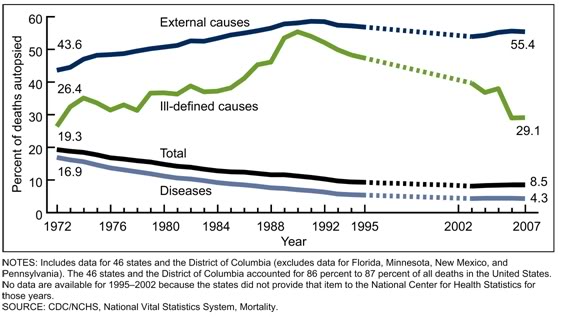 Figure 1: The autopsy rate has declined by half in the United State between 1972 and 2007, although it has shown a slight increase since these data were collected. Source: http://www.cdc.gov/nchs/data/databriefs/db67.pdf
As Figure 1 shows, the autopsy rate, which can serve as the ultimate, population wide indicator of a very bad cryopreservation, constituted 8.5% of all deaths in 2007. That percentage has risen slightly since then and is now at ~ 9%. The situation isn’t quite as grim as it might first appear if you break down the reasons for autopsy and note that 55.4% of autopsies were conducted as a result of deaths due to “external causes,” which means suicide, accident or homicide. If you think you are in a “lower risk” category for these, you may be right, in which your case your risk may be fractionally smaller. And of course, not all of these autopsies were state mandated: some were requested by the next of kin, or even the decedents themselves. Still, 9% seems a reasonable, overall unavoidable loss number currently confronting cryonicists given the culture we inhabit.
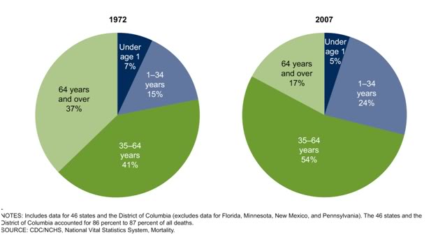 Figure 2: Since the first man was cryopreserved in 1967, the demographics of autopsy have shifted increasingly from the aged to those in younger population cohorts. Source: http://www.cdc.gov/nchs/data/databriefs/db67.pdf
If the age distribution of autopsies in the US is examined, the picture gets even more uplifting if you are, or you expect to live in into old age (which is, incidentally, medically defined as 65 years of age, or older). In this age group, the incidence of autopsy has declined dramatically from 37% of all postmortems since 1972, near the time cryonics began, to only 17% as of 2007.
However autopsy is only one of a number of factors that can and do interfere with cryonicists achieving “good,” or even “acceptable,” (forget ideal), cryopreservations. The other three factors which loom large are sudden cardiac arrest (SCA), unexpected death (UD, which is different than SCA) and brain destroying diseases ( BDDs, or dementias). While Alzheimer’s Disease is the most common of the BDDs, there are others such as Pick’s, Lewy Body, Parkinson’s and the vascular dementias, which together account for 20-30% of all age-associated BDDs.
Brain Destroying Diseases (Dementias)
Autopsy is only one of a number of factors that can and do interfere with cryonicists achieving “good,” or even “acceptable,” (forget ideal), cryopreservations. The other three factors which loom large are sudden cardiac arrest (SCA), unexpected death (UD, which is different than SCA) and brain destroying diseases (BDDs).
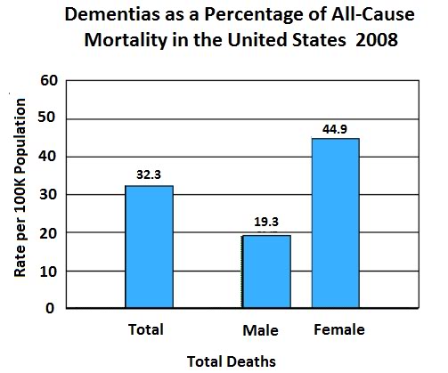 Figure 3: Incidence of dementias as a percentage of all cause mortality in males, females and the United States population as a whole. Prepared from data in the National Vital Statistics Report Volume 59, Number 10 December 7, 2011Deaths: Final Data for 2008: 2008http://www.cdc.gov/nchs/data/nvsr/nvsr59/nvsr59_10.pdf
Currently, the BDDs in aggregate (including catastrophic stroke) account for ~3.2% of all deaths in the US (Figure 3). However, insofar as cryonicists are concerned, this number is likely to be misleadingly low, because most cryonicists enter cryopreservation at or after age 65, the point at which the incidence of BDDs begin to climb exponentially. (Evans DA, 1990) This number is expected to, and in fact is exploding as a consequence of both the demographic shift due to an aging population in the West and increasingly longer life spans (Figure 4).
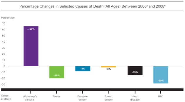 Figure 4: The large increase in Alzheimer’s Disease as a cause of death in the United States is largely a function of the increasing average age of the population and the survival of many additional individuals into advanced old age. Source: http://www.alz.org/downloads/Facts_Figures_2012.pdf
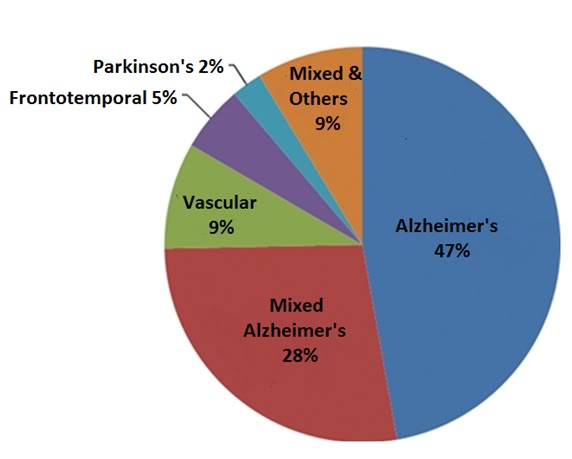 Figure 5: A breakdown of dementias by type shows that Alzheimer’s Disease accounts for 47% of the total as the sole cause of the dementia and is a major contributing factor in another 28% making it by far the most common pathological mechanism in play as the cause of dementia in the elderly. [S. Seshadri, S, Wolf, PA, Beiser, A, Au, RU, McNulty, K, White,R, et al. Lifetime risk of dementia and Alzheimer's disease: The impact of mortality on risk estimates in the Framingham Study. Neurology, 49:1498-1504,1997.]
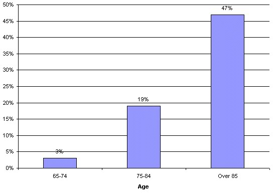 Figure 6: Incidence of Alzheimer’s Disease by age cohort in the US population as of 1988.[ Evans D, et. al. Prevalence of Alzheimer' s Disease in a community population of older persons. JAMA, 262:18;2551-6, 1989.]
In the 74-84 age cohort, 19% of that population has AD (exclusive of other dementias) and in those individuals over the age of 85, the diagnosed incidence is 47%. These numbers are almost certainly low, because many of the elderly are who are institutionalized for falls, or other issues not ostensibly related to primary brain disease, go on to develop brain disease in an institutional setting and ultimately have listed as their causes of death, pneumonia, urosespsis, sepsis secondary to decubitus ulcers, or other causes that escape epidemiological surveillance for AD. Currently, AD is responsible for 2.8% of deaths in white males men aged 65 or older and 4.7% of white males who are 85 years of age, or older. These numbers are expected to triple by the year 2050.
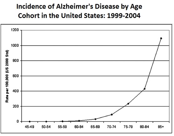 Figure 7: The incidence of Alzheimer’s Disease rises roughly exponentially with age such that over 1,100 people out of 100,000 aged 86 or older have the disease.
When cryonics was launched in the mid-1960s the problem of BDDs as a threat to the workability of cryonics was not even considered. In 1967, the year the first man was cryopreserved, the average life expectancy in the US was ~70 years and the problem of dementias was a fraction of what it currently is. Additionally, comparatively little was known about the pathophysiology of the BDDs at that time, and there was little or no awareness within the cryonics community of their potential to degrade or altogether destroy personal identity, perhaps even years in advance of the pronouncement of medico-legal death. The problem of BDDs and of age-associated destruction of the brain is arguably the foremost biomedical obstacle confronting cryonics in the long term, and it is for this reason that I will return to this topic again later in this article in the context of discussing its early detection, with a brief discussion of treatment, and ultimately, definitive interventions to halt and reverse it.
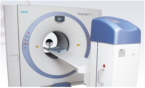 Figure 8: The Siemens Biograph mCT PET is a positron emission tomography/computed tomography (PET•CT) scanner that enables precise measurement of metabolic processes and data quantification, including the assessment of neurological disease and malignant tissues (resolution and molecular characterization of neoplasms as small 3 mm in diameter). The device can provide quantitative measurements of brain beta amyloid protein burden.
For now, I will note that because AD is by far the most common of the BDDs and because it has a unique pathophysiological feature, a remarkable advance in early diagnosis via noninvasive computerized tomography (CT) and positron emission tomography (PET) imaging has recently become clinical available. Beta amyloid is the protein found in the plaques characteristic of AD, and there has been intensive research over the past decade to identify radiolabeled tracer compounds that will safely cross the blood brain barrier (BBB) and bind to both beta amyloid and tau proteins. (Barrio 2008), (Black, 2004) In February of this year, the US FDA approved the Siemens Biograph mCT, a positron emission tomography-computed tomography (PET-CT) scanner capable of not only detecting, but of quantifying amyloid in the brain. The Biograph mCT has been very well received, and within the space of a few months the machines have appeared in most major cities in the US. The Biograph mCT in conjunction with the recently developed FDDNP, (2-(1-6-[(2-[F-18] fluoroethyl)(methyl)amino]-2-naphthylethylidene) malonitrile) tracer allows for calculation of total brain amyloid burden (Wang, 2004) and visualization of discrete amyloid containing lesions as small as ~ 3 mm in diameter (tracers for tau protein, the other primary pathological protein in AD are currently in the pipeline for FDA approval).
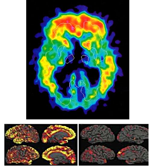 Figure 9: Top: PET scan of beta amyloid deposits in the brain of a patient with early moderate Alzheimer’s disease appear in red in the image above. The beta amyloid deposits are concentrated, as expected, in the frontal and prefrontal cortices as well as in the hippocampus. Bottom: Beta amyloid distribution in the brain of a patient with early moderate AD (L) versus normal control (R). One important consequence of this imaging is the growing realization of the global range of AD’s impact on the brain. As recently as a decade ago it was believed that the destruction of brain tissues was confined largely to the hippocampus and the prefrontal cortex, especially early in the disease. It is now understood that the histological destruction of AD is widespread and that during the end-stage of the disease few if any areas can be expected to be spared.
Very early detection of AD may turn out to be critical to achieving effective treatment, or even slowing progression of the disease, since significant beta amyloid and tau deposition seem to promote ongoing inflammation and interfere with putative therapeutic drugs. A good example of this is the recent fate (Vellas, 2010) of the investigational drug tarenflurbil ((R)-flurbiprofen ) which inhibits gamma-secretase, the enzyme that produces beta amyloid AB-42, the species of beta-amyloid that forms fibrillary plaques. (Black, 2008) Unfortunately, the drug does nothing to remove existing existing AB42 deposits, which presumably continue to exert their neuron killing and pro-inflammatory actions.
(R)-flurbiprofen is highly effective in animal models of very early AD and the drug showed significant promise in Phase I & II clinical trials. However, development of (R)-flurbiprofen was dropped when it became apparent in Phase III trials that the drug would likely only be effective in a clinical setting if it its administration was begun before clinical signs of AD developed; in other words, when beta amyloid levels were very low and would be detectable only by testing cerebrospinal fluid or, now with sensitive CT molecular imaging techniques involving the screening of subpopulations of healthy individuals at risk.
This kind of effort and application of technology and pharmacotherapy may not profitable for pharmaceutical companies, but that does not mean that it would be be worthwhile for us cryonicists. (R)-flurbiprofen is a close chemical relative of the OTC NSAID ibuprofen and it is a metabolite of the prescription NSAID flubiprofen. (R)-flurbiprofen is an enantiomer of flurbiprofen (~ 5% of (L) flubiprofen is metabolized into (R) flubiprofen by the liver after ingestion) which is completely inactive as a COX inhibitor, and is thereby free of the anti-COX side effects associated with NSAIDS. Despite it’s lack of both COX-I and COX-II activity, the drug does have strong anti-inflammatory activity by acting through inhibition of NF-κB and AP-1 activation pathways, and this may provide added benefit in controlling the inflammatory processes associated with AD. (Tegeder, 2001) As an interesting aside, (R)-Flurbiprofen has also been shown to suppress prostate tumor cells by inducing p75NTR protein expression. (Quann, 2007)
(R)-Flurbiprofen is an example of a drug with considerable therapeutic potential that will almost certainly not see clinical application due to the high cost associated with regulatory burden and the logistical hurdle of needing to start therapy years before symptoms of AD manifest themselves. (R)-Flurbiprofen might also conceivably be useful as combination therapy with the already FDA approved skin cancer drug bexarotene (Targretin), an antineoplastic, which has been shown to reverse beta amyloid deposition in a rodent model of AD as well as to improve cognitive function. Targretin rapidly cleared beta amyloid from the brains of animals in a variety of models of AD (<2 months) and while it is not a cytotoxic chemotherapeutic agent, the drug has sufficient adverse effects that it would be problematic to administer over a period of years or decades. A combination of short term therapy with Targretin to remove beta amyloid, followed by long term administration of (R)-Flurbiprofen is a possible treatment strategy that would seem attractive to explore. The ability to dynamically monitor beta amyloid levels in the brains of patients undergoing such novel therapeutic regimens, especially outside the confines of the medical-industrial establishment, is yet another advantage of this evolving singularity in medical imaging.
End of Part 1

“Currently, the BDDs in aggregate account for ~32% of all deaths in the US (Figure 3).”
Do you actually mean that dementias account for 32.3% of all deaths in the USA? This seems in conflict with the referred source cdc.gov PDF.
The PDF says:
“The 15 leading causes of death in 2008 accounted for 81.0 percent of all deaths in the United States (Tables B and 9). Causes of death are ranked according to the number of deaths; for ranking procedures, see ‘‘Technical Notes.’’ By rank, the 15 leading causes in 2008 were:
1. Diseases of heart (heart disease)
2. Malignant neoplasms (cancer)
3. Chronic lower respiratory diseases
4. Cerebrovascular diseases (stroke)
5. Accidents (unintentional injuries)
6. Alzheimer’s disease
[...]”
… With Alzheimer’s disease accounting for roughly half of all dementias (people alive with the diagnosis, not of deaths?), the above list would exceed already 90% if we take 15% for each.
Ooops, no, you caught while I was still editing on-line. That number needed a ~ and a decimal point (now added). I apologize for the errors. I will probably find many more typos and (hopefully only) minor errors. No matter how many times I proof the text, I invariably find errors every time I read over it again. Since I am dysnumeric, I tend to reverse numbers, drop or misplace decimal places and even plug in completely erroneous numbers. I do check and recheck numbers, but the problem is I cannot see the errors.
So, I really do appreciate corrections!
One thing I would point out is that the ~3.2% number for BDDs included some cases of sudden, catastrophic brain CVD. For this preliminary version of the paper, I did not take the time to define the criteria I used for selecting that number, but I believe it is low, and should probably be revised upwards – I just don’t have the time to run that data to ground right now. Those data probably merit a separate breakout along with their inclusion criteria, although the absolute number is likely to be small compared to AD and the other “chronic degenerative” dementias. — Mike Darwin