Induction of Hypothermia in the Cryonics Patient: Theory and Technique, Part 2

By Mike Darwin

External Cooling Using the Portable Ice Bath (PIB)
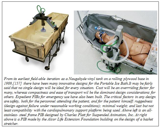
The first generation PIBs consisted of a waterproof vinyl tank which snapped to a rigid frame of 1-1/4″ OD PVC plastic pipe. The PIB could be partially broken down for transport to the patient’s location. However, these first generation PIBs were bulky, extremely fragile, impossible to move rapidly via commercial air freight, and could not be used in small aircraft such as are used by air ambulance services.
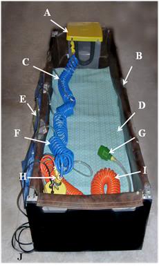
Figure 23: At left, a rugged PIB constructed from square aluminum tube stock with roller base; A) head ice positioner (HIP), B) single piece seamless puncture resistant vinyl liner, C) cold water supply line to HIP and water diffusers for the patient’s body, D) cooling blanket for chilling the dorsal surface of the patient, E) cold water diffusers, F) cold water supply line to diffusers, G) splash abating diffuser on drain line, H) pump and valve assembly (AC), I) drain line.

Early in 1995, the PIB was redesigned so that it would be collapsible, easily air transportable and extremely rugged. Since that time there have been many designs for PIBs executed around the world. The PIB design shown here can be assembled by one person in approximately 10 minutes. The lightweight aluminum construction means that it can be easily transported in a standard automobile trunk or as passenger luggage by air (both before and after patient use). This PIB was also designed to be small enough and easy enough to break down and re-deploy, so that it can be used in commercial air ambulances for air transport of patients while cardiopulmonary support or other interventions requiring access to the patient are underway.

Assembling the PIB

The PIB may be assembled using the following procedure:

NOTE: A step-by-step photographic summary of the PIB assembly procedure is presented in A Visual Guide to Assembly of the PIB immediately following the written description of the procedure.

1)   Unfold the Roller Base and lay out the components of the PIB in an organized fashion to make sure all parts are present:

Quantity            Item

1         Folding Frame

1         Roller Base

1         Vinyl Liner

2         IV Pole

8         Locking Pins

4         Attaching (Cotter) Pin assembly for securing Roller Base to Folding Frame

1         Cotter Pin for securing the Locking Bars on the underside of the Folding Frame

1         Privacy Cover

1         Records & Supplies Holster (at foot)

1         Portable Oxygen Pack Holder (POP) (and Medications Tray)

2         Heavy duty plastic tarpaulins

2)    Unfold the Folding Frame into its fully deployed configuration and insert the 8 Locking Pins (with rings on the top) into the four holes on each side of the center portion (top and bottom) of the middle section of the Folding Frame.

3)    Open the Roller Base and slide the locking bars on the underside of the Roller Base Plate into position. Secure the locking bars with the Cotter Pin.

4)    Orient the Roller Base to the Folding Frame properly. The top surface of the Roller Base and the inside of the Folding Frame are labeled “Head” and “Foot” with matching, brightly colored tape.

5)    Tilt the Roller Base so that it will fit inside the Folding Frame and the square aluminum channel on the bottom of the Folding Frame lines up with the square tubing stock of the Roller Base Plate.

6)    Push the channel on the Folding Frame up onto the bar-stock of the Roller Base.

7)    Insert the Attaching Pins into the holes on the bottom of the Roller Base.

8)    Position the Thumper Board of the MII-HLR in the correct position on the top end surface of the Roller Base.

9)    Correctly align the Vinyl Liner with the Folding Frame and secure it to the Folding Frame by mating the male and female Velcro.

10)  Insert the IV Pole into the hole in the aluminum tubing of the Folding Frame in the position most convenient as dictated by the needs of the patient (location of the IV site, position of personnel, etc.).

11)  Attach the Records and Supplies Holsters to the head end of the Folding Frame.

12)  Place the Portable Oxygen Pack (POP) Holder over the lower 1/4th of the top of the PIB and rest the POP on it.
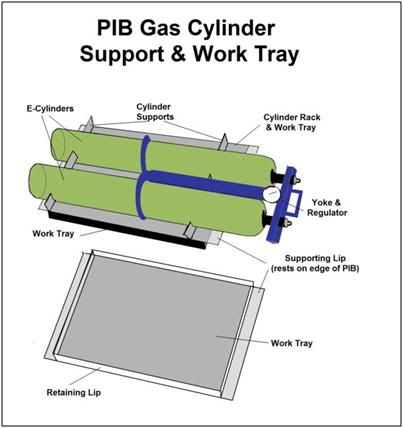
Figure 24: An essential accessory to the PIB is a combination E oxygen cylinder carrier and medications/instrument tray. Early in CPS the tray may be used to hold Transport medications. Once the medications have been administered the tray can be flipped over to allow racking and safe transport of the high pressure oxygen cylinders driving the heart-lung resuscitator (HLR).

A Visual Guide to Assembly of the PIB
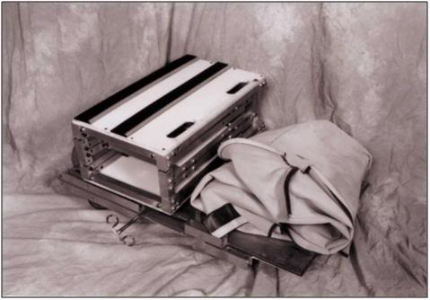
A:  Components of the Portable Ice Bath (PIB) prior to assembly.
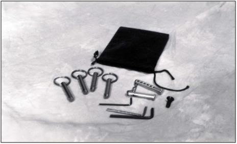
B:  Accessory Bag containing Locking and Attaching Pins, Allen wrenches and spare parts.
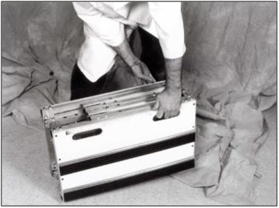
C: The Folding Frame in the collapsed configuration.
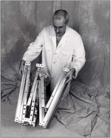
D: Open the Folding Frame by pulling apart the two endplates.
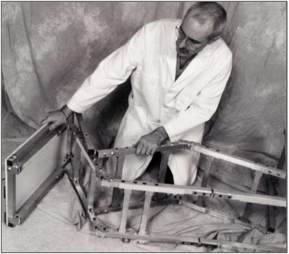
E: Fully expand the Folding Frame and make it rigid by inserting the eight locking pins.
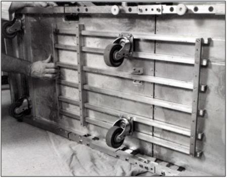
F: Rigidify the Roller Base by sliding the Locking Bars across the hinge of the Roller Base.
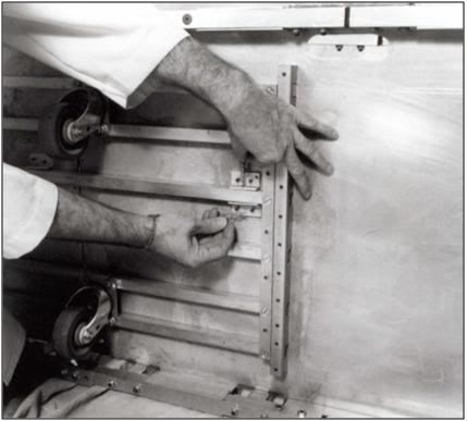
G: Secure the Locking Bars in position by placing the Cotter Pin into the securing bracket
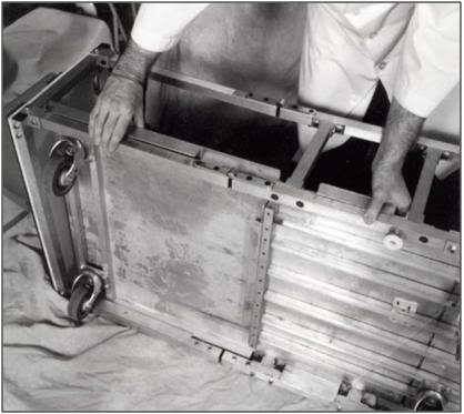
H. Insert the Roller base into the Folding Frame and snap the metal channel of the Roller Base onto the metal tubing of the Folding Frame.
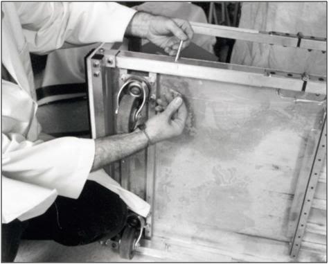
I: Secure the Folding Frame to the Roller Base with the four Attaching Cotter Pins. Be sure to have the locking end of the Cotter Pin point to the inside of the Roller Base.
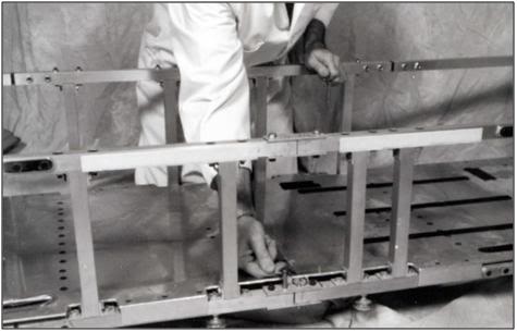
J: It is critically important that the eight Locking Pins be inserted into the Folding Frame both at the top and the bottom of the center section of the Folding Frame. Be sure to double check for proper placement of all pins prior to covering the Folding Frame with the Vinyl Liner.
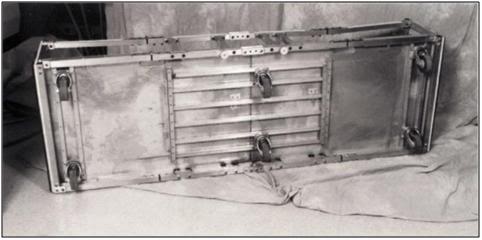
K: The Roller Base with the Folding Frame Attached should look like this after assembly.
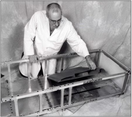
L: Place the Thumper Board onto the Roller Base being sure to anchor it into position using the Velcro on the Thumper Board and Roller Base.

M: Unfold the Vinyl Liner inside the Folding Frame on the Roller Base.
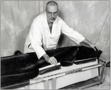
N:  Anchor the top of the Vinyl Liner to the Folding Frame using the Velcro on the lip of the Liner and the top of the square tubing of the Folding Frame.
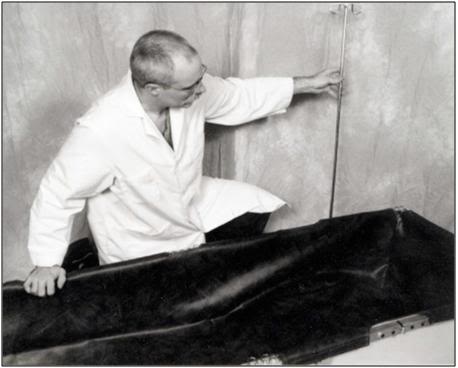
O: Insert the IV Pole into any of the four positions on the Folding Frame as convenience dictates.
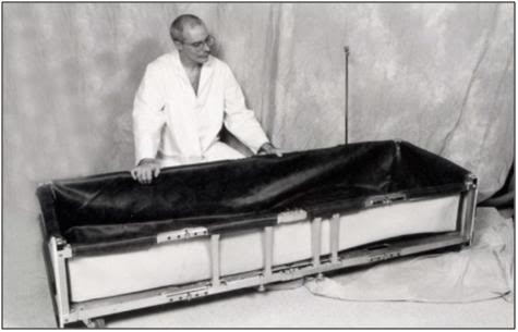
P: The fully assembled PIB shown without the POP/Medications Tray or the Records and Supplies Holster.

Initiating External Cooling with the PIB

Preparation of the PIB for Use
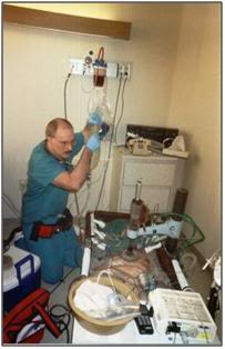
Figure 25: The PIB in use during a patient transport from a hospital in Northern California.

Upon arriving at the site where Transport is to take place the PIB should be assembled per the instructions above and, if time permits, the liner should be carefully inspected for punctures. A repair kit is included with the liner and will affect water tight repairs in ~30 minutes.

If transport is to be undertaken in a home, or in a room where there is carpeting, the heavy duty tarpaulins should be used to cover the carpeting in the work area. Use duct tape to secure the tarps to the carpeting to avoid creation of a trip hazard from folds that will develop in the tarps if they are left unsecured.

Once a place to store the PIB has been secured until the patient is pronounced the PIB should be fully outfitted with all ancillary equipment needed for induction of hypothermia, as well as with all equipment and supplies that will be immediately required to facilitate stabilization of the patient.

The dorsal cooling blanket should be positioned in the bottom of the PIB and connected to the pump that supplies cold water to the surface convective cooling device (SCCD) as well as to the cooling blanket.

If space is constrained, as it usually is in a hospital or extended care facility (ECF), ancillary equipment and supplies may be stored inside the PIB and the entire assembly covered with a sheet to avoid attracting attention, reduce the risk of pilferage of equipment and supplies, and protect the PIB and its contents from dust and fluids as shown in Figure 26.
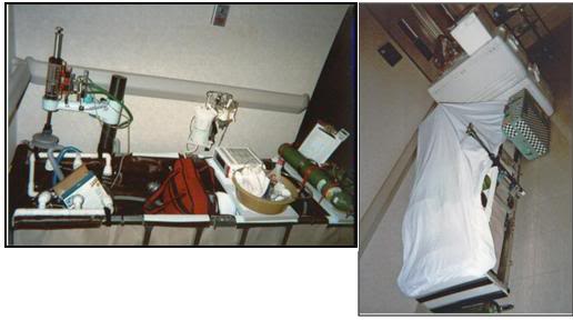
Figure 26: At left, the PIB set-up for Transport in hospital with ancillary equipment in position. At right the PIB adjacent to ice in coolers, the medications kit (light green box) and other required supplies. The PIB is covered with a Nylon sheet to protect its contents and avoid attracting attention.

As soon as legal death is pronounced, remove all clothing from the patient such as hospital gowns, undergarments, and anti-embolism stockings. The most expedient and practical way to remove clothing is to cut it off using bandage scissors or the Super Scissors contained in the RSK.  The patient’s genitals must remain covered at all times during transport and external cooling.  The genitals may be covered with a towel or small disposable drape sheet. This is an important gesture of respect and is not just a courtesy to the patient and personnel who may come in contact with the patient; it is also the law in many states. Failure to offer this respect can result in civil prosecution.

The combination fecal retention device (FRD), thermocouple probe, and colonic lavage tube should be inserted into the rectum and inflated with 30 mL of air. Easy passage of the FRD should be facilitated by lubricating it with surgical jelly before insertion (do not use oil based lubricants, including silicone lubricants, as they will destroy the retention balloon). The FRD will allow for immediate determination of the patient’s temperature, either at the time of pronouncement, or whenever Transport is permitted to begin. The FRD may also be used to irrigate the colon with cold electrolyte balanced physiologic solution (do not use water!). This will render further rectal temperature readings inaccurate, but the tradeoff in terms of speeding cooling is well worth it. Step by step instructions for insertion of the FRD and colonic irrigation are given in Colonic Lavage Cooling, below.

As soon as possible after legal death is pronounced, the patient should be rapidly transferred to the PIB and the HLR applied (manual CPR should only be used as a bridge between the time legal death is pronounced and the time it takes to organize transfer of the patient into the PIB if transfer to the PIB cannot be carried out immediately). Once the patient is positioned in the PIB and mechanical CPR has been started, the patient should be packed in ice from head to foot.  The PIB, which uses crushed ice in direct contact with the patient’s skin, will more than double the rate of cooling that can be achieved with ice-filled plastic bags. The PIB is many times more effective at reducing patient core temperature than simple air cooling such as is achieved by placing the patient in a refrigerated morgue or “reefer” unit.
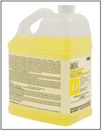
Figure 27: 300 mL Vindicator quaternary ammonium disinfectant should be added to the heat exchange water in the PIB to kill the most common transmissible human pathogens.

Concurrent with the addition of ice to the PIB, 5 gallons of water containing 300 ml of Vindicator Disinfectant (10% didecyl dimethyl ammonium chloride and 6.76% n-alkyl [C14 50%, C12 40%, C16 10%] dimethyl benzyl ammonium chloride) should be added to the PIB as a microbicide.[158] The combination of quaternary ammonium compounds present in Vindicator is effective at killing the pathogens listed in in the box below. Because of the low temperature in the PIB, microbial kill times will be longer, and it may take as long as 5 minutes after disinfectant is added until the microbial burden in the PIB water is reduced or eliminated. Quaternary ammonium compounds have very low toxicity and are generally not irritating to the oral mucosa or the conjunctiva. Used in the proper concentrations these compounds are safe for use with disposable stainless steel heat exchangers, such as those incorporated into combination hollow fiber oxygenator-heat exchanger devices used in cardiopulmonary bypass. Quaternary ammonium compounds should not be used with aluminum blood heat exchangers.

Pathogens killed by Vindicator Disinfectant

Vindicator Disinfectant  is effective against: BACTERIA: Psuedomonas aeruginosa, Staphylococcus aureus , Salmonella choleraesus, Acinetobacter calcoaceticus, Bordetella bronchispetica, Chlaymydia psittaci, Enterobacter aerogenes, Enterobacter cloacae, Enterococcus faecalis, (Vancomycin Resistant), Escherichia coli, Fusabacteria necrophorum, Klebsiella pneumonia, Leoginella pneumonia, Listeria monocytogones, Pasturella multcoccidia, Proteus mirabilis, Proteus vulgaris, Salmonella enteritidias, Salmonella typhi, Salmonella typhimurim, Serratia marcensens, Shigella flexneri, Shigella sonnei, Staphylococcus aureus – Methcillin resistant (MRSA), Staphylococcus aureus – Vancomycin resistant, Staphylococcus epidermis, Streptococcus faecalis, Streptococcus pyogenes. VIRUSES: Adenovirus Type 4, Hepatitis A virus, Hebatitis B virus, Hepatis C virus, Herpes Simplex Type I virus, Herpes Simplex Type II virus, Human Corona virus, HIV-1 (AIDS) virus, Influenza Hong Kong, Vaccinia, Rubella, SARS associated Coronavirus, Respiratory Syncitial virus. ANIMAL VIRUSES: Avian polyomavirus, Canine distemper, Feline leukemia, Feline picornavirus, Infectious bovine rhinotracheitis, Avian infectious bronchitis, Pseudorabies, Rabies, Transmissible gastroenteritis. FUNGI: Aspergilus niger, Candida albicans, Tricchyton mentagrophytes.[1]


CAUTION: Do not place ice in the PIB before the patient is transferred into it, as the presence of ice (particularly if an MII-HLR is being used) will make proper application of the HLR impossible. Exercise care to avoid wetting the piston of MII-HLR units as it will cause the piston to “lock-up” and the unit to stop cycling!
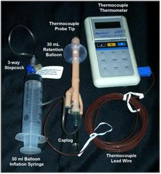
Figure 28: The Fecal Retention Device (FRD) consists of a rigid, fenestrated tube with a tough silicone rubber balloon near the tip. The FRD is outfitted with a Cu++/C thermocouple probe to allow for immediate determination of the patient’s core temperature at the time Transport begins. The silastic balloon is inflated with 50 mL of air to hold the FRD in place and prevent leakage of feces into the refrigerating water of the PIB. The FRD tube is covered with a caplug, but this may be removed and the tube connected to a closed cold physiologic solution irrigation set-up to facilitate more rapid induction of hypothermia.

The Surface Convection Cooling Device (SCCD)

Figure 29: Current implementation of the Surface Convective Cooling Device (SCCD). For use where there is no AC power, or in situations where the patient will have to be immediately relocated following the start of CPS, an Atwood self-contained battery operated marine bilge pump should be used. The SCCD above uses 4 circular diffusers to distribute cold water over the patient’s body and a Head Ice Positioner (HIP) to hold ice around the patient’s head. The HIP is also supplied with cold water from the SCCD pump and has a diffuser which may be placed on the patient’s forehead to deliver chilled water to cool the forebrain.

One of the principal barriers to efficient external cooling is the existence of “boundary layers” of insulating water which become established around the patient in the PIB. Anyone who has ever been on a camping trip and had the frustrating experience of trying to melt snow for drinking water will immediately understand this phenomenon. In the center of the kettle will be a mass of snow surrounded by tepid water while the water near the wall of the pot is boiling. Only by  stirring can such boundary layers be disrupted and efficient heat exchange achieved. One solution to this boundary-layer problem was the development by Fred Chamberlain, and consisted of a circulating pump and ice water distribution assembly that can be used in the PIB.[156]

This device, known as the Surface Convective Cooling Device (SCCD), exists in a variety of implementations. It’s most basic implementation consists of a 40 GPM (gallons per minute) output, lightweight, mains powered (AC) submersible sump pump, which is connected to a manifold of hoses and small sprinkler heads (Figure 33 ). The hosing and sprinkler heads – using quick disconnects – snap rapidly into any desired configuration. Sprinkler heads may be positioned so that a fast moving stream of ice cold water can be directed over the patient’s head, as well as to other key heat exchange areas, such as the axilla and groin. Alternative designs which employ a rigid tubing manifold with slotted cooling “fingers” have also been developed. However they operate, the principle remains the same: to rapidly move large adequate volumes of chilled water over the surface of the patient.
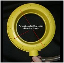
Figure 30: Close up of one of the four cold water diffuser rings used in the SCCD.

Three shortcomings of these SCCD designs is that they do not uniformly sheet or film water over the surface of the patient’s body, they do not reliably apply stirred cold water to the patient’s head, and finally, they generate splashes and aerosols that may transmit infectious disease. This has become a particular concern with the advent of multi-resistant Staphylococcus aureus (MRSA).[159], [160] Most in-hospital Staph infections are now MRSA and other, even more dangerous strains of antibiotic resistant organisms are on the way.[161]

A solution to these problems has come in the form of a more efficient SCCD which is more easily and rapidly applied. It consists of modified diffusers consisting of 4 rugged plastic rings with perforations positioned and immobilized inside two wire-reinforced fabric frames. The side of the frame that is placed facing the surface of the patient is made of an open-mesh nylon fabric that allows the free flow of water (Figures 31-32 ). The opposite side of the fabric frame is comprised of a solid fabric panel to reduce the risk of water spraying or splashing from the PIB onto staff caring for the patient.
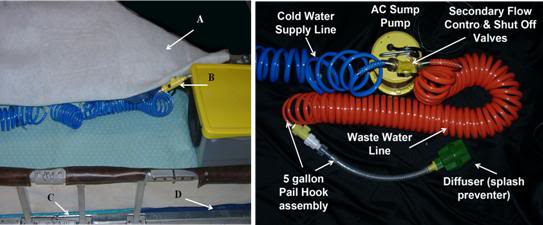
Figure 31: Above, A) cross-linked Dacron polyester wool blanket, B) Cold water delivery control valve for the HIP, C) insulating plastic covered foam mattress, D) HLR backboard.

These two frames holding the cold water diffusers are then placed atop a blanket consisting 2” thick cross-linked Dacron polyester wool which serves as a spreading medium for the cold water over the patient’s body (Figure 32). The entire assembly is then covered with a very thin sheet of nylon tricot or Dacron polyester fabric.
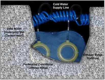
Figure 32: Cutaway looking “up” from the surface of the patient showing the relationship between the fabric frame, cold water diffusers and the Dacron wool cold water dispersing mat or spreading blanket. This assembly is covered with a nylon tricot or Dacron polyester fabric sheet to prevent splashing and aerosolization of the PIB water.

Using the SCCD

Instructions for use of both types of the SCCD are included here because a number of the older-style garden hose type units are still present in BPI field kits. Additionally, it is easy to quickly assemble a garden hose style SCCD from parts readily available at any hardware store. For these reasons instructions on use of both versions are given here.

The SCCD is simple in design and extremely easy to apply. Once the patient is in the PIB and packed in ice, add enough water to the PIB to fill it to a depth of at least 2–3 inches, typically 5-10 gallons (20-40 liters) of water. Position the SCCD pump inside the PIP at the foot end, either between the patient’s feet or to one side of them as shown in Figure 31. Snap the distribution tubing onto the pump and position the sprinkler heads as appropriate. A recommended pattern of positioning for the garden hose style SCCD is 2 sprinklers each at the head, neck, groin and axilla.
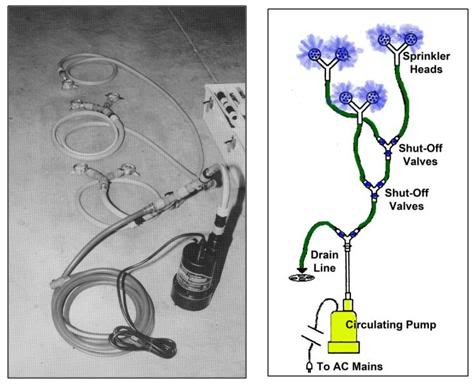
Figure 33: Garden hose style SCCD fabricated from garden hose, garden hose quick disconnects and valving and garden hose sprinkler heads; all obtainable at most hardware and home supply centers. The pump can be either a 110VAC powered submersible sump pump, or an Atwood fully submersible battery powered bilge pump powered by 4 “D-cell” batteries.
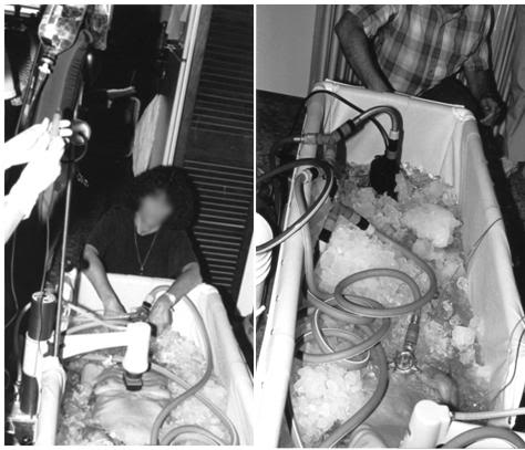
Figure 34: Two views of the garden hose style SCCD in use.

New SCCD

The new SCCD, as previously noted, uses two fabric frames containing 4 cold water diffusers. These fabric frames are connected to a cold water supply line. Connect the cold water supply line to the quick disconnect on the water circulating pump. Place the Head Ice Positioner (HIP; see discussion, below) at the head end of the PIB and connect the cold water supply line to the disperser inside the HIP. Ensure that the drain valve on the HIP is closed.
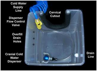
Figure 35: The Head Ice Positioner is designed to keep refrigerant in constant contact with the patient’s head. This can be accomplished with crushed or shaved ice or by a constant flow of chilled (~0oC) water flowing over the patient’s head. The HIP is equipped with a cold water disperser connected to SCCD pump allowing water to build up in the HIP and overflow from drain holes in the side of the container to return to the PIB.
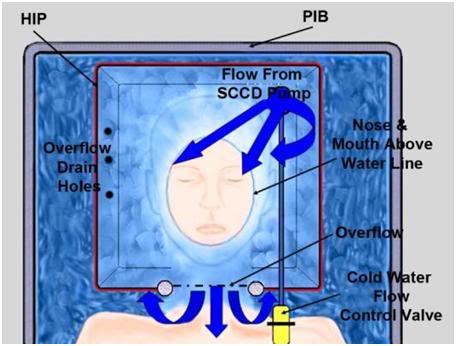
Figure 36: Relationship between patient the HIP and the PIB. Care must be taken to ensure that heat exchange water does not enter the patient’s nose or mouth.

Once the patient is in position inside the PIB (ensure that the patient’s head is properly positioned inside the HIP and that it does not interfere with ventilation) cover the patient with the Dacron wool cold water dispersing mat, and then place both fabric frames containing the cold water dispersers atop the mat; one adjacent to the AC-DC cup of the HLR, and the other over the lower abdomen and legs. Activate the circulating pump and insure that water is flowing freely from all four dispersers and that water is exhausting from the HIP around the patient’s neck, or from the overflow holes on the side of the HIP (Figure 35). Take care that the water level in the HIP is not high enough to enter the patient’s oropharynx.

Until a few years ago, the only pumps to drive the SCCD were either AC sump pumps, or 12 volt powered marine bilge pumps. While compact and lightweight, marine bilge pumps required lead-acid batteries which may not be transported by air. Attwood Marine now markets the Attwood Waterbuster™ pump, which is a self contained, fully submersible pump that runs up to 5 hours on three alkaline D batteries. It delivers a 200 gph flow at a head of 40” and measures only 6-3/8″ high x 5-1/4″ diameter. This pump is currently replacing the AC pumps in Standby Kits. However, even in situations where only an AC pump is available, the SCCD may still be very useful, particularly in situations where medications are to be administered before vehicular transport. (Administering medications typically requires 45 minutes to an hour to accomplish.) There will also be many situations where transportation (local mortician, ambulance, etc.) must be summoned after the start of CPS and external cooling and where there is likely to be a delay of 15 to 30 minutes before transportation arrives. Keep in mind that the SCCD, when used in conjunction with the PIB, can lower a thin patient’s temperature as much as 12ºC in 30 minutes. Thus, every minute that the SCCD can be used, it should be used. If use of the SCCD has to be interrupted, restart it as soon as possible.
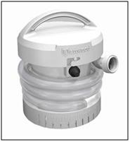
Figure 37: The Attwood Waterbuster™ pump is a self contained pump that runs for 5 hours on 3 “D-cell” batteries. It is almost completely silent and produces exactly the right amount of flow to facilitate effective convective cooling. The Waterbuster™ also greatly decreases ice consumption since, unlike it AC counterparts, it generates very little waste heat.

Limitations of External Cooling

Despite the development of immersion cooling employing a well stirred ice bath, there are fundamental limits on the amount of heat that can be moved using only the surface of the patient. These limits are a function of the patient’s mass, degree of insulating fat-covering, adequacy of circulation to the skin and surface body tissues, and the patient’s volume and surface area. In practice, the maximum rate at which low-mass emaciated patients can be cooled externally is in the range of 0.25 to 0.35ºC/min., while the maximum rate for larger mass patients with a significant layer of subcutaneous fat is in the range of 0.12 to 0.15ºC/min. Figure 38 shows the rate of cooling for a number of patients with different masses, adequacy of perfusion, and degrees of cachexia.

As Figure 38 shows, the maximum rate of cooling achievable in the first 30 minutes of CPS in a patient of average mass and with minimal subcutaneous fat is in the range of 0.5ºC/min. If this cooling rate is compared with what could be achieved starting at the same patient core temperature (~37ºC) and using extracorporeal cooling with a high efficiency heat exchanger (0.6 coefficient of heat exchange) and a “wall water” (water to the heat exchanger) temperature of 0ºC, cooling rates of two or three times that achievable with external cooling are possible (i.e., 1.5 to 2.0ºC/min for the brain in a 65 kg adult with a surface area of 2.0 square meters). As previously noted, the difficulty with this approach is that it requires a considerable amount of skill and time. Even under the best circumstances it is unlikely that cardiopulmonary bypass (CPB) can be safely established in less than 60 minutes from the time of pronouncement, even by experienced operators.

The reality is that far longer periods of time may elapse between the start of transport and the beginning of extracorporeal support. Logistic constraints, such as the need to move the patient from the home, or an acute or chronic care facility to a mortuary, the availability of skilled personnel, and pre-existing medical or anatomical complications all may greatly delay or even prevent the application of in-field CPB.
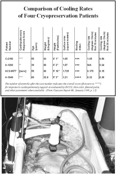
Figure 38: Comparison of the cooling rates of four cryopatients. Immediately following pronouncement of medico-legal death patients were given closed chest mechanical cardiopulmonary support and placed in a stirred ice water bath for induction of hypothermia. Epinephrine was administered as per ACLS guidelines; thus peripheral vasoconstriction would be expected to be comparable to that seen in the typical SCA patient undergoing cardiac resuscitation. The number of asterisks after the case number indicates the overall score (from zero to ****) for response to cardiopulmonary support as evaluated by EtCO2, skin color, femoral pulse, and other parameters, when available.

Ensuring Adequate Refrigeration of the Patient’s Head

Recently, experiments have been conducted that show show something that may seem surprising, principally that cooling in an unstirred water bath at 0ºC is not even twice as effective as cooling in a still (unstirred) air bath at 0ºC, as shown in Figure  50, below.

The reason for this is the limitation imposed by the very low value for the heat conductivity of the human head. This has the following important practical implications:

1)         External conductive cooling of the human head/brain is extremely slow, even under ideal conditions of maximum surface contact with ice at 0oC, where the melt water is filmed over the patient’s head.[7],[8]

2) Once the surface of the patient’s head reaches 0oC, the brain cannot be cooled any faster regardless of the type or amount of conductive media used. In other words, using more conductive refrigerating media, or delivering them at higher flow rates than necessary to keep the skin at 0oC, will not work, and may well be counterproductive (i.e., consume limited battery power and cause splashing and aerosolization of potentially biohazardous cooling bath water).[9],[10]

3)         If conditions are less than ideal because of poor contact with refrigerant (and retention of melted ice water in plastic bags) then cooling is slower still, and this is undesirable.

4)         The basic requirement of uniformly cooling the surface of the patient’s head to near 0oC is, in practice, quite difficult to achieve, because holding refrigerant in contact with the patient’s head involves problems associated with melting ice, which is messy, damaging to bedding and furnishings, and can cause a slip hazard if dripped onto the floor. Containing ice in plastic bags results in considerable loss of contact with the skin, and reduces the efficiency of cooling, by causing melt water to be retained; creating a relative convective and conductive barrier. It is also virtually impossible to keep ice bags in position around the patient’s head during movement from one location to another (or for that matter, even when the patient is not being moved.

While there is no easy solution to problems 1 and 2 above, there is a solution to problems 3 and 4: an enclosure to hold ice or another acceptable refrigerant around the patient’s head in situations where there is no portable ice bath (PIB); the Head Ice Positioner (HIP). Why is having an “ice holder” to keep ice around the patient’s head so important? Again a look at Figure 19, above, is proof that a picture is worth a thousand words. The patient in this picture is being cooled with ice bags and Kwik Kold eutectic cooling packs. As just noted, this cuts the effectiveness of ice dramatically by confining it to bags, and it is also messy, which decreases compliance and creates a real danger of slipping and falling for personnel when tile or linoleum floors become wet and slick (something that is especially likely in institutions with well waxed and polished floors; and inside ambulances). The HIP should be used inside the PIB in order to ensure uniform contact of refrigerating water with the patient’s head.

RhinoChill

In 2009, the RhinoChill, a new noninvasive method for rapid induction of MTH under development by Benechill, Inc., began clinical trials.[162] The RhinoChill uses a novel method to achieve intra-cardiac arrest cooling; transnasal evaporative cooling, wherein a liquid coolant–oxygen (or oxygen-air mixture) is sprayed into the nasal cavity and frontal sinuses where the liquid is rapidly evaporated with the high-flow compressed gas (typically O2). The heat of vaporization of the perfluorocarbon azeotrope causes cooling of the nasal passages and brain. The device is highly portable, can be used on a patient within minutes of cardiac arrest, and has been demonstrated to be safe for use in humans in the hospital setting.[162],[163]
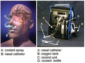
Figure 39: The RhinoChill device (R) and the anatomical areas cooled by the evaporation of the PFC refrigerant from the nasal cavity and sinuses.[163]
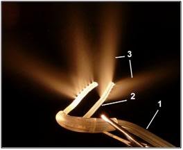
Figure 40: Photograph of RhinoChill nasal cannulae used for nasopharyngeal cooling. The perfluorochemical (PFC)-oxygen mixture is delivered from the oxygen tank and the PFC reservoir in a single tube (1) that then bifurcates into a left and right nasopharyngeal cannula (2). The perfluorochemical-oxygen spray (3) exits in dorsal and lateral direction from the distal end of the cannulae.[164]
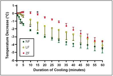
Figure 41: Change in brain temperatures from baseline (mean ± SD) during untreated cardiac arrest (ZF; n = 3), CPR (LF; n = 4) and anesthesia (NF; n = 3) over the course of 60 min of nasopharyngeal cooling. *indicates first significant decrease from baseline (<0.01).[164]

Perfluorochemical (PFC) is delivered via a proprietary cannula, the two legs of which are passed through the nares and into the nasal cavity. PFC is delivered at a rate of 1 mL/kg/min while oxygen is co-administered at a rate of 1 L/kg/min. When circulation is present, heat is removed from the brain predominantly hematogenously, through the submucosal nasal venous plexuses by the rich subepithelial vascular plexus to the deep venous sinuses of the brain, and secondarily by direct convection. The device can reduce tympanic temperature (a surrogate for brain core temperature) at the rate of 2.4°C per hour. Systemic cooling proceeds more slowly at a rate of 1°C per hour.

The liquid evaporates instantaneously, thereby removing heat. The coolant is a proprietary perfluorochemical or perfluorochemical mixture (azeotrope) the composition of which is not disclosed. No patents appear to have been filed disclosing the PFC chemical structure, or mixture of PFCs being use as the refrigerant. The PFC used by Benechill must have a temperature well below the freezing point of water since inadvertent freezing of the nasal mucosa is a complication of operation.[163] Perflourochemicals are a family of chemicals that are generally regarded as both chemically and biologically non-reactive. These chemicals are among the least acutely toxic compounds known, although they are known to be potent immunomodulators and inhibit white blood cell chemotaxis at fentogram concentrations.[165],[166],[167] They cannot reach appreciable concentrations in tissues of air-exposed animals since they have limited ability to dissolve in biological media. Many are highly volatile and have a high air–blood partition coefficient, which facilitates their rapid elimination through pulmonary expiration (more information is available via 3M Specialty Materials. Robust summaries and test plan: perfluorocompounds, C5–C18; revised summaries. EPA Report 201-14684B, Aug 2003). The cooling and safety profile associated with the specific perfluorochemical used in the coolant was determined by Wolfson et al., in an ovine model, where no damage to the epithelial surface was noted.[168]

The nasal cavity with its proximity to the cerebral circulation, basal brain regions, hippocampus and the brain stem, offers an approach that allows for preferential cooling of some of the most selectively vulnerable areas of the brain.[169] The device has been tested in a porcine model of prolonged ventricular fibrillation cardiac arrest both with and without cardiopulmonary resuscitation (CPR). In the CPR group, jugular venous temperature, which was used as surrogate for brain temperature, dropped from 38.1oC to 34.2oC within 5min of the onset of CPR and cooling. Importantly, the rate of brain cooling as measured by a temperature probe placed in the center of the right frontal lobe was almost the same at 60 min in the zero flow (no CPR) group as it was in the groups with spontaneous circulation and low flow (CPR) circulation (see Figure 38).[164]  When perfusion is absent, cooling of the brain is by conduction, via the cribiform plate and frontal sinuses.

The RhinoChill device (Figure 39) consists of the tubing set, the control unit, and the coolant bottle. The tubing set delivers oxygen and coolant to the patient. The cooling cannulae rest in the nasal cavity adjacent to the chonchae and have spray ports on their dorsal surface (Figure 40). The coolant is nebulized by turbulent mixing with oxygen at the spray ports. A battery operated control unit controls coolant flow rate and acts as an over-pressure shut-off valve. The patient pressure safety circuitry switches the system to a standby mode if the pressure in either nasal cavity exceeds 60 cm H2O. Coolant delivery is maintained at a constant ratio to oxygen flow such that cooling level is controlled by setting the oxygen flow rate between 0 and 80 L/min. The patient’s mouth is kept open to provide venting of the coolant vapor. Duration of nasopharyngeal cooling in the clinical trial has been 60 minutes (50;90; range 25–195 min), and the amount of  refrigerant per-patient used was 3.5 liters (2.0; 4.0 L).[163]

Figure 42: Time to target temperature (tympanic) of 34°C in minutes (median) from the cardiac arrest in the treatment and control groups among those patients admitted to the hospital.

In the latest human trials a tympanic temperature of 34°C was achieved by a median of 102 minutes (interquartile range 81 to 155 minutes) in the treatment group compared with 291 minutes (interquartile range 183 to 416 minutes, P0.03) in control patients (Figure 42). Median time to target temperature (core) of 34°C in the treatment group was 155 minutes (interquartile range 124 to 315 minutes) versus 284 minutes (interquartile range 172 to 471 minutes) in control patients (Figure 42). The time required to apply the device was ~2 min. The improvement in outcome in neurologically intact survival is shown in Figure 43.

These cooling rates may not seem impressive, and the relevance of this device to human cryopreservation may seem questionable. Undoubtedly the cost of the device and the PFC refrigerant will be prohibitive if the device is FDA approved for widespread clinical application. However, neither the likely high cost nor the modest cooling rate should obscure the fact that this technology demonstrates that a substantial increase in the rate of brain cooling can be achieved by using the heretofore unutilized surface area of the nasopharynx and frontal sinuses. Whether exploited by evaporative PFC cooling, or by the use of an aqueous heat exchange medium, this surface area should be used in inducing hypothermia in cryopatients, and in particular for cooling the brain. The advantages that the RhinoChill system has of not requiring bulky, heavy equipment and of leaving the nasooropharynx (NOP) devoid of liquid are also substantial. Introducing saline or other liquids into pharynx carries with the risk of aspiration in the mechanically ventilated patient.
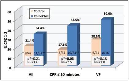
Figure 43: Rates of neurologically intact survival (defined as having a cerebral performance category [CPC] of 1 or 2) in the treatment and control groups among those patients admitted to the hospital for the entire group, those who received rescuer CPR within 10 minutes, and those with a presenting rhythm of VF. RR indicates relative risk.

*Unadjusted 2 test.

**In one  admitted patient, outcome data were missing.[163]

While the RhinoChill uses compressed oxygen and a fairly sophisticated delivery device, it is easily possible to substitute compressed air for oxygen, fabricate a less expensive delivery system, and presumably find an acceptable azeotrope of PFCs to use as the refrigerant. Perfluropropane and one or more of the 3M Fluorinert liquids would seem to be a good starting place. Alternatively, chilled saline can certainly be used as it was in the RhinoChill swine pilot study. While liquid assisted pulmonary cooling (LAPC) may seem an attractive alternative to transnasal cooling, there are many problems with this approach including the risk of serious systemic embolization with PFC during closed chest CPS in patients with friable or seriously injured lungs.

Finally, it is important to keep in mind that, with due consideration to cost and logistics, the various approaches to cooling discussed here are complementary and synergistic rather than opposing or exclusive. As is the case with cold IV saline and external cooling, the RhinoChill, as a standalone method for rapid induction of hypothermia (-3oC in ≤ 15 min) will not suffice. However, in combination with other easily applied and non- or minimally-invasive modalities, it may prove the long sought answer to the problem of cooling the brain by 3oC in (ideally) 10 minutes.

Liquid Assisted Transpulmonary Cooling

As has been noted many times before, the most powerful moderator of ischemic injury at the disposal of the Transport Technician is induction of hypothermia.[170],[171],[172] In the absence of the ability to provide adequate perfusion, reduction of the patient’s core temperature will be even more important. As the discussion of liquid ventilation in Chapters 4 and 7 makes clear, rates of cooling using this technique approach those achievable with CPB (Figure 44). However, due to current technological limitations it is not possible to continuously deliver and aspirate the PFC to/from the lungs through a heat exchanger. Thus, the initial load of PFC will only cool the patient approximately 2º to 4ºC during the first 10 minutes of cooling, and thereafter heat exchange will decrease as the PFC comes to thermal equilibrium with the patient.[173] The theory and technique of liquid assisted transpulmonary cooling (LATC) will not be discussed in detail here since they are complex enough to merit treatment in a separate Chapter (Chapter 7).
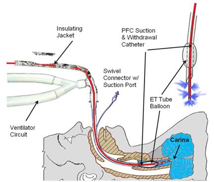
Figure 44: LATC PFC delivery and withdrawal patient tubing configutation.

Intraperitoneal Cooling

The  lungs of the patient are not, of course, the only spaces into which cold fluid can  be introduced. In theory, any body cavity accessible via a natural orifice, or rapidly accessible surgically, could be used as a reservoir for cold heat exchange fluid. In particular, the peritoneum, the stomach and the colon all have a capacity for loading with a significant amount of refrigerant.
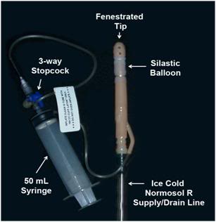
Figure 45: Self retaining and sealing peritoneal irrigation catheter. The catheter is inserted through a small mid-ventral incision in the abdomen into the peritoneal space at which time the silastic balloon is inflated with air to hold the catheter in position and make a reasonably water tight seal. This eliminates the need for a purse string suture. The body of the catheter and the Normosol-R supply line must be affixed to the abdomen with Backhaus forceps to prevent the catheter from becoming dislodged during use.

The optimal refrigerant for intraperitoneal cooling is a physiological saline-ice slurry,[174],[175] but owing to logistic constraints in preparing and pumping such slush on-site, this is not a viable option for the immediate future.[176] The next most effective heat exchange medium would be ice-chilled balanced electrolyte solution. An off-the-shelf, sterile product such as Normosol-R pH 7.4 would be suitable, since it is inexpensive, readily available, ruggedly packaged, and can be refrigerated by packing in ice. Consideration of the amount of heat that could be removed with gastric, peritoneal and colonic lavages of reasonable amounts of Normosol-R indicate that approximately 0.50º to 0.75ºC/min heat removal should be possible in a 65 kg man.[177],[178],[179]

Peritoneal lavage has the added advantage of being repeatable: the peritoneal cavity can be repeatedly filled and drained of cold Normosol to exchange heat in the same fashion that peritoneal dialysis is used to exchange mass. Colonic lavage may be similarly repeated. Application of 0ºC colonic and peritoneal Normosol lavages in addition to standard PIB cooling has been undertaken in one human cryopreservation patient to date with a resultant threefold increase in the cooling rate over that anticipated with external cooling alone. On the basis of this data and data obtained from animal experimentation, colonic and peritoneal lavage has been added to the standard protocol for induction of hypothermia in cryopatients.

Even with an endotracheal tube in place, gastric lavage cooling is not recommended at this time due to concerns about possible compromise of the airway as a result of reflux and aspiration. Experimentation is currently underway to develop a device with a cuffed infusion tube, similar in design to the Esophageal Gastric Tube Airway, which can be passed blindly into the stomach via the esophagus. The cuff would then be inflated to achieve a seal between the tube and the esophagus, preventing the possibility of stomach contents being aspirated.

Peritoneal Lavage Cooling

Intraperitoneal cooling is far more efficient than colonic cooling but requires surgical opening of the abdomen. This is neither a complex procedure, nor one fraught with complications if the procedure outlined below is scrupulously followed.  Basic surgical experience on animals or humans is required, and it is preferable that the operator has previous experience entering the abdominal cavity, and is familiar with basic anatomy of the body wall from first-hand experience.
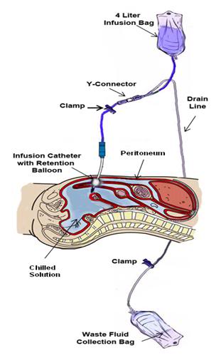
Figure 46: The peritoneal lavage assembly consists of two 4 liter peritoneal dialysis bags and a peritoneal dialysis (PD) infusion and drain line set. One bag is filled with Normosol-R™ using aseptic technique and attached to infusion leg of the PD set. A second bag is emptied of PD fluid (again using aseptic technique) and attached to the drainage leg of the PD set and the entire assembly is placed inside double ZipLoc bags and refrigerated on ice. Additional 4 liter bags of Normosol may be prepared in advance as needed and the entry port plugged with a sampling site connector. The bag containing the Normosol may be raised above the patient on an IV pole to facilitate gravity infusion of cold solution. Optimum dwell time is ~5 minutes. The Y-connector on the PD set connects the catheter to the waste fluid collection bag when the clamp on the drain line is opened.

Technique for Peritoneal Catheter Insertion

1)      Prepare the Peritoneal lavage set-up for use by aseptically unwrapping the infusion/drainage set, waste fluid collection bag and peritoneal catheter. Be sure clamps are present on both the infusion and drainage legs of the infusion/drain line. Spike a bag of chilled Normosol-R and prime the infusion leg of the infusion/drain line. Once the peritoneal catheter is in position and secured a fresh (cold) bag of Normosol R should be used to replace the bag employed to prime the lines.

2)      Select an entry site in the mid-line of the abdomen one third of the distance from the umbilicus to the pubis. Do not deviate from the midline unless scarring from prior surgery requires you to do so. Staying on the midline is important because, as shown in Figure 47, the epigastric arteries lie 3-4 cm to either side of the midline.
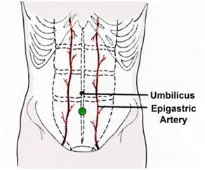
Figure 47: Site of incision for placement of peritoneal lavage catheter is in the midline of the abdomen well away from the epigastric arteries. The optimum site is marked with a ● on the drawing above.

3)      3) Use sterile technique, protective clothing and a face shield (See Chapter 16 for details on Universal Precautions and infection control).

4)      Disinfect the skin and apply a 3M adhesive drape to the incision site.

5)      Using a #10 scalpel blade make a 1 cm incision in the skin using the broad part of the blade.

6)      Deepen the incision using sharp dissection until the muscular body wall is encountered.  Mobilize the overlying tissues to expose a window of muscle approximately 2 cm square using blunt dissection with the index finger.

7)      Reflect the margins of the wound in the skin to expose the body wall with a self-retaining Weitlaner retractor.

8)  Using the broad cutting surface of the scalpel, divide the muscle taking care not to cut too deeply through the body wall and injure the underlying viscera. As the muscle is divided, pause and explore the incision with the finger tips to gauge the remaining thickness of the body wall to be cut.
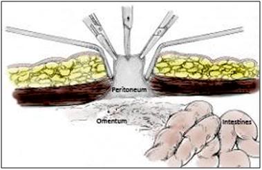
Figure: 48: Once the ventral abdominal wall has been opened the peritoneum will be visible as a grayish membrane with a smooth consistency. The peritoneum is then grasped and tented with forceps and opened with a #11 scalpel blade or Metzenbaum scissors.

9)  Continue sharp dissection until the muscle is penetrated and the peritoneum can be seen. The peritoneum can then be incised by careful use of sharp dissection using the scalpel and Metzenbaum scissors. Be careful to keep the opening in the muscle and peritoneum to no wider than 0.5 cm.

10)  Upon opening the peritoneum, insert a finger into the abdominal cavity and expand the wound using blunt dissection. The size of the opening sought should be roughly one-half to three quarters the diameter of the peritoneal lavage catheter. Avoid making the incision too large as it will prevent a tight seal of the catheter and will allow leakage of lavage fluid out of the peritoneum and entry of non-sterile and hypotonic water from the PIB into the abdominal cavity.

11)  While grasping the catheter and supporting it with your hand, push it through the opening in the body wall and peritoneum until the balloon on the distal end of the catheter is fully past the peritoneal membrane. A twisting motion may be necessary to achieve this. It may also be necessary to expand the diameter of the muscular and peritoneal incisions to facilitate entry of the catheter. Entry into the peritoneum with the catheter through a tight incision is indicated by a “pop” and sharp decrease in resistance to insertion.

12)  Inflate the catheter balloon with 50 cc of air.

13)  Secure the infusion/drain line to the skin of the abdomen using a large Backhaus towel clamp.  The clamp should be used to pierce the full thickness of the skin and firmly anchor the line near the catheter to the patient. Adhesive tape or clear 3-M adhesive drape may be used to cover the wound site and the body of the catheter where it exits the abdomen.

Procedure for Peritoneal Cooling Fluid Exchanges

1)      Once the catheter is placed and secured, spike the 4 liter bag of ice-cold Normosol-R and hang it for infusion.

2)      Unclamp the clamp on the infusion leg of the infusion/drain line and insure that the drainage leg of the line leading to the waste reservoir bag is clamped. Fluid should start to flow from the reservoir into the abdomen under the force of gravity. Adjust the reservoir height to achieve rapid filling. Infuse l liter of ice-cold Normosol-R for every 20 kilos (44 lbs) of pre-morbid body weight to a maximum of 4 liters.

3)      When the bag is empty, clamp the infusion leg of the infusion/drain line. Optimum dwell time in humans of chilled Normosol-R in the peritoneum under transport and cool down conditions is unknown but is thought to be in the range of 3-5 minutes.

4)      As soon as the infusion is completed, unclamp the drainage leg of the infusion/drain line and allow the lavage solution to drain into the waste reservoir bag which should be positioned on the floor below the patient at least 1 meter above the level of the patient’s abdomen.

5)      As drainage slows, the next bag of hilled chilled Normosol-R should be spiked and hung. After drainage has stopped, the drainage leg of the infusion/drain line should be clamped and the infusion leg unclamped and another lavage carried out.

The crude thermal transfer proportionality constant k, for cooling of dogs with chilled fluorocarbon (FC-77) in the lungs, and Normosol-R administered intraperitoneally, according to the rough empirical relation is:

dT/dt = -k [delta T]     which seemed to fit the data reasonably well (although there was evidence of  a “ring” or oscillation in warming of the lung lavage fluid, reflecting the dynamic rather than passive nature of the heat flows).

To a first approximation, for one liter of coolant at 0 C administered to a 20 kg dog,  in both cases the proportionality constant k is approximately the same: ~ 1oC per minute (warming rate), per oC difference in coolant and body temp (i.e., k  in both cases was 1 minute^ -1).   (That both these numbers were the same for FC-75 and Normosol R is coincidence.  The heat capacity of FC-75 is about half that of water, on a volume basis and the blood flow and exposed vascular surface area to lungs and peritoneum are very different.  Half-times to reach equilibrium temp for this system are ~ k/ln2 = 1/ln2 = 1.44 minutes.

It is not anticipated that the k values for further boluses of fluid into lungs or peritoneum will be nearly as favorable as those seen in first exchange.  The first exchange is unique in that the coolant enters spaces to transfer heat that subsequent exchanges will not (i.e., the terminal airways and the many invaginated spaces in the abdomen will initially fill with fluid, but will not completely drain during subsequent exchanges and will therefore represent dead space and added mass which heat must be removed from.  Continuous peritoneal lavage, perhaps with a cyclic component (as is used in machine delivered peritoneal dialysis) to reduce laminar flow, seems the optimum way to achieve rapid in-field cooling of cryopatients via peritoneal cooling in the future.

Colonic Lavage Cooling

In order to carry out internal lavage cooling with multiple exchanges of fluid it is necessary that the PIB be elevated on two or three sawhorses, a sturdy folding table, or some other stable work surface, so that gravity dependent drainage can be carried out after each infusion of chilled solution.

Once CPR and medication administration is underway and the patient is situated in the PIB, the Fecal Retention Device (FRD) should be placed if it has not been already. The balloon on the FRD is inflated, and an initial temperature reading taken.  Connection of the infusion/drainage set to the FRD is made and lavage of the colon with 1 liter of buffered, ice-cold Normosol-R is then carried out per the following procedure:

1)      Spike a pre-chilled bag of Normosol-R with the infusion leg of the infusion/drain line set.  Prime the infusion leg of the line till fluid exits the FRD.

2)      Lubricate the FRD with K-Y or other water soluble surgical lubricant. Do not use Vaseline, silicone oil based or any other oil based lubricants because they will cause failure of the retention balloon on the FRD.

3)      Insert the FRD well into the rectum through the anus. Be sure the balloon on the FRD is well beyond the anal sphincter.

4)      Inflate the balloon of the FRD with 50 cc of air.

5)      Elevate the bag of chilled Normosol-R 20-30 cm above the patient’s abdomen.

6)      Unclamp the infusion leg of the infusion/drain line and insure that the drainage leg of the line leading to the fluid waste bag is clamped. Fluid should start to flow from the Normosol bag into the colon under the force of gravity. Fill the colon with 20 cc of Normosol-R per kilogram of the patient’s pre-morbid (i.e., healthy) weight to a maximum of 1.5 liters. In the case of patients who are chachectic and weigh far under their pre-morbid weight at the time of cardiac arrest, their healthy body weight should be used as the index in determining the lavage volume. Thus, in the case of a man who in health weighed 79.5 kg (175 lbs) but due to end-stage adenocarcinoma of the lung weighs only 50 kg (110 lbs) a full 1.5 liter exchange of lavage should be used.

7)      After 10 minutes, remove the clamp from the drainage leg of the infusion/drain line and allow the lavage solution to drain into the waste reservoir bag which should be positioned on the floor below the patient. For optimum drainage the patient should be at least 1 meter above the bag. Be careful to place the waste bag so that it is not stepped on or punctured.

In the event that colonic lavage is going to be used prior to peritoneal lavage, lavage of the colon should be carried out prior to or during surgery to place the peritoneal lavage catheter. Colonic and peritoneal lavage may not be used at the same time. Once the peritoneal catheter is in place, the colon should be drained while the peritoneum is lavaged; this is best accomplished by leaving the drain line to the fluid waste bag open during peritoneal lavages in order to allow any retained fluid in the colon to escape. This increases the available space in the abdomen for the cold peritoneal lavage solution.

Optimum Number of Lavages

The optimum number of colonic and peritoneal exchanges is not known. While infusion of fluid is quite rapid, it is anticipated that drainage will take considerably longer.  In the only human case in which Intraperitoneal cooling has been used to date each exchange of 4 liters took approximately 10 minutes, with most of the time required for drainage before refilling the abdomen.  In the absence of extracorporeal cooling the limitation on the number of exchanges is likely to be the availability of Normosol-R.

Termination of Lavages

Whether the reason for discontinuing exchanges is the beginning of bypass or the exhaustion of the supply of chilled heat exchange fluid, the colon and peritoneum should be drained of fluid as completely as possible at the conclusion of the lavages. The reason for this is that the continued presence of “thermally spent” lavage solution during extracorporeal cooling or external iced cooling during subsequent transport or air shipment will act as thermal ballast, in effect increasing the patient’s total mass, and will thus impede further cooling.

Once drainage is complete the infusion leg of the infusion/drain lines of both the peritoneal catheter and the rectal tube are double-clamped within 2-3 cm of the peritoneal catheter or rectal tube with the clamps placed 3-4 cm apart. The lines are then cut with scissors, and a tubing plug inserted into the stub of the line attached to the peritoneal catheter and rectal tube. A sterile plug must be used in the case of the peritoneal line, and the peritoneal line should be divided using aseptic technique.

The infusion reservoirs are emptied of any fluid, the infusion/drain lines are clamped near the exit port from the infusion reservoirs, the infusion/drain lines are disconnected from the reservoirs and the tubing and drainage bag assembly are properly disposed of as biohazardous waste in accordance with local regulations.

Ice Bags and Alternative Methods

If a standard PIB is not available, use a makeshift PIB by using a mortuary air transfer case lined with plastic sheeting, an inexpensive casket similarly waterproofed a heavy-duty mortuary body pouch (“disaster bag”), or even a plastic tarpaulin or plastic sheeting under the patient on the bed.

If the patient is in the morgue and the morgue tray has drainage capability, mound ice up over the patient in direct contact with the skin. Whenever possible use ice in contact with the skin, as it is a far more efficient refrigerant than ice in plastic bags.
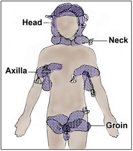
Figure 49: If ice is in limited supply, the head should be completely packed in ice first. If CPS is underway the next areas to pack in ice are the vascular areas of the body; those places where large caliber, high flow blood vessels lie close to the skin surface; the neck, axilla and groin.

If the PIB or a reasonable facsimile is unavailable, pack the patient in crushed ice contained in high quality Zip-Loc plastic bags per Figure 49. (Bags with the Zip-Loc brand name and manufactured by Johnson & Johnson, Co., are preferred.) Leaking bags present a serious safety hazard in the form of water on the floor, and an electrical hazard if the patient is in an electrically operated bed. Special attention should be paid to packing the head, neck, axilla (armpits), and groin in ice, since large vessels which carry a significant fraction of the cardiac output lie close to the skin in these areas, and are thus available for heat exchange.

Cooling Absent Cardiopulmonary Support

Unfortunately, in many cases Transport of the patient will occur under conditions where CPS is not possible or is contraindicated due to the presence of too much warm ischemic time. Under such circumstances the only option for inducing hypothermia will be external cooling. As has been previously noted, the first requirement for maximally effective external cooling is that all of the surface area of the patient be continuously in contact with refrigerant that is as near to 0ºC as possible. It is not possible to go lower than 0ºC because the patient would freeze in the absence of cryoprotection. This constraint on the temperature of the refrigerant has profound implications for the rate at which cooling is possible. To understand why this is so, and to understand the importance of uniform and continuous contact of the patient’s skin with the refrigerant, it is first necessary to understand how cooling occurs.

A good place to start is with the simplest situation, and the one most frequently encountered in sudden and unexpected cardiac arrest, where CPS is not possible. This situation is simple in the sense that the only kind of cooling that will be possible is external cooling with ice (or another appropriate refrigerant) and cooling of the head/brain will be by conduction alone. This is so because the human head is a solid consisting of gels (skin, muscle, brain) and bone. Convection does not occur in solids, and the kind of cooling experienced such a situation is called non-Newtonian cooling or non-convective cooling.[180] Conduction cooling is comparatively straightforward mathematically because the parameters which determine its behavior are fewer than in convective cooling (where there is a complex interplay between conduction, convection and radiation). However, while cooling within the patient’s head is purely conductive, cooling at the interface between the scalp and other skin of the head will typically be convective, since it will involve not only conduction, but also convection (air movement in a morgue cooler, or liquid movement from melting ice, or even convection of water in an “unstirred” ice water bath).

Fortunately, this problem is of concern to forensic pathologists who are interested in determining the time of death of persons who die under different conditions (including being immersed in cold water) and lying undisturbed either in room temperature or cold ambient air. This has historically been a virtually impossible problem to apply any precision to because most of the human body is usually covered in varying amounts of insulating clothing and fat. People also have widely varying surface to volume ratios depending upon their body type, height, weight and body composition (i.e., 50 kg of muscle conducts heat far better than 50 kg of fat). The exception to this is the human head which is typically not covered with such variable amounts of insulating clothing and fat, and which has a reasonably uniform shape, size and mass; it has an approximate radius of 10 cm, a typical mass of 4.5 kg, and is more or less spherical.

Very recently, the French forensic pathologists Baccino, Cattaneo, and Jouineau, et al.,[181] conducted a series of experiments using pig heads to empirically determine the rate of cooling under a variety of conditions, all of which are importance to cryonicists. While pig heads are not human heads, the data from this study map the spotty and less rigorous data obtained in human cryopreservation cases. These data show something that may seem surprising, principally that cooling in an unstirred water bath at 0ºC is not even twice as effective as cooling in still (unstirred) air at 0ºC as shown in Figure 50, below.
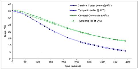
Figure 50: Cooling rate observed in porcine heads subjected to unstirred air or water cooling at 0oC from data by Baccino, et al. [181]

The reason for this is the limitation imposed by the very low value for k (heat conductivity) of the human head. This has the following important practical implications:

1)            External conductive cooling of the human head/brain is extremely slow even under ideal conditions of maximum surface contact with ice at 0oC where the melt water is filmed over the patient’s head.

2)            Once the surface of the patient’s head reaches 0oC, the brain cannot be cooled any faster regardless of the type or amount of conductive media used. In other words, using more conductive refrigerating media or delivering them at higher flow rates than necessary to keep the skin at 0oC will not work and may well be counterproductive (i.e., consume limited battery power and cause splashing and aerosolization of potentially biohazardous cooling bath water).

3)             If conditions are less than ideal because of poor contact with refrigerant and retention of melted ice water in plastic bags then cooling is slower still and this is undesirable.

4)            The basic requirement of uniformly cooling the surface of the patient’s head to near 0oC is, in practice, quite difficult to achieve because holding refrigerant in contact with the patient’s head involves problems associated with melting ice which is messy, damaging to bedding and furnishings, and can cause a slip hazard if dripped onto the floor. Containing ice in plastic bags results in considerable loss of contact with the skin and reduces the efficiency of cooling by causing melt water to be retained; creating a relative convective and conductive barrier. It is also virtually impossible to keep ice bags in position around the patient’s head during movement from one location to another (or for that matter, even when the patient is not being moved).

OroChill Oronasopharyngeal Cooling
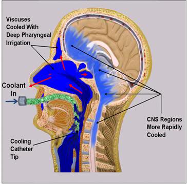
Figure 51: The OroChill oronasopharyngeal (ONP) cold saline irrigation system consists of a fenestrated catheter that is introduced oropharynx at the level of the vallecula, or deeper to allow for irrigation of the oropharynx and sinuses with ice cold saline. This results in ~30% improvement in cooling time to ~5 oC in porcine heads placed in a stirred normal saline bath chilled to ~1-2 oC.

While there is no easy solution to problems 1 and 2 above, there is a solution to problems 3 and 4: an enclosure to hold ice or another acceptable refrigerant around the patient’s head allowing for maximal contact of the patient’s head with the refrigerant. This is why the HIP was invented. Figure 52 shows just how bad conductive cooling is, even in water at 0 oC both in absolute terms; 3.5 hours after the start of cold water cooling the brain core temperature is still ~20 oC! The situation is worse for ice bag cooling (see Figure 38) where brain temperature will still be ~20oC 6.5 hours after the start of cooling. At a minimum the HIP must be used with ice in direct contact with the patient’s skin and with the ice being replenished before any part of the patient’s head becomes exposed. A stirred ice water bath will confer additional advantage, but the most effective way of cooling is to continuously irrigate the ONP with ice cold solution.
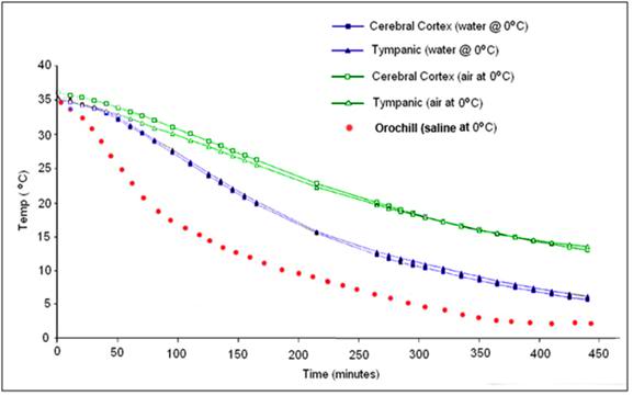
Figure 52: Cooling curve obtained using the OroChill ONPcooling device in a porcine head as compared to conductive cooling in air and water at 0 oC. The TC probe was placed in the cerebral cortex at a depth of ~70 mm from the skin on the dorsal surface of the head. Modified from Baccino, et al.

The OroChill kit contains 4 packets of mannitol and sodium chloride designed to be added to the PIB ice and water. This will yield a hypertonic solution that will decrease in osmolality as the ice in the PIB melts. The purpose of these added osmolytes is to prevent edema and ‘water logging’ of the nasopharyngeal mucosa which can significantly impede heat exchange. Isoosmolality is not important; the important thing is to avoid edema and, if possible, to induce dehydration in soft tissues overlying the brain. This has the effect of decreasing the thickness of the insulating gel layer surrounding the brain and thus improving heat exchange.

In the event rigor is present and sufficiently advanced, the pharyngeal irrigating tube may be replaced with the OroChill nasal cannula. The nasal cannula is ~20% less effective at cooling the brain than the pharyngeal irrigating tube.

Procedure for Using the OroChill
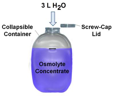
Figure 53: OroChill osmolyte concentrate is prepared by adding 3L of warm water to the osmolyte powder and blue food dye present in the flexible plastic mixing container. Once the osmolyte powder is fully dissolved it is added to the water of the PIB.

1) The OroChill osmolyte powder is packed in collapsible, heavy walled, screw cap containers. Unfold the container and add ~3 L of warm tap water. Agitate the contents by shaking the closed container until all of the powdered components have dissolved in solution.

2) Add the OroChill osmolyte concentrate to the water of the PIB and ensure that it is well distributed. It is not necessary for it to be evenly dispersed. The Osmolyte mix contains an innocuous blue food dye which serves both as indicator of the degree to which the concentrated has become well distributed in the water of the PIB, and to mask the presence of any blood or urine in the PIB water, the latter of which is disturbing to medical and mortuary personnel who may be unaware of the presence of antimicrobials in the PIB water. A color indicator strip is attached to the concentrate mixing bottle and it should be used to determine when distribution of the concentrate has reached a level sufficient to initiate ONP irrigation.

3) In order to prevent the OroChill heat exchange medium from entering into and accumulating in the lungs or the stomach, both the patient’s airway and esophagus must be sealed. These two objectives may be met in a variety of ways; an endotracheal tube may be used in combination with the obturator from a DEGTA (or EGDTA), or a DEGTA or D-Combitube airway may be used.
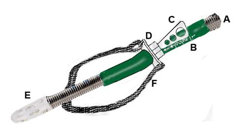
Figure 54: Oral wire reinforced OroChill catheter(A) for delivery of refrigerated cold solution to the oronasopharynx.The catheter rests within a sleeve that allows it to be advanced to retracted as necessary (B). The catheter is contained within a bite block assembly (D) with a positioning handle (C). The catheter has a fenestrated tip (E) for more even dispersion of coolant in the patient’s oropharynx. The entire assembly is held in place with a Velcro head strap (F).

4) Once the airway is secured, the patient’s head is positioned in the OroChill Head Cooling Positioner (OCHCP) and the oral irrigation cannula is placed. Advance the cannula until the tip is positioned in the vallecula.

5) Secure the OCHCP to the patient’s head using the integral Velcro strap. Exercise care not to excessively tighten the strap; it should be just tight enough to prevent the oral cannula from becoming dislodged. It should be possible to easily slip two fingers between the securing strap and the patient’s face.
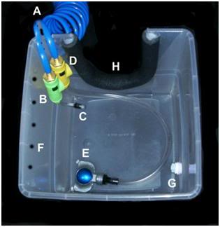
Figure 55: The Orochill catheter line is attached to the quick disconnect fitting (C) on the OroChill pigtail line issuing from the SCCD cold water supply line (A) to the HIP (H). Coolant flow to the OroChill catheter is adjusted with green control valve (B).

6)  Using the quick disconnect fitting on the OroChill pharyngeal or nasal catheter (as appropriate) attach the male quick disconnect fitting on the end of the catheter supply line to the female end of the connector on the green flow control valve of the SCCD supply line in the HIP.

7) Open the flow control valve and adjust the flow until a steady stream of heat exchange liquid can be seen to be issuing from both nares (and alternatively, if a nasal cannula has been used, from the mouth).

8) As additional ice is added to the PIB, or as ice melt occurs, re-check to color of the PIB bath liquid (osmolyte solution) and add additional osmolyte concentrate as needed to maintain the concentration of the osmolyte in the PIB bath in a acceptable range.
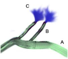
Figure 55: The OroChill nasal cannula may be used if the patient’s jaw is in rigor, or it is otherwise impossible to place the oral catheter.  Flows are considerably lower through the nasal cannula and a significant amount of back flow may occur from the nares. For these reasons, the use of the pharyngeal catheter is preferred, when possible.


Monitoring Temperature Descent

As is hopefully clear from much ofthe foregoing discussion, it is critical that cooling data be obtained from every patient where it is possible to do so, as soon as it it possible to do so. Not only is documenting the patient’s temperature descent an important element of care, it also serves as a surrogate marker for the quality of that care, as well as providing valuable scientific data to allow for improvements to the care of future patients. Thus, it is essential that temperature probes be placed, and data acquisition be started as soon as possible after stabilization begins. Where possible, the patient’s agonal temperatures should be recorded at 15 minute intervals up until the time of pronouncement, using whatever method the attending medical staff are using, or are comfortable with being used.
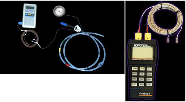
Figure 56: Two examples of remote sensing thermometers that can be used to monitor patient cool down. The device on the left is a non-recording thermocouple thermometer from which temperature measurements must be recorded by hand. It is shown connected to a probe attached to a gastric medication and esophageal (intrathoracic) pressure monitoring balloon. The gastric tube TC probe may be used in cases where an endotracheal tube is in place or will be used.

The DEGTA and the Darwin Rectal Tube (DRT) are both outfitted with copper (CU++)/Constantan (C+) thermocouples (TCs) so that as soon as they are placed, temperature monitoring and data acquisition may be initiated.This is best accomplished by either pre-connecting the TC probes to an automatic datalogging thermocouple thermometer, such as the Digi-Sense DualLogger, or to the data acquisition laptop computer which is collecting other data from the patient’s stabilization and transport. Detailed instructions for the use of the DualLogger, including how to interface it with its waterproof perspex case, are present as Appendix 2 (NOTE: Not included in this article). If the DualLogger is used, it must be not be water proofed in an expedient way, such as by placing it in Ziploc bags, because this still leaves the data entry/setting keys susceptible to being accidentally depressed. This has proved a major source of data loss in the past; the device must be placed in a waterproof hard case to protect the keys from accidentally being depressed while the patient is being moved or air shipped.
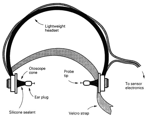
Figure 57: Bilateral tympanic temperature monitoring device.

If the patient has an endotracheal tubbe (ET) or will be intubated, tympanic temperature probes may be placed using a device such as one shown in Figure 57. This earphone-like assembly contains two wells of a fast setting silicone sealant which is injected into the ear canal using syringes that are pre-attached to each probe holder (not shown). Once the TC probes have been advanced into the auditory canal to abut the eardrum, the sealant is injected to prevent the ingress of cold water from the HIP, and to secure the probes against accidental dis-lodgement. These, and all other probes, should be left in place at the conclusion of Transport. If such a device is not available, a soft, flexible vinyl TC probe may be advanced into the auditory canal until it abuts the eardrum, where it may be sealed in place using silione swimmer’s ear canal sealing putty, as shown in Figure 58, below.
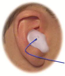
Figure 58: Silicone swimmer’s ear sealing putting may be used to seal and secure TC probes into the auditory canal. If this method is to be used, it is important to clean the external auditory meatus with alcohol swabs to remove waxy secretions which may prevent the putty from adhering well and creatina good seal. The probe line should be looped over the top of top of the pinna (after it is secured in the ear canal ) and taped firmly in place to prevent it from accidentally become dislodged.

Where automatic logging equipment is available, temperatures should be collected at 5 second intervals, with consideration given to limitations that may present as a result of the available memory in the data collection device. The lowest acceptable rate of temperature data acquisition is intervals of 5 minutes.
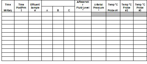
Figure 58: Sample data collection sheet for manual collection of temperature data in the field. This sheet also includes columns for the entry of total body washout/perfusion data. Note that there is a space for recording the time post-pronouncement; this allows for immediate feedback as to how how effectively (rapidly) the patient is cooling. The exact anatomical or physical location in the extracorporeal circuit of thetemperature probes must be entered in the shaded boxes.

At the conclusion of stabilization, the temperatures probes should be left in place and the lead wires carefully separated, coiled up, and placed atop the patient’s thorax. Vinyl coated wire twist ties are ideal for both separating and holding the probes in the coiled position.

Only Cu++/C- TC probes since these may, if necessary, be left in position and used for data acquisition throughout cooling to liquid nitrogen temperature. This eliminates the requirement for replacing the probes with different probes for the subzero parts of the operation, and also helps to ensure continuity of data acquisition, since the same probe should be in the same position from the stat to the finish of the patient’s cryopreservation.

References

1.             Hillyard Industries I: Vindicator+ Technical Data Sheet #168. In. St. Joseph, MO: Hillyard Industries, Inc; 2010.

2.             Greeley W, Kern, FH, Ungerleider, RM. et al.: Cerebral metabolic suppression during hypothermic circulatory arrest in humans. The Annals of Thoracic Surgery 1999, 67(6):1895-1899.

3.             Hixon H: The question column: How cold is cold enough? Cryonics 1985, 6(1):19-25.

4.             Bellinger DC WD, duPlessis AJ, Rappaport LA, Jonas RA, Wernovsky G, Newburger JW.: Neurodevelopmental status at eight years in children with dextro-transposition of the great arteries: the Boston Circulatory Arrest Trial. J Thorac Cardiovasc Surg 2003, 126(5):1385-1396.

5.             Wypij D, Newburger, JW, Rappaport, LA, duPlessis, AJ, Jonas RA, Wernovsky, G, Lin, M, Bellinger, DC.: The effect of duration of deep hypothermic circulatory arrest in infant heart surgery on late neurodevelopment: the Boston Circulatory Arrest Trial. J Thorac Cardiovasc Surg 2003, 126(5):1397-1403.

6.             Ungerleider R, Gaynor,  JW.: The Boston Circulatory Arrest Study: An analysis. J Thorac Cardiovasc Surg 2004, 127:1256-1261.

7.             Lorentzen G: Food preservation by refrigeration, a general introduction. International Journal of Refrigeration 1978, 1(1):9-12.

8.             Ross H MV, Escott ML.: 72-hr canine kidney preservation without continuous perfusion. Transplantation 1976, 21(6):498-501.

9.             Sung D, Woods, JE.: Forty-Eight-Hour Preservation of the Canine Liver. Ann Surg 1974, 199(4):422-426.

10.          Monden M, Fortner, JG.: Twenty-four- and 48-hour canine liver preservation by simple hypothermia with prostacyclin. Ann Surg 1982, 196(1):38-42.

11.          Todo S, Hamada, N,  Zhu, Y,  Zhang, S,  Subbotin, V, Nemoto, A,  Takeyoshi, I,  Starzl, TE.: Lazaroid U-74389G for 48-hour canine liver preservation. Transplantation 1996, 61(2):189-194.

12.          Belzer F, Southard, JH.: Principles of solid-organ preservation by cold storage. Transplantation 1988, 45(4):673-676.

13.          Michenfelder J, Milde, JH.: The effect of profound levels of hypothermia (below 14 degrees oC) on canine cerebral metabolism. J Cereb Blood Flow Metab 1992, 12(5):877-880.

14.          Haneda K, Thomas, R, Sands, MP, Breazeale, DG, Dillard, DH.: Whole body protection during three hours of total circulatory arrest: an experimental study. Cryobiology 1986, 23(6):483-494.

15.          Drabek T, Fisk, JA, Dixon, CE, Garman, RH, Stezoski, J, Wisnewski, SR, Wu, X, Tisherman,, SA K, PM.: Prolonged deep hypothermic circulatory arrest in rats can be achieved without cognitive deficits.

. Life Sci 2007, 8(7):543-552.

16.          Goldzveig S, Smith, AU.: A simple method for reanimating rats and mice. J Physiol 1956, 132(2):406-413.

17.          Benjamin-Cummings T: Chemical Kinetics, Third Edition. Weinheim: Wiley-VCH Verlag GmbH & Co. KGaA; 1997.

18.          Irias J, Olmsted, MR.: Pyruvate carboxylase. Reversible inactivation by cold. Pyruvate carboxylase. Biochemistry 1969, 19:91-98.

19.          Avery S, Lloyd, D, Harwood, JL.: Temperature-dependent changes in plasma-membrane lipid order and the phagocytotic activity of the amoeba Acanthamoeba castellanii are closely correlated. Biochem J 1995, 312((Pt 3)):811-816.

20.          Zakim D, Kavecansky, J, Scarlata, S.: Are membrane enzymes regulated by the viscosity of the membrane environment? . Biochemistry 1992, 31(46):11589-11594.

21.          Perry R: Towards a measure of ischemic injury. . Cryonics 1996, 17(2):21.

22.          Harris S: Initial cooling in cryonics from body temperature to ice temperature: Physiologic and physics theory, quality control proposals, historical cryonics case analysis examples, lab experimental results, literature review, numerical recipe examples, and practical summaries and recommendations for the future.

. In. Rancho Cucamonga, CA: Critical Care Research; 2003.

23.          Kim H, et al.: Amelioration of impaired cerebral metabolism after severe acidotic ischemia by tirilazad post-treatment in dogs. . Stroke 1996, 27:114-121.

24.          Iwatsuki N, et al.: Hyperbaric oxygen combined with nicardipine administration accelerates neurologic recovery after cerebral ischemia in a canine model. . Crit Care Med 1994, 22:858-863.

25.          Cervantes M, Moralı´, G, Letechipı´a-Vallejo, G.: Melatonin and ischemia reperfusion injury of the brain. J Pineal Res 2008, 45:1-7.

26.          Krep H, Bernd W, Bottiger, BW, et al.: Time course of circulatory and metabolic recovery of cat brain after cardiac arrest assessed by perfusion- and diffusion-weighted imaging and MR-spectroscopy. Resuscitation 2003, 58:337-348.

27.          DeGraba T, Pettigrew, C.: Why do neroprotectivedrugs work in animals but not in humans? Neurologic Clinics 2000, 18:475-493.

28.          Ginsberg M: Adventures in the Pathophysiology of Brain Ischemia: Penumbra, Gene Expression, Neuroprotection. The 2002 Thomas Willis Lecture. Stroke 2003, 34:214-223.

29.          Cheng J, Al-Khoury, L, Zivin, JA.: Neuroprotection for Ischemic Stroke: Two Decades of Success and Failure. NeuroRX 2004, 1:36-45.

30.          Roine RO, Kaste M, Kinnunen A, Nikki P, Sarna S, Kajaste S: Nimodipine after resuscitation from out-of-hospital ventricular fibrillation. A placebo-controlled, double-blind, randomized trial. JAMA 1990, 264(24):3171-3177.

31.          Longstreth WT, Jr., Fahrenbruch CE, Olsufka M, Walsh TR, Copass MK, Cobb LA: Randomized clinical trial of magnesium, diazepam, or both after out-of-hospital cardiac arrest. Neurology 2002, 59(4):506-514.

32.          Landau WM, Schneider S, Machado C, Longstreth WT, Jr., Fahrenbruch CE, Olsufka M, Walsh TR, Copass MK, Cobb LA: Randomized clinical trial of magnesium, diazepam, or both after out-of-hospital cardiac arrest. Neurology 2003, 60(11):1868-1869.

33.          Halstrom A, Rea, TD, et al.: Manual chest compression vs use of an automated chest compression device during resuscitation following out-of-hospital cardiac arrest: a randomized trial. JAMA 2006, 295:2620-2628.

34.          Lafuente-Lafuente C, Melero-Bascones, M.: Active chest compression-decompression for cardiopulmonary resuscitation. Cochrane Database Syst Rev 2004, (2):CD002751.

35.          Aung K, Htay, T.: Vasopressin for cardiac arrest: a systematic review and meta-analysis. Arch Intern Med 2005, 10:17-24.

36.          Callaham M, Madsen, CD, Barton, CW, Saunders, CE, Pointer, J.: A randomized clinical trial of high-dose epinephrine and norepinephrine vs standard-dose epinephrine in prehospital cardiac arrest. JAMA 1992, 268:2667-2672.

37.          Rincon F, Mayer, SA.: Therapeutic hypothermia for brain injury after cardiac arrest. . Semin Neurol 2006, 26:387-395.

38.          Bernard S, Gray, TW, Buist, MD, et al.: Treatment of comatose survivors of out-of-hospital cardiac arrest with induced hypothermia. N Engl J Med 2002, 346:557-563.

39.          Liu L, Yenari MA: Therapeutic hypothermia: neuroprotective mechanisms. Front Biosci 2007, 12:816-825.

40.          The-Hypothermia-after-Cardiac-Arrest-Study-Group: Mild therapeutic hypothermia to improve the neurologic outcome after cardiac arrest. N Engl J Med 2002, 346:549-556.

41.          Nolan J, Morley, PT, Vanden Hoek, TL, Hickey, RW.: Therapeutic Hypothermia After Cardiac Arrest: An Advisory Statement by the Advanced Life Support Task Force of the International Liaison Committee on Resuscitation. Circulation 2003, 108:118-121.

42.          Arrich J: Clinical application of mild therapeutic hypothermia after cardiac arrest. Crit Care Med 2007, 35(4):1041-1047.

43.          Sandroni C, Nolan J, Cavallaro F, Antonelli M: In-hospital cardiac arrest: incidence, prognosis and possible measures to improve survival. Intensive Care Med 2007, 33(2):237-245.

44.          Safar P: Resuscitation from clinical death: Pathophysiologic limits and therapeutic potentials. Crit Care Med 1988, 16:923-941.

45.          Safar P, Xiao F, Radovsky A, Tanigawa K, Ebmeyer U, Bircher N, Alexander H, Stezoski SW: Improved cerebral resuscitation from cardiac arrest in dogs with mild hypothermia plus blood flow promotion. Stroke 1996, 27(1):105-113.

46.          Polderman KH: Hypothermia and neurological outcome after cardiac arrest: state of the art. Eur J Anaesthesiol Suppl 2008, 42:23-30.

47.          Busto R, Dietrich, WD, Globus, MY, et al.: Small differences in intraischemic brain temperature critically determine the extent of ischemic neuronal injury. J Cereb Blood Flow Metab 1987, 7:729-738.

48.          Safar P, Behringer, W. (ed.): Brain resuscitation after cardiac arrest. Philadelphia: WB Saunders; 2001.

49.          Shimohata T, Zhao H, Steinberg GK: Epsilon PKC may contribute to the protective effect of hypothermia in a rat focal cerebral ischemia model. Stroke 2007, 38(2):375-380.

50.          Zhao H, Steinberg GK, Sapolsky RM: General versus specific actions of mild-moderate hypothermia in attenuating cerebral ischemic damage. J Cereb Blood Flow Metab 2007, 27(12):1879-1894.

51.          American-Heart-Association: AHA CPR Guidelines: Part 7.2: Management of Cardiac Arrest. Circulation 2005, 112:IV-58-IV-66.

52.          Abella B, Rhee, JW, Huang, KN, et al.: Induced hypothermia is underused after resuscitation from cardiac arrest: A current practice survey. Resuscitation 2005, 64:181-186.

53.          Merchant R, Soar, J, Skrifvars, MB, et al.: Therapeutic hypothermia utilization among physicians after resuscitation from cardiac arrest. . Crit Care Med 2006, 34:1935-1940.

54.          Kennedy J, Green, RS, Stenstrom, R.: The use of induced hypothermia after cardiac arrest: A survey of Canadian emergency physicians. CJEM 2008, 10:125-130.

55.          Suffoletto B, Salcido, DD, Menegazzi, JJ.: Use of prehospital-induced hypothermia after out-of-hospital cardiac arrest: A Survey of the National Association of Emergency Medical Services Physicians. Prehosp Emerg Care 2008, 12:52-56.

56.          Alam H: Just do it. Critical care medicine, 2008, 36:2456-2457.

57.          Skulec R, Kovarnik T, Dostalova G, Kolar J, Linhart A: Induction of mild hypothermia in cardiac arrest survivors presenting with cardiogenic shock syndrome. Acta Anaesthesiol Scand 2008, 52(2):188-194.

58.          Hay AW, Swann DG, Bell K, Walsh TS, Cook B: Therapeutic hypothermia in comatose patients after out-of-hospital cardiac arrest. Anaesthesia 2008, 63(1):15-19.

59.          Wolfrum S, Pierau C, Radke PW, Schunkert H, Kurowski V: Mild therapeutic hypothermia in patients after out-of-hospital cardiac arrest due to acute ST-segment elevation myocardial infarction undergoing immediate percutaneous coronary intervention. Crit Care Med 2008, 36(6):1780-1786.

60.          Bernard S: Therapeutic hypothermia after cardiac arrest: now a standard of care. Crit Care Med 2006, 34(3):923-924.

61.          Wolff B, Machill K, Schumacher D, Schulzki I, Werner D: Early achievement of mild therapeutic hypothermia and the neurologic outcome after cardiac arrest. Int J Cardiol 2008.

62.          Nagel S, Papadakis M, Hoyte L, Buchan AM: Therapeutic hypothermia in experimental models of focal and global cerebral ischemia and intracerebral hemorrhage. Expert Rev Neurother 2008, 8(8):1255-1268.

63.          Nozari A, Safar P, Stezoski SW, Wu X, Kostelnik S, Radovsky A, Tisherman S, Kochanek PM: Critical time window for intra-arrest cooling with cold saline flush in a dog model of cardiopulmonary resuscitation. Circulation 2006, 113(23):2690-2696.

64.          Reich H, Angelos M, Safar P, Sterz F, Leonov Y: Cardiac resuscitability with cardiopulmonary bypass after increasing ventricular fibrillation times in dogs. Ann Emerg Med 1990, 19(8):887-890.

65.          Angelos M, Safar P, Reich H: A comparison of cardiopulmonary resuscitation with cardiopulmonary bypass after prolonged cardiac arrest in dogs. Reperfusion pressures and neurologic recovery. Resuscitation 1991, 21(2-3):121-135.

66.          Radovsky A, Safar P, Sterz F, Leonov Y, Reich H, Kuboyama K: Regional prevalence and distribution of ischemic neurons in dog brains 96 hours after cardiac arrest of 0 to 20 minutes. Stroke 1995, 26(11):2127-2133; discussion 2133-2124.

67.          Thoresen M, Wyatt, J.  [see comments]: Keeping a cool head, post-hypoxic hypothermia: an old idea revisited. Acta Paediatr 1997, 86:1029-1033.

68.          Nozari A, Safar P, Stezoski SW, Wu X, Henchir J, Radovsky A, Hanson K, Klein E, Kochanek PM, Tisherman SA: Mild hypothermia during prolonged cardiopulmonary cerebral resuscitation increases conscious survival in dogs. Crit Care Med 2004, 32(10):2110-2116.

69.          Kuboyama K, Safar, P, Radovsky, A, Tisherman, SA, Stezoski, SW, Alexander, H.: Delay in cooling negates the beneficial effect of mild resuscitative cerebral hypothermia after cardiac arrest in dogs: a prospective, randomized study [see comments]. 21 1993, Crit Care Med:1348-1358.

70.          Takata K, Takeda, Y, Sato T, Nakatsuka, H, Yokoyama, M, Morita, K.: Effects of hypothermia for a short period on histologic outcome and extracellular glutamate concentration during and after cardiac arrest in rats. . Crit Care Med 2005, 33:1340 -1345.

71.          Abella B ZD, Alvarado J, Hamann K, Vanden Hoek T, Becker L.: Intra-arrest cooling improves outcomes in a murine cardiac arrest model. Circulation 2004, 109:2786 -2791.

72.          Dietrich W, Busto, R, Alonso, O, et al. 993: Intraischemic but not postischemic brain hypothermia protects chronically following global forebrain ischemia in rats. J Cerebr Blood Flow  Metab 1993, 13:541-549.

73.          Maier CM, Sun GH, Kunis D, Yenari MA, Steinberg GK: Delayed induction and long-term effects of mild hypothermia in a focal model of transient cerebral ischemia: neurological outcome and infarct size. J Neurosurg 2001, 94(1):90-96.

74.          Marion D, Leonov Y, Ginsberg, M, Katz, LM, Kochanek, PM, Lechleuthner, A, Nemoto, EM, Obrist, W, Safar, P, Sterz, F, Tisherman, SA, White, RJ, Xiao, F, Zar, H.: Resuscitative hypothermia. Crit Care Med 1996, 24:S81-89.

75.          Zivin J: Editorial comment. Stroke 1999, 30:1899.

76.          Wake-County-Government: Wake County Emergency Medical Services. In.: Wake County Government; 2008.

77.          Hinchey P, et al.: Out-of-hospital cardiac arrest survival after the sequential implementation of 2005 AHA guidelines for compressions, ventilations, and induced hypothermia. In: SAEM Meeting. vol. Abstract 167. Washington, D.C. ; 2008.

78.          Olson J, Hinchey, P, Myers, B.: Wake County EMS Cools Down: System begins induced hypothermia of ROSC patients. In: JEMS. vol. 31: Elsevier Public Safety; 2006.

79.          Bayegan K, Janata A, Frossard M, Holzer M, Sterz F, Losert UM, Laggner AN, Behringer W: Rapid non-invasive external cooling to induce mild therapeutic hypothermia in adult human-sized swine. Resuscitation 2008, 76(2):291-298.

80.          Kurz A, Sessler, DI, Birnbauer, F, Illievich, U, Spiss, C.: Thermoregulatory vasoconstriction impairs active core cooling. Anesthesiology 1995, 82:870-876.

81.          Stolwijk J, Hardy, JD. (ed.): Control of body temperature: Reactions to environmental agents. Bethesda: American Physiological Society; 1977.

82.          Doufas A: Consequences of inadvertent perioperative hypothermia. Best Pract Res Clin Anaesthesiol 2003, 17:535-549.

83.          Sheffield C, Sessler, DI, Hunt, TK.   : Mild hypothermia during isoflurane anesthesia decreases resistance to E. coli dermal infection in guinea pigs. Acta Anaesthesiol Scand 1994, 38:201-205.

84.          Lee S, Battistella, FD, Go, K.: Hypothermia induces T-Cell production of immunosuppressive cytokines. J Surg Res 2001 100(4):150-153.

85.          Reed R, Bracey, AW, Hudson, JD, Miller, TA, Fisher, RP. : Hypothermia and blood coagulation: dissociation between enzyme activity and clotting factor levels. Circ Shock 1990, 32:141-152.

86.          Nirula R, Gentilello, LM. (ed.): Detrimental Effects of Hypothermia New York: Springer US; 2007.

87.          Flores-Maldonado A, Medina-Escobedo, CE, Rios-Rodriguez, HM, Fernandez-Dominguez, R. : Mild perioperative hypothermia and the risk of wound infection. Arch Med Res 2001, 32:227-231.

88.          Kurz A, Sessler, DI, Lenhardt, R.: Perioperative normothermia to reduce the incidence of surgical wound infection and shorten hospitalization. N Engl J Med 1996, 334:1209-1215.

89.          Sheffield C, Sessler, DI, Hopf, HW, Schroeder, M, Moayeri, A, Hunt, TK, West, JM. : Centrally and locally mediated thermoregulatory responses alter subcutaneous oxygen tension. Wouns Rep Reg 1997, 4:339-345.

90.          Wenisch C, Narzt, E, Sessler, DI. Parschalk, B, Lenhardt, R, Kurz, A, Graninger, W.: Mild intraoperative hypothermia reduces production of reactive oxygen intermediates by polymorphonuclear leukocytes. Anesth Analg 1996, 82:810-816.

91.          Beilin B, Shavit, Y, Razumovsky, J, et al. : Effects of mild perioperative hypothermia on cellular immune responses. Anesthesiology 1998, 89:1133-1140.

92.          Jyung R, Mustoe, TA.: Role of cytokines in wound repair. New York: Oxford University Press; 1993.

93.          Baigrie R, Lamont, PM, Kwiatkowski, D, Dallman, MG, Morris, PG.: Systemic cytokine response after major surgery. Br J Surg 1992, 79:757-760.

94.          Qwarnstrom E, Page, RC, Gillis, S, Dower, SK.: Binding, internalization and intracellular localization of interleukin-1 beta in human diploid fibroblasts. B Biol Chem 1988;, 263::8261-8269.

95.          Peterson K, Carson, S, Carney, N.: Hypothermia treatment for traumatic brain injury: a systematic review and meta-analysis. J Neurotrauma 2008, 25(1):62-71.

96.          Hammersborga S, Brekkea, KB, Haugena, OM, et al.: Surface cooling versus core cooling: Comparative studies of microvascular fluid- and protein-shifts in a porcine model. Resuscitation 2008, 79:292-300.

97.          Merchant R, Abella,BS, Peberdy, MA, Soar, J, et al.: Therapeutic hypothermia after cardiac arrest: Unintentional overcooling is common using ice packs and conventional cooling blankets. Crit Care Med 2006, 34[Suppl.]:S490-S494.

98.          Chen R, Chien, S.: Hemodynamic functions and blood viscosity in surface hypothermia. Am J Physiol Lung Cell Mol Physiol 1978, 235:H136-143.

99.          Zarins C, Skinner, DB.: Hemodynamics and blood flow distribution following prolonged circulation at 5 degrees C. Am J Physiol 1975, 229:275-278.

100.        Hammersborga S, Farstad, M, Haugen, O, Kvalheim, VL, Onarheim, H, Husby, P.: Time course variations of haemodynamics, plasma volume and microvascular fluid exchange following surface cooling: an experimental approach to accidental hypothermia. Resuscitation 2005, 65:211-219.

101.        Lewis T: Swelling of the human limbs in response to immersion in cold water. Clin Sci 1939, 4.

102.        Farry P, Prentice, NG, Hunter, AC, Wakelin, CA.: Ice treatment of injured ligament: an experimental model. N Z Med J 1980, 91:12-14.

103.        Matthew C, Sils, IV, Bastille, AM.: Tissue-specific extravasation of albumin-bound Evans blue in hypothermic and rewarmed rats. Can J Physiol Pharmacol 2002, 80(3):233-243.

104.        Kruuv J, Glofcheski, D, Cheng, KH, Campbell, SD, Al-Qysi, HMA, Nolan, WT, Lepock, JR: Factors influencing survival and growth of mammalian cells exposed to hypothermia. I. Effects of temperature and membrane lipid perturbers. J Cell Physiol 1983, 115:179-185.

105.        Rule G, Frim, J, Thompson, JE, Lepock, JR, Kruuv, J.  : Theeffect of membrane lipid perturbers on survival of mammalian cells to cold. Cryobiology 1978, 15:408-414.

106.        Marsh D, Lindell, SL, Fox, LE, Belzer, FO, Southard, JH. : Hypothermic preservation of hepatocytes. I. Role of cell swelling. Cryobiology 1989, 26:524-534.

107.        McAnulty J, Ametani, MS, Southard, JH, Belzer, FO.: Effect of hypothermia on intracellular Ca21 in rabbit renal tubules suspended in UW-gluconate preservation solution. . Cryobiology 1996, 33:196-204.

108.        Breton S, Brown, D.: Cold-induced microtubule disruption and relocalization of membrane proteins in kidney epithelial cells. J Am Soc Nephrol 1998, 9:155-166.

109.        Stefanovich PE, RM, Sheehan, SJ, Tompkins, RG, Yarmush, ML.: Effects of hypothermia on the function, membrane integrity, and cytoskeletal structure of hepatocytes. Cryobiology 1995, 23:389-403.

110.        Zieger M, Glofcheski, DJ, Lepock, JR, Kruuv, J. : Factors influencing survival of mammalian cells exposed to hypothermia IV. Effects of iron chelation. . Cryobiology 1990, 27:452-464.

111.        Rauen R, DeGroot, H.: Cold induced release of reactive oxygen species asa decisive mediator of hypotherrmiainjury to cultured liver cells. Free Rad Biol & Med 1998, 24(7/8):1316-1323.

112.        Hochachka P: Defense strategies against hypoxia and hypothermia. Science 1986, 231:234-241.

113.        Endrich B, Hammersen F, Messmer, K.: Microvascular ultrastructure in non-freezing cold injury. Res Exp Med 1990, 190:365-379.

114.        Verrey F, Groscurth, P, Bolliger, UI.: Cytoskeletal disruption in A6 kidney cells: Impact on endo-exocytosis and NaCI transport regulation by antidiuretic hormone. J Membr Bio 1995, 145:193-204.

115.        Hall S, Komai, H, Reader, J, Haworth, SG. : Donor lung preservation: Effect of cold preservation fluids on cultured pulmonary endothelial cells. . Am J Physiol Lung Cell Mol Physiol 1994, 267: L508-L505 I 507.

116.        Breton S, Brown, D.: Cold-induced microtubule disruption and relocalization of membrane proteins in

kidney epithelial cells. J Am Soc Nephrol 1998, 9:155-166.

117.        Reed C, Clark, D, editors (ed.): Heat exchangers and hypothermia. Houston TX: Texas Medical Press, Inc.; 1975.

118.        Furuse M, Ohta, T, Ikenaga, T, et al.: Effects of intravascular perfusion of cooled crystalloid solution on cold-induced brain injury using an extracorporeal cooling-filtration system. Acta Neurochir 2003, 145:983-993.

119.        Behringer W, Safar P, Wu X, Nozari A, Abdullah A, Stezoski SW, Tisherman SA: Veno-venous extracorporeal blood shunt cooling to induce mild hypothermia in dog experiments and review of cooling methods. Resuscitation 2002, 54(1):89-98.

120.        Overlie P: Emergency Use of Cardiopulmonary Bypass. Journal of Interventional Cardiology 1995, 8:239-247.

121.        Kurusz M, Zwischenberger, JB.: Percutaneous cardiopulmonary bypass for cardiac emergencies. Perfusion 2002, 17:269-277.

122.        Holzer M, Behringer, W, Janata, A, Bayegan, K, Schima, H, Deckert, Z, Losert, U, Laggner, AN, Sterz, F.: Extracorporeal venovenous cooling for induction of mild hypothermia in human-sized swine. Crit Care Med 2005, 33:1346-1350.

123.        Bernard S, Buist, M, Monterio, O, Smith, K.: Induced hypothermia using large volume, ice-cold intravenous fluid in comatose survivors of out-of-hospital cardiac arrest: a preliminary report. Resuscitation 2003, 56:9-13

124.        Bernard S, Rosalion, A.: Therapeutic hypothermia induced during cardiopulmonary resuscitation using large-volume, ice-cold intravenous fluid. Resuscitation 2008, 76:311 – 313.

125.        Kamarainen A, Virkkunen I, Tenhunen J, Yli-Hankala A, Silfvast T: Prehospital induction of therapeutic hypothermia during CPR: a pilot study. Resuscitation 2008, 76(3):360-363.

126.        Haslam D, James, WP. : Obesity. Lancet 2005, 366:1197-1209.

127.        Feldschuh J, Enson, Y.: Prediction of the normal blood volume. Relation of blood volume to body habitus. Circulation 1977, 56:605 – 612.

128.        Plaisier BR: Thoracic lavage in accidental hypothermia with cardiac arrest–report of a case and review of the literature. Resuscitation 2005, 66(1):99-104.

129.        Xiao F, Safar P, Alexander H: Peritoneal cooling for mild cerebral hypothermia after cardiac arrest in dogs. Resuscitation 1995, 30(1):51-59.

130.        Neuropreservation of Alcor patient A-1068. [http://www.alcor.org/Library/html/casereport8504.html]

131.        Cryopreservation Patient Case Report: Arlene Francis Fried, A-1049 [http://www.alcor.org/Library/html/fried.html]

132.        Jones T: Cryopreservation case report: the cryopreservation of patient A-2063. Cryonics 2005, 26(1):10-12.

133.        Leaf J, Quiafe, A.: Case study of neuropreservation. Cryonics 1981, 2(16):21-28.

134.        Leaf J, Darwin, MG, Hixon, HL.: Case report: Two consecutive cryopreservations, a comparative study in experimental suspended animation in humans. Cryonics 1985, 6:13-37.

135.        Darwin M, Leaf, JD, Hixon, HL.: Case report: Neuropreservation of Alcor patient A-1068. Cryonics 1986, 7(2-3).

136.        Darwin M: Alcor member placed in suspension. Cryonics 1985, 6(4):10-19.

137.        Darwin M: The journey begins: Alcor member enters biostasis. Cryonics 1987, 8(8):14-22.

138.        Bridge S: The cryonic suspension of Alice Black. Cryonics 1988, 9(11):15-25.

139.        Edwards N, et al.: Survival in adults after cardiac arrest due to drowning. Intensive Care Med 1990, 16(5):336-337.

140.        Husby P, et al.: Accidental hypothermia with cardiac arrest: complete recovery after prolonged resuscitation and rewarming by extracorporeal circulation. Intensive Care Med 1990, 16(1):69-72.

141.        Sekar T, et al.: Survival after prolonged submersion in cold water without neurologic sequelae.Report of two cases. Arch Intern Med 1980, 140(6)):775-779.

142.        Martin T: Near drowning and cold water immersion. Ann Emerg Med 1984, 13(4):263-273.

143.        Ornato J: The resuscitation of near drowning victims. JAMA 1986, 256:75-77.

144.        JB. R: Hypothermia: pathophysiology, clinical settings, and management. Ann Intern Med 1978 89(4):519-527.

145.        Conn A, Barker, GA, Edmonds, JF, et al. (ed.): Submersion hypothermia and near drowning. Minneapolis: University of Minnesota Press.; 1983.

146.        Bolte R, Black, PG, Bowers, RS, et al.: The use of extracorporeal rewarming in a child submerged for 66 minutes. JAMA 1988, 260(3):377-379.

147.        Alexander L: The Treatment of Shock from Prolonged Exposure to Cold, Especially in Water. Item No. 24, File No. XXVI. In. Edited by Subcommittee CIO. Washington, D. C.: United States Congress; 1945 (July): 1-228.

148.        Mitscherlich  A MF: Doctors of Infamy: The Story of Nazi Medical Crimes. New York: Henry Schuman; 1949.

149.        Vogelaere P: Cardiac output variations in supine resting subjects during head-out cold water immersion. . Int J Biometeorol 1995, 39(1):40-45.

150.        Giesbrecht G: Inhibition of shivering increases core temperature afterdrop and attenuates rewarming in hypothermic humans. Appl Physiol 1997, 83(5):1630-1634.

151.        Bristow G (ed.): Clinical aspects of accidental hypothermia. New York: Elsevier; 1985.

152.        Hayward J, et al.: Physiological responses and survival time prediction for humans in ice-water. Aviat Space Environ Med 1984, 55(3):206-211.

153.        Griepp R, et al.: Prosthetic replacement of the aortic arch. J Thorac Cardiovasc Surg 1975, 70(6):1051-1063.

154.        Hayward J, et al.: Thermal and cardiovascular changes during three methods of resuscitation from mild hypothermia. Resuscitation 1984, 11(1-2):21-33.

155.        Toner M, et al.: Effects of body mass and morphology on thermal responses in water. J Appl Physiol 1986, 60(2):521-525.

156.        Darwin M: Cryopreservation case report: Arlene Francis Fried, A-1049. In. Scottsdale: Alcor Life Extension Foundation; 2004.

157.        Darwin M, Pizer, D.: Alcor outstanding support award nominee, Coordinators column: http://www.alcor.org/cryonics/cryonics8907.txt Cryonics 1989, 10(7):11-14.

158.        Borlaug G, Fox, BC, Davis, JP.: Community associated methcillin resistant staphylococcus aureus (CA MRSA). Guidelines for Clinical management and control of transmission. In. Edited by Wisconsin Division of Public Health bdswu, 608-267-7711. Madison; 2005.

159.        Fridkin S, Hageman, JC, Morrison, M, et al.: Methicillin resistant Staphylococcus aureus disease in three communities. NEJM 2005, 352:1436-1444.

160.        Johnson L, Saravolatz, LD.: Community acquired MRSA: current epidemiology and management issues. Infect Med 2005, 22:16-20.

161.        Pichichero M, Casey, JR.: Emergence of a multiresistant serotype 19A pneumococcal strain not included in the 7-valent conjugate vaccine as an otopathogen in children. JAMA 2007, 298:1772-1778.

162.        Buscha H, Eichwedeb, F, Födischc, Tacconed, MFS.: Safety and feasibility of nasopharyngeal evaporative cooling in the emergency department setting in survivors of cardiac arrest. Resuscitation 2010, 81:943-949.

163.        Castren M, Nordberg, P, Svensson, L., et al.: Intra-arrest transnasal evaporative cooling a randomized, prehospital, multicenter study (PRINCE: Pre-ROSC IntraNasal Cooling Effectiveness). Circulation 2010, 122:729-736.

164.        Bollera M, Lampea, JW,  Katza, JM, Barbutc, D,  Beckera, LB.: Feasibility of intra-arrest hypothermia induction: A novel nasopharyngeal approach achieves preferential brain cooling. Resuscitation 2010, 81:1025-1030.

165.        Edwards C, Lowe, KC, Röhlke, W, Geister, U, Reuter, P, Meinert , H.: Effects of a novel perfluorocarbon emulsion on neutrophil chemiluminescence in human whole blood in vitro. . Artif Cells Blood Substit Immobil Biotechnol 1997, 25:255-260.

166.        Flaim S: Pharmacokinetics and side effects of perfluorocarbon-based blood substitutes. Artif Cells l 1994, 22:1043-1054.

167.        Bucala R, Kawakami, M, Cerami, A.. Cytotoxicity of a perfluorocarbon blood substitute to macrophages in vitro. Science 1983, 27:965-967.

168.        Wolfson M, Malone, DJ, Wu, J, Hoffman, J, Rozenberg, A, Shaffer, TH, Barbut D.: Intranasal perfluorochemical spray for preferential brain cooling in sheep. Neurocrit Care 2008, 8:437- 447.

169.        Guan J, Barbut, D, Wang, H, Li, Y, Tsai, M, Sun, S, Inderbitzen, B, Weil, MH, Tang, W.: Rapid induction of head cooling by the intranasal route during cardiopulmonary resuscitation improves survival and neurological outcomes. Crit Care Med 2008, 36(suppl):S428 -S433.

170.        Leonov Y, et al.: Mild cerebral hypothermia during and after cardiac arrest improves neurologic outcome in dogs. J Cereb Blood Flow Metab 1990, 10(1):57-70.

171.        Walpoth B, et al.: Accidental deep hypothermia with cardiopulmonary arrest: extracorporeal blood rewarming in 11 patients. Eur J Cardiothorac Surg 1990, 4(7):390-393.

172.        Piepgras A, et al.: Rapid active internal core cooling for induction of moderate hypothermia in head injury by use of an extracorporeal heat exchanger. Neurosurgery 1998, 42(2):311-317; discussion 317-318.

173.        Harris S, Darwin, MG, Russell, SR, O’Farrell, JM, Fletcher, M, Wowk, B.: Rapid (0.5 degrees oC/min) minimally invasive induction of hypothermia using cold perfluorochemical lung lavage in dogs. Resuscitation 2001, 50(2):189-204.

174.        Gill I, Abreu, SC, Desai, MM, et al.: Laparoscopic ice slush renal hypothermia for partial nephrectomy: the initial experience. J Urol 2003, 170(52-6).

175.        Ames C, Ramakrishna, V, Weld, KJ, et al.: Laparoscopic renal parenchymal hypothermia with novel ice-slush deployment mechanism. Urology 2005, 66:33-37.

176.        Kasza K: Method and apparatus for producing phase change ice particulate saline slurries. In. Edited by Office USP. United States; 2001.

177.        Roberts J, Hedges, JR, Chanmugan, AS, et al. (ed.): Procedures pertaining to hyperthermia.: WB Saunders; 2004.

178.        Levitt M, Kane, V, Henderson, J, Dryjskimd, M.: A comparative rewarming trial of gastric versus peritoneal lavage in a hypothermic model. The American Journal of Emergency Medicine

1990, 8(4):285-288.

179.        Vella J, Farrell, J, Leavey, S, Magee, C, Carmody, M, Walshe, J.: The rapid reversal of profound hypothermia using peritoneal dialysis.

. Ir J Med Sci 1996 165(2):113-114.

180.        Bezinger T, Taylor, GW.: Temperature: Its Measurement and Control in Science and Industry., vol. 111. New York: Rheinhold; 1963.

181.        Baccino E, Cattaneo, C, Jouineau, C, Poudoulec, J, Martrille, L.: Cooling rates of the ear and brain in pig heads submerged in water implications for postmortem interval estimation of cadavers found in still water. Am J Forensic Med Pathol 2007, 28:80-85.


Appendix 1

Qualifications to the Q10 Rule

‘Reaction rates double for every 10oC rise in temperature’ – it is almost axiomatic in biology in medicine and it is in the pages of learned articles that the the Q10, the ratio of reaction rate at temperature T to that at T + 10. The doubling is usually offered as a fact of chemical (or biochemical) life; unfortunately, it isn’t true.

As originally promulgated in university texts it was “Reaction rates roughly double or triple…”; but in the rapidly proliferating number of papers in biology and medicine a number of fairly important words were omitted such as ‘roughly’, “double or triple.” A number of other important caveats disappeared as well. In fact, the first hint of trouble comes when the diligent scholar tries to cite the Q10 rule. What is then discovered is that while there are many references to the Q10 rule, there is no proper citation. Even searches for the vant Hoff Q10 rule yield disappointment. There is a reason for this, and that is that no such rigorous citation exists. Further consideration of the Arrhenius equation is now in order.

The Arrhenius Equation

The rate of a chemical reaction is described by an empirically-determined equation of the form

rate = k [A]x [B]y

where k is the rate constant, [A] and [B] are the molar concentrations of the species A and B, and x and y are the orders of reaction with respect to A and to B. There may, of course, be only A, or there may be more than two species involved.

The concentrations do not depend significantly upon temperature; the term that changes with a change in temperature  is k. That is why all experiments on reaction rates must be done at constant temperature. The temperature-dependence of k is described by the Arrhenius equation (S Arrhenius, 1889, building on earlier work of J H van’t Hoff (1884)):

ln (k2/k1) = (Ea/R) (1/T1 – 1/T2)

where k1 and k2 are the rate constants at (absolute, i.e. Kelvin) temperatures T1 and T2, Ea is the activation energy for the reaction in J mol-1, and R is the gas constant, 8.314 J K-1 mol-1.

The term that is Q10 is k2/k1, and this is how I shall refer to it from now on.

Two Important Points

What is not usually stated by biologists and physicians using the “Q10 rule” kinetics:

    • k2/k1 depends on the value of Ea;
    • k2/k1 depends on temperature – Q10 is not constant. Increasing the temperature decreases k2/k1 .

A Few Calculations

A value for Ea:

Firstly we shall find the value of Ea that would give a doubling of reaction rate between 0oC and 10oC, i.e. for k2/k1 = 2. Then with this value of Ea we shall see what the effect is on k2/k1 at two different 10oC ranges. Remember that the temperatures in the Arrhenius equation must be in K.

For the calculation of Ea we take k2/k1 = 2, T1 = 273 K, T2 = 283 K, R = 8.314 J K-1 mol-1:

Arrhenius: ln (k2/k1) = (Ea/R) (1/T1 – 1/T2)
Therefore: ln (k2/k1) / (1/T1 – 1/T2)  = Ea/R
Substituting the values given: (8.314 x ln 2) / (1/273 – 1/283)  = Ea
thus (8.314 x 0.693) / (1/273 – 1/283) = Ea

and it is only arithmetic to show that Ea = 44,500 J mol-1 = 44.5 kJ mol-1.


The effect of increasing the temperature on k2/k1:
If we now use this value of Ea and find the value of k2/k1 at different temperatures for this hypothetical reaction, i.e. evaluate

ln (k2/k1) = (44,500/8.314) (1/T1 – 1/T2)

more arithmetic shows the following:

T1/K T2/K k2/k1
273 283 2.00
373 383 1.45
473 483 1.26

Indeed, the value of k2/k1, our old – but now unreliable – friend, Q10, falls as the temperature increases.

Finally,  the rate of a reaction seems to be determined by Ea. This is true to a good approximation (unlike Q10); but in fact it is the free energy of activation that is the important factor. However, that’s another mini-dissertation: Ea is pretty good until then.


The first person in cryonics to consider this problem was the mathematician and cryonicist Art Quaife, who did extensive mathematical modeling of this problem in the late 1970s.

Posted in Cryonics Technology (General), Ischemia-Reperfusion Injury | 1 Comment

Induction of Hypothermia in the Cryonics Patient: Theory and Technique, Part 1

By Mike Darwin

Understanding Hypothermia

The Q10 Rule and Protective Hypothermia

Hypothermia is widely understood to protect against ischemia by virtue of its ability to slow metabolic rate. In man, each 10oC decrement of temperature reduction (below 37oC) results in an approximate halving of metabolic rate, or to be more precise, a reduction of metabolic activity by a factor of ~ 2.2. This consistent reduction in metabolic rate as a function of temperature is known as the “Q10 rule” where Q is oxygen consumption (O2 used per unit of time) which decreases by 1/2.2 with each 10oC drop in body temperature).[2] The Q10 is calculated as:

Where:

R is the rate

T is the temperature (oC)

(Q10 is a unit-less quantity, as it is the factor by which a rate changes, and is thus a useful way to express the temperature dependence of a process.)

In point of fact, the Q10 rule (or more accurately, the Arrhenius equation from which it was derived) serves as one of the three pillars upon which human cryopreservation rests; [1] i.e., continued reduction in temperature eventually results in the slowing of metabolic and catabolic activity to the extent where, at approximately the boiling point of liquid nitrogen (-196 oC ), all biochemical change is arrested, more or less indefinitely.[3]

The Q10 rule shows surprising constancy across species, with the value being typically between 1 and 3 and, under conditions of hypothermia, has been verified as operational in the brains of rats, dogs and men to ~5oC, at a value of ~2.2.[2] It is important to understand that the decrease in metabolic rate predicted by the Q10 rule is exponential; thus, a decrease in body temperature from 37oC to 17oC results in a decrease in metabolic rate by a factor (1/2.2)2 = 1/4.8. If the Q10 rule is applied to the human brain, using the tolerable limit of cooling before ice formation occurs inflicting freezing damage (~0oC), the predicted slowing of catabolism during ischemia would be such that each hour spent at 0oC would be the equivalent of approximately three and a quarter minutes spent under conditions of normothermic ischemia ([60 min]* 2.2-3.7 =  3.24).

The Q10 rule has important implications for surgery employing deep hypothermic circulatory arrest (DHCA) where there is the need to bound the safe period of cold ischemia with a high degree of confidence. In 1991 Greeley, et al.,[2] derived an equation for approximating the safe circulatory arrest time at any temperature; the Hypothermic Metabolic Index (HMI) which is written as follows:

HMI (min) = (37oC / CRMO2 x XoC)

Where:

CRMO2 = cerebral minute O2 consumption

X = patient cold ischemic temperature

Two important caveats accompany the HMI and they are that the HCT and pH be taken into consideration when making the calculation. HCT determines the hemoglobin decay curve that will take place during the period of hypothermic circulatory arrest (in essence the stored oxygen available in the blood at the time that circulation is interrupted). The pH strategy management strategy management employed during cardiopulmonary bypass (CPB) will affect cerebral blood flow and thus may impact brain metabolic housekeeping. Use of pH stat management[2] results in higher cerebral blood flow (CBF) during CPB and thus, typically, better brain oxygenation and overall metabolic status at the time circulatory arrest begins.[4] Applying the HMI equation to humans yields the spectrum of times vs. temperatures shown in Figure 1 below, and these are in close agreement with what would be predicted on the basis of the Q10 rule. The advantage that the HMI enjoys over the Q10 rule is that it has been empirically “proved” in humans via the Boston Circulatory Arrest Trial.[5],[6]

Figure 1: Probable safe circulatory arrest time vs. temperature for humans, as calculated using the Hypothermic Metabolic Index (HMI).

Applying the Q10 rule to cryopatients, or to dogs or rats for that matter, would suggest that 3 hours of cold ischemia is the limit beyond which recovery (absent reparative therapies) would be impossible, and this is indeed the case. Application of the Q10 rule to cryopatients who experience prolonged periods of cold ischemia during Transport, on the order of 24 to 72 hours, would suggest a grim situation pertains; indeed one where decomposition has begun. However, there are problems in extending the Q10 rule over long periods of time at temperatures close to 0oC; with apparent contradictions surfacing in the form of successful preservation of meat and other foodstuffs by simple refrigeration (~4-10oC) for prolonged periods of time,[7] and of even more relevance, the successful storage of human organs (which are comparably sensitive to the brain in terms of cold ischemic injury), for periods of 48 to 72 hours at 1-4 oC[3].[8],[9],[10],[11]

Preservation of ischemia-intolerant organs such as the liver and kidney is made possible not by any sophisticated interruption of metabolism, but by the use of intracellular organ preservation solutions which act primarily by inhibiting cellular edema and scavenging free radicals.[12]  So, while the Q10 rule predicts the mammalian brain’s response to ischemia (at least to ~5oC) reasonably well,[13],[14],[15],[16] it does not predict the behavior of other ischemic mammalian organs under the conditions of cold storage for transplantation. Preservation of foodstuffs by refrigeration and prolonged storage of organs near 0oC are possible because the Q10 rule does not take into account several important facts; the first, and probably most important of which, is that much of  the metabolic and catabolic activity characteristic of biological systems is facilitated by the catalytic action of enzymes. In fact, biology as we know it is largely an artifact of the greatly accelerated speed of chemical reactions made possible by enzymes, as compared to the rate of reaction predicted on the basis of the Arrhenius equation.[17]

Enzymes are proteins with complex shapes – shapes that are essential to their action as facilitators of chemical reactions – and these shapes are critically dependent upon the structure of the enzymes – in particular, their folding pattern. Profound and ultraprofound hypothermia can destabilize the folding of proteins resulting in a loss of stereospecificity in the case of many enzymes. This phenomenon was first described during cooling of enzymes to below 10oC by Irias and Olmstead in 1969, who referred to it as “cold scission,” or “cold lability,” and noted its effectiveness in halting their biochemical activity.[18] Additionally, phase changes in the non-aqueous lipid components of cells, brought on by deep cooling, can also relieve these molecules of their normal physical mobility and thus their availability for biochemical activity.[19] Additionally, enzymes embedded in lipds that undergo phase change upon cooling to below room temperature may be spatially inhibited by being confined in the solidified membrane.[20]

Figure 2: The  Gibbs-Donnan Equilibrium; An unstable situation occurs in a solution if one side (I) of the semi–permeable membrane contains a solution consisting of a permeable cation such as K+ with an impermeable anion (Pr), whereas the other side (II) contains a solution of K+ and Cl, both of which are permeable to the membrane. The K+ concentrations are equimolar on both sides I and II.
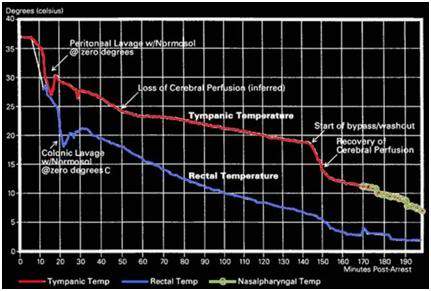
Since side I does not contain Cl, the Cl from side II will diffuse along the Cl concentration gradient from side II to side I. This results in a negative charge on side I relative to side II due to the excess concentration of anions ( Pr I + ClI > ClII ) on side I. The Cl concentrations will not become equal on sides I and II because the negative charge on side I will repel Cl movement from side II to side I so that side II will have the higher concentration of Cl. The presence of excess anion in the form of Cl on side I establishes a negative electrical gradient between side I and side II. The negative electrical gradient attracts K+ to migrate from side II to side I. However, the [K+] on side I now exceeds that of side II , hence K+ will move back from side I to side II along a K+ -concentration gradient.

The net result at equilibrium is that K+ and Cl move in equimolar amounts together from one side to the other. Each side is electrically neutral within itself due to equimolar concentrations of cations and anions, but there is an unequal concentration of the diffusible ions with [K+] highest on side I and [Cl-] highest on side II.

The effectiveness of intracellular organ preservation solutions provides a clue that, at least near 0oC it may be the case that much of the cold ischemic injury predicted by the Q10 rule (and which is in fact observed to occur) results from not from biochemical activity, per se, but rather from biophysical changes which proceed in the absence of metabolism or catabolism. Under normal metabolic conditions approximately 1/3rd of resting cellular energy expenditures are on ion homeostasis. The protein rich intracellular milieu is positively charged, and the sodium chloride (NaCl- ) rich extracellular milieu is negatively charged (Figure 2). Because NaCl- is osmotically active, movement of NaCl- from the extra- to the intracellular space across the cell membrane (to balance the charge difference represented by the positively charged intracellular protein; the Gibbs-Donan Equilibrium), the result is cellular edema. It is cellular edema, and the biophysics of the Gibbs-Donan Equilibrum, that appear to be a major driver of cold ischemic injury. This is antagonized by intracellular organ preservation solutions by removing most of the offending sodium from the extracellular spaces and replacing it on a roughly equimolar basis with cell membrane impermeable osmotically active species; typically sugars such as lactobionate and raffinose or the sugar-alcohol, mannitol.

Developing a Performance Score for Cryopatient Transport

The Hypothermic Metabolic Index

While thoracic surgeons and neurosurgeons are interested in knowing what the safe limits of DHCA, human cryopreservation professionals have an interest in the reverse problem; how much ischemic injury occurs at a given temperature over a given period of time. This is an important question to answer, even if just approximately, because it offers a quantifiable and reproducible surrogate measure for the quality of care a given patient receives. Absent feedback in the form of neurological deficit or other adverse effects, there is no way for those delivering care to cryopatients to know how well or how poorly they are performing. Such feedback is essential in any enterprise or human undertaking. Absent feedback, quality inevitably deteriorates and science vanishes from the picture.

Measure of Ischemic Exposure (MIX)

By inverting the HMI, or more simply using the Q10 rule in a straightforward calculation of injury vs. ischemic time, it is possible to generate a number for ischemic injury, as opposed to for ischemic protection. In fact, this is exactly what has been proposed by Perry and Harris with their Measure of Ischemic Exposure (MIX)[21] and Equivalent Homeothermic Ischemic Time (E-HIT).[22] Harris’ E-HIT proposal was laid out in an unpublished paper in 2003and is a complex, but potentially very useful mathematical analysis of both cooling in cryopatients, as well as consideration of ischemic injury versus temperature. While neither Harris nor Perry consider the confounding problem of cold inactivation of enzymes in trying to bound ischemic injury, both proposals will serve adequately as a  first approximation for surrogate markers (as opposed to reliable objective indicators) of ischemic injury in cryopatients.

Because of its simplicity and ease of use, I have chosen the MIX as the surrogate marker for use here. The mix is a straightforward adaptation of the Q10 rule which normalizes the measurement such that a MIX score of 1 corresponds to 1 hour of cold ischemia at 0oC. Thus, the MIX for 1 hour of ischemia at 10oC would be 2, for 1 hour at 20 oC, 4, at 10 oC, and so on. The MIX for fixed temperatures scales linearly with time so that twice as much ischemia at any given temperature would double the MIX score. This can be expressed by the equation:

MIX  =  2T/10t

Where:

t = hours at a constant temperature (oC)

T = patient temperature (oC)

Since patient temperature during cooling is dynamic and continuously changing as a function of time T(t) it is necessary to integrate the expression 2 T/110 over the time period of the ischemic interval; t0 to t0 in which case the MIX iscalculated usingthe following equation:

Where:

t = hours at a constant temperature

T = patient temperature (oC)

dt = change in temperature (oC)


The MIX has a simple form for cases involving a constant cooling rate, r degrees per minute starting at the patient’s measured body temperature at the timeof cardiac arrest. For a patient being cooled from 37oC to 0 oC the MIX would be 2.88/r. Thus, for a patient cooled at a rate of 15 oC per hour (T = 2.5 hours to reach 0oC) the MIX would be ~14. For patients who experience peri-arrest cerebral hypoxic or ischemic exposure, this time, beginning with loss of pupillary responsiveness, would be added to the MIX score using the same calculation and using the patient’s  body temperature(s) (T) at which the insult was experienced.

The lower the MIX score, the less ischemic injury the patient can be expected to have experienced. Abstracting a performance MIX score from the raw MIX score involves a subjective assessment of what segments of the ischemic insult were avoidable via actions of the human cryopreservation personnel caring for the patient. Unfortunately, this where the opportunity for self delusion, or the delusion of others (unintentional or otherwise), may enter the equation.  This is worth exploring, briefly, by using the following scenario.

A terminally ill cryopatient experiences cardiac arrest 45 minutes before Standby personnel arrive on the scene. During that interval he receives no CPS and no refrigeration. The human cryopreservation organization (HCO) may be either blameless, or fully responsible, depending upon a great many factors that were, or were not, under their control. For instance, did the HCO provide proper advice, counseling and support to deal with the possibility of cardiac arrest occurring in their absence (deployment of a field kit, instructions for caregivers on how to perform effective CPS, counseling about the need for ice or other refrigerant to be at hand, etc.)? If not, are they responsible for the extra MIX hit, or some fraction of it? Was their late arrival due to lack of proper medical assessment of the patient that, were it available, would have lead to deployment of the Standby team hours or even days earlier? Or, did the late arrival result from poor logistics by the HCO personnel responsible for booking the flight(s) for the Standby team? If the MIX is to be used as a feedback tool to hold the HCO and the Standby team personnel accountable, then many factors (aside from the fact of the ischemic insult) must be considered, and considered objectively. This is unlikely to happen, absent a set of objective standards for HCO performance, and an outside agency being responsible for evaluating HCO performance with respect to such standards.

Nevertheless, the raw MIX scores for all of a given HCO’s patients for any reasonable given period of time (say on a yearly basis) will necessarily provide a fairly strong indication of the quality of care a member or patient can expect from a given cryonics organization or service provider. For instance, HCOs that provide no Standby, Transport or CPS will have very high MIX scores compared to those that do.

Therapeutic Hypothermia

Figure 3: Cooling curve of a cryopatient given excellent cardiopulmonary support (CPS) (and who responded well to that support) who was cooled using external cooling via submersion in a stirred ice water bath, intraperitoneal cooling using ~1-2C o Normosol lavages and, at 145 minutes post arrest, extracorporeal cooling and blood washout.
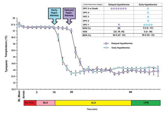
Today, yet another factor is confounding the ability of the Q10 rule to delineate the limits of ischemic tolerance (or at least warm ischemic tolerance). That factor is therapeutic, as opposed to preservative, hypothermia. Therapeutic hypothermia may seem to have little relevance to the cryopatient since the primary objective of cooling during Transport is to arrest metabolism and induce a state of preservative hypothermia. However, given current legal and logistic constraints rapid induction of protective hypothermia is not possible for cryopatients. Cooling times to below 20oC are typically in the range of 2-4 hours under good circumstances, and in patients who do not receive intraperitoneal or extracorporeal support may exceed 6 hours, even with AC-DC HI mechanical CPS. The fastest rate at which a cryopatient has been cooled is shown in Figure 3 above, and serves to illustrate that, absent the therapeutic effects of mild and moderate hypothermia, ischemic injury would be far worse in cryopatients. Therefore, a review of the mechanics of therapeutic hypothermia is very much in order.

Over the past 25 years a vast number of therapeutic interventions have shown great promise in animal models of regional and global cerebral ischemia-reperfusion injury (RCIRI & GCIRI) in the laboratory.[23],[24],[25],[26] In the last 6 years alone, over 1000 experimental papers and over 400 clinical articles on pharmacological neuroprotection have been published.[27],[28] However, with one exception, none of these interventions has been successfully applied clinically despite many attempts. [29],[30],[31],[32],[33],[34],[35],[36] The sole exception to this frustrating debacle has been the introduction of mild therapeutic hypothermia (MTH) as the standard of care for a select (and very small) minority of  sudden cardiac arrest (SCA) patients.[37],[38],[39],[40],[41],[42],[43]

Since the demonstration by Safar, et al., of the neuro-salvaging effects of mild systemic hypothermia after prolonged cardiac arrest in dogs [44],[45] there has been an explosion of translational research which has lead to a revolution in the understanding and application of mild hypothermia.[46],[47] Once seen only as a protective tool which conferred benefit solely by reducing metabolism, it has become clear that mild hypothermia (33°C–35°C) [48] has therapeutic effects which appear to be primarily anti-inflammatory and anti-apoptotic in nature, and which operate independently of hypothermia’s effect on metabolic rate.[49],[50] Table 1-1 reviews some of the known pro-inflammatory factors inhibited or moderated by mild therapeutic hypothermia (MTH) and documents the supporting literature.

Table 9-1: Inhibition of Injury Cascades by Mild Therapeutic Hypothermia (MTH)


Reference Model Species T (oC) Factors
Takeda et al (2003) Global Gerbil 31 and 34 Anoxic depolarization
Busto et al (1989b) Global Rat 30 and 33 Glutamate
Dietrich et al (1990) Global Rat 30 and 33 BBB
Kawanishi (2003) Hemorrhage Rat 35 Edema; BBB; PMNL
Kawai et al (2000) Focal Rat 33 ICAM-1 mRNA; PMNL
Wang et al (2002) Focal Rat 30 ICAM-1; neutrophil and monocyte; microglia
Hamann et al (2004) Focal Rat 32 and 34 MMP-2; MMP-9; m-PA; t-PA
Karibe et al (1994a) Focal Rat 33 Ascorbate; glutathione
Kader et al (1994) Focal Rat 33 NOS; nitrite
Toyoda et al (1996) Focal Rat 30 Neutrophil
Chopp et al (1992) Global Rat 30 HSP-70
Mancuso et al (2000) Focal Rat 33 HSP-70; C-fos
Tohyama et al (1998) Focal Rat 30 PKC
Shimohata et al (2007a) Focal Rat 30 ePKC
Harada et al (2002) Global Rat 32 CaM kinase II; PKC-a,b,g synaptosome
Tsuchiya et al (2002) Global Mouse 33 Zn2+
Phanithi et al (2000) Focal Rat 33 Fas; caspase-3
Zhao et al (2007) Focal Rat 33 Cytochrome c and AIF
Karabiyikoglu et al (2003) Focal 


Rat 


33 intra or 

post

iNOS; nNOS 


Wagner et al (2003) Focal Rat 33 post BBB; MMP-9
Inamasu et al (2000) Focal Rat 34.5 post Neutrophil infiltration; microglia
Horstmann et al (2003) Stroke Human 33 post MMP-9
Horiguchi et al (2003) Global Rat 32 post Hydroxyl radical
Han et al (2003) Focal Rat 33 post NF-kB; iNOS; TNF-a
Van Hemelrijck et al (2005) Focal Rat 34 post Caspase-3; nNOS
Inamasu et al (2000) Focal Rat 34.5 post Bax
Friedman et al (2001) Global Rat 30 intra/post GluR1A; GluR2B; GluR3C; NMDAR1
Ohta et al (2007) Focal Rat 35 post Inflammatory genes: osteopontin, early 

growth response-1, and macrophage inflammatory protein-3a

Luo et al (2007) Focal Rat 33 post Base-excision repair pathway
Preston & Webster (2004) Global Rat 32 post BBB
Liebetrau et al (2004) Focal Rat 32 post Calpain
Hu et al (2008) Global Rat 32 pre/post of GluR6-PSD95-MLK3 signaling module
Deng et al (2003) Focal Rat 33 post ICAM-1
Karabiyikoglu et al (2003) Focal Rat 33 post nNOS; iNOS and peroxynitrite
AIF, apoptosis-inducing factor; BBB, blood–brain barrier;; HSP-70, heat-shock protein-70; iNOS, inducible nitric oxide synthase; intra, intraischemic hypothermia; MMP-9, matrix metalloprotease-9; M, mouse; NF-kB, nuclear transcription factor kB; NOS, nitric oxide synthesis; nNOS, neuronal nitric oxide synthase; PKC, protein kinase C; PMNL, polymorphonuclear leukocytes; post, postischemic hypothermia; R, rat; S, species; T(1C), intraischemic temperature, unless specified; TNF-a, tumor necrosis factor-a.

Reproduced with modifications from Zhao, H., Steinberg, GK, Sapolsky, RM., General versus specific actions of mild-moderate hypothermia in attenuating cerebral ischemic damage. J Cerebr Blood Flow Metab, 2007. 27: p. 1879-1894.

Table 1-1: Intraischemic hypothermia delays or attenuates both ATP depletion (Ibayashi et al, 2000; Sutton et al,1991; Welsh et al, 1990) and anoxic depolarization (Bart et al, 1998; Nakashima and Todd, 1996; Takeda, et al, 2003), it also blocks glutamate release (Busto et al, 1989b; Patel et al, 1994; Winfree et al, 1996), suppresses inflammation (Kawai et al, 2000; Wang et al, 2002), maintains the integrity of the BBB (Dietrich et al, 1990; Huang et al, 1999; Kawanishi, 2003), reduces free radical production (Maier et al, 2002), inhibits protein kinase C translocation (Cardell et al, 1991; Shimohata et al, 2007a, b; Tohyama et al, 1998), inhibits matrix metalloproteinase expression (Hamann et al, 2004), and blocks both necrosis and apoptosis. Intraischemic hypothermia also preserves the base-excision repair pathway, which repairs oxidative damage (Luo et al, 2007). In addition to those cascades directly associated with neuronal injury, hypothermia further blocks astrocyte activity, and inhibits white matter injury. (Colbourne et al, 1997; Dempsey et al, 1987; Kimura et al, 2002). Similarly, postischemic hypothermia blocks free radical generation (Horiguchi et al, 2003), attenuates inflammation (Horstmann et al, 2003; Ohta et al, 2007), prevents BBB permeability (Preston and Webster, 2004), and suppresses caspase activities (Van Hemelrijck et al, 2005). Indeed, a browse through the literature gives an overwhelming impression that hypothermia seems to block every damaging event associated with necrosis or apoptosis. One reason for this impression of pan-inhibition may lie in the causality of ischemic damage.  For example, is the inflammatory response the cause of tissue damage or is it induced by brain injury? If it is the latter, then since hypothermia prevents tissue damage, it certainly also prevents the inflammatory response.

The journey from the laboratory to the clinic for MTH has been long and difficult. Seven years after the publication of the prospective randomized trials clearly showing that MTH improves survival and neurological outcome in out-of-hospital cardiac arrest patients, and 6 years after the ILCOR and AHA Guidelines  [51] recommended that: “Unconscious adult patients with spontaneous circulation after out-of-hospital cardiac arrest should be cooled to 32°C to 34°C for 12 to 24 hours when the initial rhythm was ventricular fibrillation (VF),” [41] only a minority of SCA patients are being treated with MTH. In surveys of emergency and critical care physicians conducted in 2005 and 2006, 74% of those responding in the US [52] and 64% of the international respondents indicated they had never used MTH.[53],[54] The use of pre-hospital, in-field MTH, is virtually nonexistent.[55]

No doubt, the commonly cited ‘obstacles’ of lack of institutional protocols, lack of physician education about the benefits and guideline changes, as well as the inevitable inertia that accompanies any paradigm shift in treatment are playing a significant role in the failure of MTH to become the practiced standard of care for the post resuscitation syndrome.[56],[52] However, what is not being said, or considered, is that while MTH as currently practiced represents a large relative improvement in outcome, the benefits are still modest in absolute terms. Only a miniscule subgroup of SCA patients currently can benefit from MTH; and even in its best clinical implementation MTH still fails to rescue ~60% of that sub-group of SCA patients to whom it is applied.[57],[58],[59],[60],[61] This is in stark contrast to what can be achieved with MTH in ameliorating post-ischemic encephalopathy in the laboratory, where post-resuscitation MTH consistently provides rescue with stunning efficacy.[62],[63]

Figure 4: The impact of a delay of 10 min in inducing MHT is a dog model of cardiac arrest followed by 3 min of systemic ischemia, 7 minutes of mechanical CPR and 50 minutes of advanced life support. Hypothermia to 34oC was induced beginning at 10 min post arrest in the early hypothermia group˜ and at 20 min post arrest in the delayed hypothermia group¢.  In the early hypothermia group, 5 of 7 surviving dogs were functionally normal (OPC 1 or 2), 1 had OPC 3, and 1 had OPC 4 (coma) at 96 hours of recovery. Histologically, 4 of 8 dogs in this group were normal (HDS 0), 1 had HDS 16, 1 had 22 and 1 had 98. The only surviving dog in the DH group was functionally normal at 96 hours (OPC 1, NDS 0) with an HDS of score of32 (mild injury) Due to early mortality only two other dogs in the delayed hypothermia group were evaluated histologically and their HDS scores 38 and 45, respectively. Dogs in this study were scored by ‘overall performance categories’ (OPC; 1=normal, 2=moderate disability, 3=severe disability but conscious, 4=coma, and 5=death) Neurological function and neurological deficit scores (NDS; 0% to 10%=normal, 100%=brain death). [64],[65] Histological damage scores were obtained by neuropathological examination of 19 discrete brain regions for severity and extent of ischemic neuronal changes, infarcts, and edema.  A total brain histological damage score (HDS)  >40 represented moderate damage, and HDS >100 represented severe damage.[66] Redrawn from Nozari, A., et al., Critical time window for intra-arrest cooling with cold saline flush in a dog model of cardiopulmonary resuscitation. Circulation, 2006. 113(23): p. 2690-6.
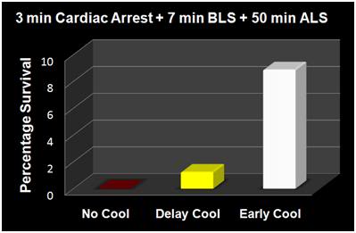
The primary obstacle to realizing this bonanza in translation research has been the practical impossibility of achieving systemic cooling within the narrow therapeutic window demonstrated in animal models of SCA and resuscitation.[67],[59],[57],[58],[59],[60],[61] If the clinical outcome of MTH was even half that achievable in the laboratory, widespread application would likely have been rapid and uniform; there is rarely resistance to the ‘miraculous’ if it is simple, easy to understand, biophysically well characterized and highly cost-effective. MTH applied immediately post ROSC would be all of these things.[4]

Figure 5: The results of the Nozari, et al., [63] study on the effects of delayed MTH are presented graphically at above, with the addition of historical controls from the literature treated similarly, but with no hypothermia (no survivors). This graphic illustrates the potency of truly rapid post arrest hypothermia increasing survival.
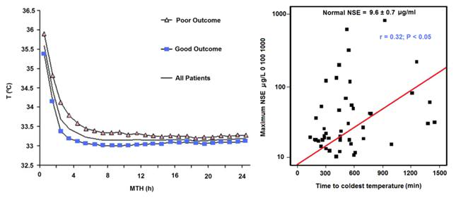
The data in Figures 4 and 5 exemplify what is possible when MHT is induced within its optimum therapeutic window of 0-15 min post ROSC versus a delay of even 10 minutes. In this study by Nozari, et al., of Peter Safar’s group, [63] VF was electrically induced in 17 dogs all of whom were subjected to a period of 3 minutes of no flow beginning when the MAP dropped below 30 mm Hg, followed by 7 minutes of mechanical CPR and 50 minutes of advanced life support during which time VF was maintained and mechanical CPR was continued. Nine animals were treated with rapid (early) induction of MTH to 34oC starting at 10 min post arrest (EH group) (concurrent with the start of ALS to simulate the time course of arrival of EMS paramedics) using a combination of cold IV saline and veno-venous heat exchange. Induction of hypothermia was not begun until 20 min post arrest in the delayed hypothermia group (DH group) which consisted of 8 dogs. Target core temperature was achieved at 6.0±2.7 minutes after the initiation of cooling (3.5 minutes after the start of veno-venous cooling) in both groups. The delay from arrest to reaching ~34oC was 16.6 min in the EH group and 25.4 minutes in the DH group.

Figure 6: Results of a study of 48 cardiac arrest patients treated with MTH via endovascular cooling. A strong correlation was found between rapidity of cooling and both neurological outcome and serum neuron specific enolase levels. Left: Time course of MTH among patients with good and poor neurological outcome. The curves indicate the course of mean body core temperature during MTH among patients with good (¢) and those with poor (p) outcome as well as in the entire (▬) patient group. Right: Correlation between time to coldest temperature (minutes) and the maximum NSE values (μg/L). Normal serum NSE is 9.6±0.7 μg/L. Redrawn from Wolff B, et al., Early achievement of mild therapeutic hypothermia and the neurologic outcome after cardiac arrest, Int J Cardiol. (2008).
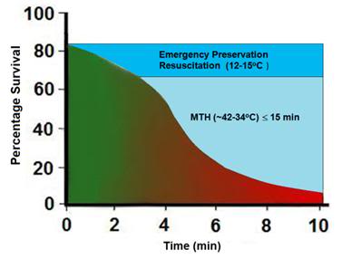
After 60 minutes of VF, ROSC was achieved with cardiopulmonary bypass for 4 hours, and intensive care was given for 96 hours. In the early hypothermia group,

7 of 9 dogs survived to 96 hours, 5 with good neurological outcome. By contrast, in the delayed hypothermia group 7 of 8 dogs died of multiple organ failure within 37 hours (P=0.012); 3 animals in secondary VF that was resistant to CPR with antiarrhythmic treatment and repeated defibrillations. Only one dog in the EH group died, and that animal succumbed to single organ failure; pulmonary edema with hemoptysis. This study extends the previous work by this group documenting an optimum therapeutic window for MHT (in dogs) of ~10-15 min.[68],[68],[69] The therapeutic window of MTH after cardiac arrest has been demonstrated to be similarly short in other species.[70],[71],[72],[73]

The dramatic efficacy of MTH in the laboratory made quick converts of the pioneering researcher-clinicians who forged ahead with the application of MTH to SCA in the clinic precisely because it was dramatic; indeed it was as close to the miraculous as interventions in medicine come. The real barrier to translating that ‘miracle’ to everyday practice has been the seemingly intractable problem of achieving cooling over the same time course that has proven so effective in the research setting.

The problem is that the optimum therapeutic window for the treatment of cerebral ischemia-reperfusion injury appears to be in the range of 0 to 15 minutes post ROSC. One of the first follow-up studies on MTH carried out by Safar, et al., demonstrated that in a standardized model of cardiac arrest in dogs a delay in the application MTH of as little as15 min after ROSC abolished most of the benefit.[69],[74] While the work of Bernard, et al., [38] and that of the Hypothermia after Cardiac Arrest Study Group [40] demonstrated that delays in cooling of up to 2-3 hours post ROSC in humans still have sufficient clinical utility to justify the routine application of MTH in a selected group of SCA patients, this benefit is marginal when contrasted to that achievable in the laboratory when MTH is rapidly induced during the first 15 minutes after ROSC.[69] Thus, the optimum clinical benefit of MTH in ischemic and very likely traumatic, CNS injury requires the ability to achieve very rapid core cooling.[75]

Figure 7: Survival after cardiac arrest declines rapidly as a function of time to ROSC, exhibiting the sigmoidal curve shown at left, above (¢>¢), with essentially all patients failing to survive with normal mentation after arrest intervals of @ 10 min. Application of MTH (¢) within the window of @ 15 min offers the promise of squaring the survival curve in SCA of @ 10 min duration yielding a survival rate of ~65-70% with little or no neurological deficit. Application of deep (10-22oC) or profound hypothermia (5-9oC) (¢) may allow survival after intervals of as long as 1-2 hours of CPR.
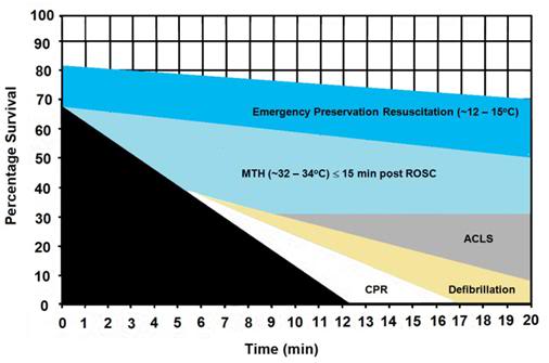
In the clinical arena the time to reach the target core temperature under ‘good’ circumstances is in the range of 3-4 hrs; not 10 to 30 min, as is the case in the laboratory. Even with such long delays in cooling the adverse effect of delay is still present. Wolf, et al., recently published a study of 49 out of hospital cardiac arrest patients who were treated with MTH (32.0-34.0°C; with a target temperature of 33.0°C) of 24 h duration using endovascular cooling.[61] The study endpoints were neurological outcome on discharge from hospital and serum neuron specific enolase (NSE) levels (a sensitive and specific marker of neuroinjury) at 24 h intervals to 3 days (Figure 6).

Figure 8: The graph above shows the hypothesized relative effect on survival of effectively administered CPR started at 5 min post-arrest followed by defibrillation at 6 min and ACLS at 8 min post arrest (~30% survival). The light blue shaded area of this graph shows the expected improvement in survival if MTH is induced at the start of ACLS (8 min post-arrest) and target temperature is reached by 15 min post ROSC. The dark blue shaded area shows the potential of Emergency Preservation resuscitation (EPR) using moderate (10-22oC ) or profound (5-9oC) hypothermia to not only square the curve of survival with CPR, but to facilitate survival in patients who would otherwise not benefit from either BCLS or ACLS (i.e., refractory to defibrillation, hypovolemic, etc.). Graphic by M.G. Darwin
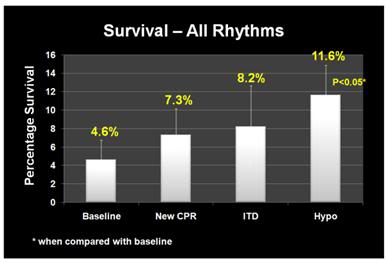
As is the case in laboratory studies of MTH in cardiac arrest in dogs, Wolff, et al., found that neurological outcomes were binary, with no patients who survived experiencing moderate degrees of disability; patients either recovered well with no or /mild neurological impairment, or experienced severe disability (n = 1) coma or PVS (n = 6). Twenty-eight patients were discharged with a good outcome and a strong correlation was found between good outcome and the time interval from the start of cooling to the lowest temperature (p =.035) and a less robust correlation with the time to reach target temperature (p=.071).

Similarly, NSE levels were found to correlate well with the time required to reach the lowest temperature achieved in each patient (Figure 6).

Even with delays in the start of cooling that averaged 2.5 hrs; and a mean time to reach target core temperature of 6.8 hrs, additional injury accruing from slowness in cooling was still clinically and biologically apparent. Despite the homogeneity of the patients, their arrest times, and their course of treatment, ~60% of the patients in this study either did not survive, or were comatose or PVS.

The Benefits and Limits of ‘Delayed’ MTH: Real World Experience

To understand the benefits and limits of MTH when it is aggressively and competently implemented with currently available technology, it would be hard to find a better example than that of Wake County, SC. Wake County, is located in the northeast central region of North Carolina and is part of the Research Triangle metropolitan area, which consists of Raleigh, Durham, Chapel Hill, and surrounding urban and suburban areas. The area serviced by the Wake County Emergency Medical System (Wake EMS) has a population of 832,970 (as of 2007). The Wake County EMS operates 35 ambulances from 23 locations with 825 ALS personnel; ambulances are staffed with two paramedics 95% of the time and there is always one paramedic responding.[76] In 2002 the Wake EMS answered more than 50,000 medical requests for service. Based on the latest national data Wake County ranks third in the US for recovery from “survivable” cardiac arrests (primarily ventricular fibrillation-ventricular tachycardia). Nationally, the average survival rate is 17% for patients presenting with these arrhythmias.

Figure 9: (right): Improvement in overall survival of patients in cardiac arrest in response to the phased introduction of CPR per AHA 2005 Guidelines, use of an impedance threshold device (ITD) and in-field induction of MTH.[77]
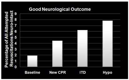
Beginning in January 2004 Wake EMS initiated a study to evaluate the efficacy of CPR as they then practiced it, and to evaluate the effectiveness of the impending change in the American Heart association (AHA) guidelines for CPR, the introduction of an impedance threshold valve (the ResQPod™) and Wake EMS’ planned implementation of the ILCOR Guidelines for post-arrest MTH.[77],[78] From January 2004 until April of 2005, Wake EMS personnel employed the then extant AHA guidelines, which mandated an emphasis on intubation and a 15:2 compression-to-ventilation ratio; with interruption of chest compressions for ventilation. This period constituted the baseline of the study, and data were collected per protocol; not gathered retrospectively.

During the baseline period survival to discharge from hospital was 2.4% for all patients given CPR and 12.1% for patients with ventricular fibrillation-ventricular tachycardia (VF-VT) arrhythmias. In April 2005 Wake EMS implemented continuous cardiac compression CPR with a 30:2 compression-to-ventilation ratio with emphasis on no, or very minimal, interruption of chest compressions.  After 12 months, the overall survival rate had risen to 4%; and had more than doubled to 21.8% for patients who presented in VF-VT.

In April 2006, Wake EMS added the use of an impedance threshold device (ITD) to improve cerebral and coronary perfusion during CPR. Introduction of the ITD resulted in an increase in overall survival to 4.5% and an increase in the survival of patients with VF-VT to 28.5%.

Figure 10: (right): Dramatic improvement in neurologically intact survival as a result of the phased introduction of CPR per AHA 2005 Guidelines, use of an impedance threshold device (ITD) and in-field induction of MTH.[77]
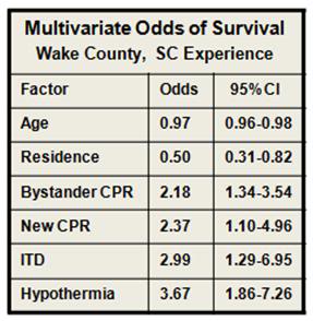
The final phase of the investigative protocol began in October of 2006 when Wake EMS added in-field induction of MTH to the two previous interventions. MTH was induced using a combination of external cooling employing ammonium nitrate-water eutectic ‘instant cold packs’ applied to the axilla and groin, and  cold IV saline (1-2oC, 30mL/kg to a maximum of 2 liters) given rapidly via two large bore catheters and/or intraosseous infusion. Criteria for induction of hypothermia were that the patient have ROSC and show no return of consciousness (Glasgow Coma Score (GCS) <8).

Induction of hypothermia was initiated two to three minutes after ROSC. There was heavy emphasis on avoiding over-ventilation and on attempting to maintain end-tidal CO2 (EtCO2) at a minimum of 40 mm Hg.  Patients undergoing MHT were sedated with etomidate, paralyzed with vercuronium and given a titrated dopamine drip to maintain mean arterial pressure (MAP) between 90-100 mm Hg. The mean time to target temperature (34oC) in this study was extraordinarily short: 68 minutes (95% CI 47 to 88); compared to the 2-3 hours typically required to induce MTH.

With the combination of continuous compression CPR, use of the ITD and prompt application of MTH, survival rates for the 12 months from October of 2006 to October 2007 had increase to 6.7% overall and 37.4% for patients with VF-VT. The odds of overall survival increased three-fold (95% CI 1.7 to 5.0) and the odds of survival for patients in VF-VT increased 4.3-fold (95% CI 2.2 to 8.6) from the beginning of the study (Figures 9 and 10).The probability of a good neurological outcome increased from 20% at baseline to 80% at the conclusion of the study (Figure 11). In a multivariate analysis, the odds ratios for survival for each phase of implementation were as follows:

Figure 11: (right): Multivariate odds for all factors in outcome evaluated during the Wake EMS study. MTH was by far the most powerful intervention. As in most previous studies of survival factors associated with CPR age and residence (home versus extended care or assisted living facility) had only modest impact on survival.[77]
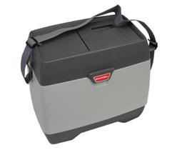
  • New CPR protocol: 2.13 (95% CI 1.12 to 4.04)
  • Addition of impedance threshold device: 2.33 (95% CI 1.09 to 5.00)
  • Addition of early hypothermia: 3.99 (95% CI 2.19 to 7.27)
  • Patients who received bystander CPR 1.79-fold (95% CI 1.18 to 2.72) more likely to survive.

Figure 12: (right): The Engel-15 portable, (compressor-type) refrigerator/freezer has a 14 L capacity, weighs 11.5 kg and can maintain 12-13 liters of saline at 1-2oC at ambient temperatures as high as 40oC. It retails for ~$380 US. [Photo courtesy of Engel, Ltd., Australia]
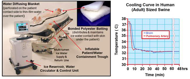
 

Interestingly, all three elements of the Wake EMS protocol were implemented at a cost of less than $200 per patient. Due to budget constraints, Wake EMS chose simple, inexpensive commercial products for refrigeration of IV fluid and implementation of external cooling, as opposed to more costly products developed specifically for medical application, such as the EMCOOLS surface cooling system (Emergency Medical Cooling Systems, AG, Austria). Saline was kept at the requisite temperature of 1-2oC with a compact, 12V operated, consumer travel refrigerator/freezer (Figure 9-12, Engel-15: http://www.i-m-d.com/) and surface cooling was with generic ammonium nitrate cooling packs.

The Wake County EMS program is extraordinary in every way. It represents the best application of the best available technology by arguably some of the best medical and paramedical personnel in the US. The mean time to target temperature of 68 minutes is unprecedented in any clinical study employing MTH. Of the 359 patients who participated in the study (all comers) after MTH was in place; 25 survived. In the subgroup of 93 patients who presented with VF-VT; 34 survived, with 78% or 27 patients being discharged with a good neurological outcome. Put another way 92% of patients who presented under the most favourable circumstances (VF-VT), treated with the best currently available interventions, at the fastest rate of cooling so far reported, failed to survive or did so with profound neurological debility.

The primary difference between the survivors and the profoundly disabled or dead was the development of the post-resuscitation syndrome and the primary reason for this complication was not comorbidity, or delay in paramedical assistance, but rather delay in the rapidity of cooling which, if achieved within the first 15 min post ROSC, would have offered the prospect of neurologically intact survival in the range of 70-80% in patients presenting with VF-VT, and 30-40% in all comers.

These interventions, remarkable achievements that they are, do not escape from the harsh reality that the 400% increase in survival from cardiac arrest in Wake County, when expressed in absolute terms, means that the number of lives saved increased from ~5 to 25 – out of 395 SCA patients; a huge relative gain, but a comparatively small increase in the absolute number and percentage of lives saved, and minds salvaged. The true life saving potential of MTH remains elusive by virtue of its exceedingly small therapeutic window.

The Problem of Heat Exchange

Because of this minute therapeutic window, there is a pressing need to achieve rapid and durable core cooling of patients during CPR by simple, easily accessible means.  External cooling is only effective at reducing core temperatures by 0.15 to 0.25ºC/min in the average patient undergoing CPR (Figure 38) and this is achieved only by complete immersion of patients in a stirred ice water bath.

The most effective external cooling achieved by a commercial, FDA approved system using direct, whole body surface cooling employing circulation of ice water (ThermoSuit,™ Life Recovery Systems, Kinnelon, NJ)  is probably the work of Janata, et al., using human human-sized swine.[79]

They were able to achieve core cooling at a rate of 0.3oC/min; however it is important to note that the animals in this study were not in cardiac arrest while undergoing CPR in the presence of profoundly peripherally vasoconstricting agents, such as epinephrine or vasopressin; as would usually be the case during ACLS in humans [51] and which is known to further slow surface cooling.[80]

Figure 13: The Life Recovery Systems ThermoSuit™ employs direct ice water contact with the patient’s skin to achieve the maximum possible rate of cooling by external means. The system consists of an inflatable insulating and water containment patient enclosure inside of which the patient rests on a mat of Dacron bonded polyester ‘wool’ which acts to diffuse and film water pumped over the dorsal surface of the patient’s body. Water at 2-4oC is thin-filmed over the ventral surface of the body by a thin, transparent blanket with many hundreds of small perforations through which water under pressure pours out and over the patient. Cold water is recirculated over crushed or cubed ice in an insulated reservoir containing a disposable liner and pumps. Cooling is computer controlled via a thermistor which can be placed in any desired anatomical location. All patient contact items are single-use and disposable (including, as previously mentioned, the pumps) http://www.life-recovery.com/.
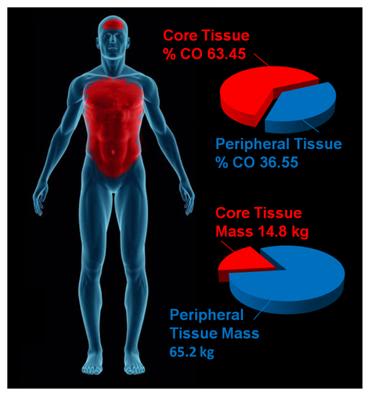
The obvious problems with this system are its bulk (Figure 13), its high cost ($1,800 per patient for disposables), lack of ease in field deployment (again related to its bulk and weight) and the intrinsic physiological problems associated with the induction of hypothermia via external cooling. As extensively discussed in Section Two, the mammalian body consists of multiple thermal compartments transiently ‘isolated’ from each other by differences in blood flow and heat conductivity.[81] Broadly, these compartments can be classified as strongly and weakly circulated (perfused); corresponding to the body core and periphery. The core tissues receive ~63% of the resting cardiac output (CO) but constitute only ~19% of the total body mass. By contrast, the peripheral tissues receive ~37% of the basal CO and constitute ~81% of the body’s mass (Figure 14).

Figure 14: The parenchymatous organs that comprise the visceral core of the body receive an aggregate of ~63% of resting the cardiac output while comprising only ~19% of the body mass. By contrast, the peripheral tissue mass which accounts for ~81% of body mass receive only ~19% of the cardiac output. External cooling profoundly chills peripheral tissues before significantly reducing core temperature. Values for organ and tissue masses were obtained from: IAEA. Compilation of anatomical, physiological and metabolic characteristics for a Reference Asian Man. Volumes 1 and 2. Report IAEA-TECDOC-1005, (Vienna, Austria: International Atomic Energy Agency) (1998), Boecker, BB. References values for Basic Human Anatomical and physiological characteristics for use in radiation protection. Radiation Protection Dosimetry.105(1–4): 571–574;2003, de la Grandmaison GL, Clairand I, Durigon M. Organ weight in 684 adult autopsies: new tables for a Caucasoid population. Forensic Sci Int. 2001 Jun 15;119(2):149-54 and Heymsfield SB, Gallagher D, Mayer L, Beetsch J, Pietrobelli A. Scaling of human body composition to stature: new insights into body mass index. Am J Clin Nutr 2007;86:82–91.Values for organ and tissue blood flows were obtained from: Williams, LR, Leggett, RW. Reference values for resting blood flow to organs of man. Clin Physiol Meas. 10:187-212;1989. Graphic by M.G. Darwin
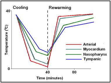
Pathophysiology and Biophysical Limitations of External Cooling

The objective of MTH is to provide protection against ischemia-reperfusion injury to the brain, heart, kidneys and liver; the visceral organs that constitute the strongly circulated core of the body. The peripheral tissues (skin, skeletal muscle, connective tissues and bone) are at once much more resistant to ischemia and less well perfused.  External cooling rapidly chills the ischemia-resistant peripheral tissues cooling them profoundly, while failing to provide protection to the vulnerable parenchymatous organs in the body core. This is not only undesirable in terms of its inefficiency; it also poses a number of hazards and risks.[82] Hypothermia is therapeutic in ischemia-reperfusion because it down-regulates the immune-inflammatory response; a response that is vital for host defense, wound healing and hemostasis. Hypothermia, like any major medical intervention that perturbs fundamental physiological processes, carries with it serious risks, as well as benefits. In both animals and humans, hypothermia is markedly immunosuppressive.[83],[84] and interferes with the both the biochemistry of the clotting cascade and the production of platelets and clotting proteins.[85],[86]

In humans perioperative minimal hypothermia (MinH) (36oC) increases the rate of wound infections [87] and prolongs hospitalization. [88] These effects occur in part due to the regional thermoregulatory vasoconstriction MinH induces; which in turn leads to reduced oxygen delivery to injured  tissues, [89] inhibition of oxidative killing by neutrophils,[90] and reduced collagen deposition.[88] MinH induces significant suppression of mitogenic responses to Concanavalin A (con A), phytohemagglutinin (PHA), and pokeweed mitogen (PWM) and these changes are known to persist for at least 48 h. The mitogens PHA and ConA activate T cells, whereas PWM stimulates both T and B cells, thus indicating that the suppressive effects of MinH involve a variety of lymphocyte subpopulations. Hypothermia of as little as 1oC significantly inhibits production of  interlukins (IL-1б, IL-2, IL-6) and TNF-α in post-surgical patients, [91] and this suppression of cytokine production persists for least 24 hours after even a brief post-operative hypothermic interval.[88] The inhibition of pro-inflammatory cytokine production by IL-1б and TNF-α induce tissue factor which is critical to angiogenesis, collagen elaboration and fibroblast activation; all essential processes in wound repair and hemostasis.[92],[93],[94] Significantly, many of these of adverse effects of post-operative MinH can be prevented by maintaining normothermia in the perioperative period.[88]

In the Hypothermia After Cardiac Arrest Study Group, patients treated with MTH experienced twice the incidence of sepsis. This finding is consistent with other studies where MinH and MTH were found to double the rate of post operative wound infection.[88] A recent meta-analysis of MTH for traumatic brain injury (TBI) found that the incidence of pneumonia was also doubled for patients undergoing MTH.[95]

External cooling causes greater perturbation in hemodynamics than does central cooling, [96] resulting in increased systemic vascular resistance and decreased cardiac index; a phenomenon observed in all of the patients in the Bernard, et al., study that employed MTH for post-arrest cerebral resuscitation.[38] This is particularly undesirable in the setting of MI, CHF and cardiogenic shock. For these reasons, as is the case with any potent therapy, careful attention must be paid to the dose-response curve, and overshoot or excessive regional cooling must be minimized or avoided.[97]

When the tissues of non-hibernating (or unprepared hibernating mammals) are cooled to £20ºC a wide range of deleterious changes occur. The saturated fats which comprise cell membrane lipids undergo phase change, resulting in red and white blood cell rigidity; with accompanying inability to deform and pass through capillaries. Red cell aggregation also occurs and this, in association with reduced flow as a result of vasoconstriction, results in blood sludging and failure of the microcirculation.[98],[99] Profound hypothermia, either local or systemic, results in hemoconcentration as a consequence of translocation of vascular water and electrolyte to the interstitial space.[100],[96],[101],[102],[103] This hemoconcentration further exacerbates regional ischemia in deeply chilled tissues.

Independent of injury from the freezing of water, moderate, profound or ultraprofound hypothermia is known to cause cellular damage which is referred to as ‘chilling injury.’ Chilling injury appears to be a multifactorial process in which alterations of membrane structure (reorganization of lamellar lipid sheets with lateral phase separation between regions of gel phase and regions of liquid crystal phase result in loss of membrane integrity), [104],[105] failure of ion pumping (with consequent disruption of cellular ionic homeostasis), [106],[107] depolymerisation of some elements of the cytoskeleton, [108],[109] generation of free radicals, [110],[111] and metabolic disruption due to selective inactivation of critical enzymes [112] all appear to play a role.

Cooling of tissues to 5ºC for as little as 1 hour has been shown to cause microvascular endothelial damage similar to that observed in ischemia-reperfusion injury; loss of endothelial cell tight junctions, infiltration of capillary and venule walls with leukocytes, and frank extravasation of red cells from injured vessels.[113] A possible reason for the similarity in the histological appearance of chilling injury with ischemia-reperfusion injury may be due to the fact that both types of injury appear to be caused, at least in part, by reactive oxygen species and by disruption of the cytoskeleton.

The molecular changes induced by moderate, profound and ultraprofound hypothermia may also directly compromise endothelial cell integrity. For example, chilling of several types of epithelial cells has been shown to result in disassembly (depolymerisation) of the intracellular microtubules resulting in compromise of the polarized membrane expression and function of some transport proteins in these cells.[114] These functions are slow to return to normal (@20 hr) and are associated with prolonged dysfunction of allografts that have undergone cold preservation storage.[115],[116]

Deep cooling of the peripheral tissues may also result in immunosuppresion in chilled limbs and skin, and possibly impaired hematopoiesis due to localized moderate hypothermia in bone marrow in the cranium, sternum, vertebrae, and to a lesser extent, in the pelvis. [In children, the long bones are the principal repository of hematocytoblasts, and this marrow would also be disproportionately chilled during external cooling.] In contrast to the anti-inflammatory effect of MTH, systemic hypothermia to £28ºC, either accidental or induced has been shown to increase levels of pro-inflammatory cytokines.

Figure 15: (right): Typical (idealized) cooling and re-warming curve achievable with maximum extracorporeal (cardiopulmonary bypass) cooling.
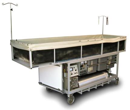
Core cooling is thus the gold standard for the induction of systemic hypothermia (mild, moderate, profound or ultraprofound) and external (peripheral) cooling should be used only where there is no other alternative for achieving truly rapid cooling, or maintaining it for the required 24-48 hours following induction.

Consideration of Invasive Core Cooling Methods

Figure 16: An example of a mobile extracorporeal membrane oxygenation (ECMO) cryopatient support cart. This unit was fabricated by the Alcor Life Extension Foundation and was designed for extended (12-24) hour extracorporeal support of patients in profound or ultraprofound hypothermia.
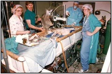
Extracorporeal cooling via cardiopulmonary bypass (CPB) [117] or high flow veno-venous heat exchange [118],[119] allows for cooling rates of 0.8ºC/min to 1.0ºC/min (Figure 15) However, it cannot be applied rapidly enough given existing logistic and regulatory constraints. CPB requires complex hardware and highly skilled personnel who must maintain their clinical reflexes by practicing perfusion on a regular (preferably daily or at least weekly) basis. Even in centers of excellence, with a highly skilled, rapid-response CPB team at the ready (including a well-practiced surgeon or cardiologist), the soonest CPB can be initiated after cardiac arrest is typically 15-20 minutes.[120],[121]

The use of emergency CPB applied in this time frame is largely confined to patients undergoing cardiac catheterization and/or revascularization (i.e., angioplasty or stent placement) who arrest in the cardiac catheterization lab. When such patients experience cardiac arrest that is refractory to treatment with drugs and defibrillation – they are usually otherwise healthy – they have normally functioning lungs, normal fluid balance and fluid distribution (are neither dehydrated nor edematous from fluid overload), and have failure of a only a single organ –  the heart. Even under such ‘ideal’ conditions, CPR is often inadequate to maintain brain viability during the brief interval between cardiac arrest and the start of CPB.

Figure 17: Cryopatient undergoing ECMO and blood washout in the home. Even under ideal circumstances cardiopulmonary bypass takes in excess of 30 minutes to initiate
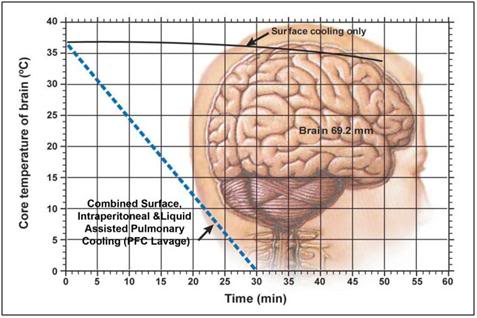
While veno-venous extracorporeal cooling is less technically demanding, it still requires skill-intensive vascular access under field conditions, and reported rates of cooling are modest; ~0.012ºC/min. [122],[79] Both of these techniques may require anticoagulation and certainly require coagulation monitoring; which again is a barrier to in-field application.

Administration of large volumes (40mL/kg) of chilled intravenous fluid has also been used to reduce core temperature in the field in patients undergoing CPR. However this method is extremely slow (~0.058ºC/min),[123],[124],[125] and is sharply constrained by the maximum volume of fluid that can be administered.

The effectiveness of intravascular fluid administration in achieving durable core cooling is also a function of body composition. Obesity, which has become epidemic in the developed world, is now rapidly becoming a global problem with 1.1 billion adults worldwide classified as overweight, and 312 million of them as obese by the World Health Organization (WHO).[126] Vascular volume does not increase proportionally to increase in body weight; in fact, the vascular volume to body weight ratio falls toward an asymptotic value of approximately 45 ml/kg in the obese human.[127] Thus, the use of cold intravenous infusions will necessarily be even less effective in the obese than might be expected since the maximum volume of fluid that may be safely given does not increase linearly with body weight. Fat is also an extraordinarily good insulator, and obesity often unfavorably alters body surface to volume area. Both of these factors combine to dramatically reduce the speed and overall effectiveness of external cooling.

The limits of efficiency of core cooling that can be achieved by irrigating the peritoneal or pleural spaces with cold liquid are in the range of 0.05 to 0.3ºC/min [128],[129] and both of these techniques are invasive, require considerable technical skill, and carry with them risks of serious iatrogenic consequences.

As noted earlier, the ideal way to address these needs would be extracorporeal membrane oxygenation (ECMO) and cooling. However, the practicality of rapidly bringing this demanding technology to bear in the EMS setting is negligible. Surgery (or time-consuming percutaneous vascular access; at or beyond the optimum therapeutic window of 15 min post ROSC) is required to access the circulation and this cannot be accomplished under field conditions. Experience with emergent initiation of ECMO in patients presenting for experimental cryopreservation (Figure 38) are probably representative of the time required to achieve extracorporeal support under field conditions for patients with SCA. Such patients typically experience a delay of 60 to 80 minutes (and not infrequently longer) after the initiation of closed chest CPR before the application of CPB, even under optimum circumstances.[130],[131],[132]

Thus, what is needed for cryopatients, patients who have suffered prolonged (³5 min) cardiac arrest, or in whom ROSC cannot be effectively established, is the ability to rapidly induce hypothermia via core cooling simply and noninvasively.  In addition to moderating the injury cascade initiated by ischemia-reperfusion, mild (33-35oC) or moderate (28-32oC) hypothermia can indirectly act to improve gas exchange and hemodynamics by bringing the patient’s cerebral and systemic metabolic demands closer to those that can be delivered by CPR.

The clinical experience of the Wake EMS and in-house research into accelerated non-invasive cooling methods will be combined in the next section to allow far greater rates of cooling of cryopatients than have been possible in the past.

Induction of Hypothermia in the Cryopatient

Figure 18: Idealized optimal core brain cooling curve. The solid black line (___) indicates the brain core (69.2 mm depth) cooling curve achieved using external cooling with ice water immersion during CPS. The broken blue line (—-) shows the theoretical cooling curve that might be possible by combining surface cooling with either cardiopulmonary bypass (CPB) cooling, or with liquid assisted pulmonary cooling (LAPC) and intraperitoneal cooling using ~1-2oC physiologic salt solution, such as Normosol-R.™
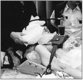
Figure 19: Maintaining contact between the patient and the refrigerating ice bags was virtually impossible in the early days of human cryopreservation since the bags continually slide off the patient during Transport.
In the early days of cryonics, post-cardiac arrest cooling was exclusively “external” cooling which was achieved by covering the patient with plastic bags containing crushed or small cubed ice (Figure 19).[133], [134],[135] This approach had the obvious advantage of being simple, straightforward to implement, and very inexpensive. Unfortunately, years of actual field experience with this approach have disclosed a number of serious limitations and problems:

1)      Heat exchange is greatly attenuated by both the insulating properties of the plastic and the reduction of convective transfer due to containment of the ice water generated from the melting ice.

2)      It is difficult to properly and completely pack the patient in ice because the bags are constantly falling off or not staying properly positioned, i.e. in good contact with the patient’s skin. Maintaining ice bags around the head and neck is a particular problem.

3)      It is virtually impossible to get ice packs under the patient, which means that 35% to 45% of the patient’s surface area is unavailable for heat exchange.

4) Ice bags leak and sweat which cause water to wet the patient and drip off the cot or gurney during transport. This presents both an immediate safety hazard (creating slippery floors and a potential electrical hazard) and also serves to contaminate staff and the working environment with potentially infectious fluids.

The PIB was developed after in-field core temperature measurements during the transport of a human cryopreservation patient employing ice bags to facilitate external cooling disclosed that this method was grossly unsatisfactory resulting in a core cooling rate of approximately 0.05°C/min during the first 4 hours of cooling.[134], [136], [137], [138] Subsequent cases confirmed very slow rates of cooling using externally applied plastic bags filled with water ice ranging between a high of 0.12°C/min to a more typical 0.064°C/min.[134] For these reasons, a lightweight, Portable Ice Bath (PIB) was developed by the author to allow for direct contact of ice water with essentially the entire surface area of the patient to simulate rates of heat exchange presumably encountered in cold water drowning where recovery of adults without neurological deficit after 20-40 minutes or more of submersion in ice water has been repeatedly clinically documented.[139],[140], [141],[142] The rate of heat-loss for an adult human in water is 32 times that of in air5. Indeed, in such cases of cold water drowning, it is sometimes possible to successfully resuscitate adults who have been chilled in the absence of any cardiopulmonary support for up to 30 minutes.[143],[144],[145] Children have been known to recover following up to 66 minutes of cold water drowning.[146]

Determining the rate of cooling likely to be achieved with the PIB, particularly with the addition of convection cooling by stirring or circulating the PIB water around the patient was important prior to expending the considerable resources required and logistic difficulties to be overcome if these techniques were to be implemented routinely during Transports. A careful examination of the literature was undertaken by the author circa 1987 to determine if there were any published data on the rate(s) of core cooling likely to be feasible with these modalities.

Unfortunately, the only published data were those of the SS (Schutzstaffel) Nazi physicians Holzloehner, Finke, and Rascher, et al., conducted in 1942 on prisoners in the German concentration camp Dachau as part of an effort to understand the mechanisms and time course of hypothermia (as well as methods of  safe re-warming) in Luftwaffe pilots downed in cold North Sea water (Figure 20). Most of the actual work was conducted by Sigmund Rascher, M.D. and was summarized after its post-war recovery by Major Leo Alexander of the U.S Army Medical Corps in 1946.[147] This document, now referred to as the “Alexander Report,” contains detailed information in both graphic and tabular form on the effects of convective cold water external cooling on human prisoners.

Figure 20: Sturmbannfuerher Dr. Sigmund Rascher (R) and Dr. Ernst Holzhoen (L) experimenting on a prisoner from Dachau using cold-water immersion to induce hypothermia followed by attempted re-warming after loss of consciousness. (Art from photo via Vad Yashem, Jerusalem, Israel.)
At this time, the citation and use of this data are extremely controversial and their relevance, integrity, and scientific usefulness have been called into question.[148] There is no question in the author’s mind that the work of Holzloehner, et al., constitutes an atrocity and a crime against humanity of the worst kind. One of the principal criticisms of the scientific validity of this work has been the observation that many of the more than 300 victims of this research were chachectic and weakened from malnutrition and frank starvation making their physiological responses non-representative of that of the healthy German aviator. Ironically, it is just this criticism that makes the data obtained in this study uniquely valuable to human cryopreservation.

Data from the Alexander Report must be interpreted with caution and within the context of both the typical conditions under which human cryopreservation takes place and more recent data on the effects of cold water immersion on human subjects which is unquestionably both ethically and scientifically sound.[149],[150],[151]

Several important caveats apply: Cooling curve data obtained by Holzloehner, et al., must be evaluated with the understanding that non-paralyzed, conscious individuals subjected to immersion in water cooled to between 2°C and 12°C will shiver either until exhaustion or until the temperature of the skeletal muscle drops below 30°C at which point shivering is impossible.[151] Until either or both of these events happen core temperature will either transiently increase or will not decrease appreciably. Thus, use of these data to evaluate the efficacy of convective cold water immersion must subtract the first 10 to 20 minutes of core temperature data when shivering and elevated oxygen consumption are effectively maintaining homeostasis in the face of profound external cooling.[152]

From the graphic data it is very apparent when this point is reached, typically at about 15 minutes post-immersion, for the unclothed emaciated subject. At this point the cooling curve begins to decline markedly and within 5 minutes achieves a steady rate of descent of approximately 0.26°C/min to 28°C after which it slows appreciably to 0.13°C/min until cardiac arrest typically occurs at ~27°C. This slowing is probably due to deteriorating hemodynamic status (BP was typically 40 mmHg to 50 mmHg near the terminal portion of the cooling curve) and a decrease in the ∆T between subject core temperature and bath temperature. In one series of 7 subjects the mean time from immersion to ventricular fibrillation (VF) was 66 minutes with a mean rectal temperature at the point of VF of 26.94°C. The fastest rate of cooling observed was 0.36°C/min in malnourished females.

One potentially very important observation made by of Holzloehner, et al., was the lack of any difference in the rate of early core temperature drop between subjects immersed in stirred water baths at 2°C or 12°C. This would seem to imply that the rate limiting factor in cooling, at least to ~27°C, is not the volume of water flowing over the subject, nor the ∆T, but rather peripheral vasoconstriction resulting in decreased blood flow to tissues in contact with cold water. Stirring of the PIB water in excess of that required to achieve optimum heat exchange is also undesirable for the following reasons:

  • Given the modest amount of stirring employed in the experiments of Holzloehner, et al., it is unlikely that movement of large volumes of water over the patient will increase the rapidity or efficiency of heat exchange.
  • Excessive stirring may result in unnecessary heat exchange with surrounding air, and unnecessarily waste heat generated by stirring pumps.
  • Smaller more efficient pumps will allow for the use of household alkaline or lithium batteries facilitating initiation of SCCD cooling in the field and during vehicular transport where 110VAC wall current is unavailable.
  • Increasing the efficacy of external cooling further will require improvements to either or both cardiac output and peripheral perfusion by either pharmacologic and/or mechanical means.

It is important to consider that temperature was measured rectally in the subjects of these experiments as opposed to esophageally or tympanically and thus cardiac and brain core temperatures were probably 2- 4°C lower than those reported since rectal temperature is known to lag significantly behind esophageal temperature during induction of hypothermia during both external cooling and extracorporeal cooling.[153],[154],[155]

Third, comparison between subjects is difficult due to the lack of body mass or surface area measurements, or any determination of fat cover; this is a problem which must also be resolved in human cryopreservation cases to facilitate comparisons of the efficacy of cooling methods among cases.

Finally, quantification of hemodynamic status during cooling was not done. Blood pressure was measured only after the subject was removed from the cooling bath; not during active cooling. Similarly, measurement of cardiac output and peripheral and systemic vascular resistance were not techniques available at that time. However, it seems reasonable to presume that as the investigators reported, most subjects were maximally peripherally vasoconstricted. This can be inferred from the gruesome clinical descriptions contained in the data which report facial skin becoming immediately pale and then cyanotic after the first 45 to 50 minutes of cold water immersion (i.e., near the point of cardiac arrest).

The data also disclose the importance of cooling both the neck and the occiput of the head if the maximum rate of temperature descent was to be achieved, presumably reflecting both the rich perfusion of the scalp and the cooling of the high flow and relatively superficial jugular and carotid blood flows. Failure to cool the neck and occiput were sufficient to reduce core temperature drop by approximately 0.16°C/min.

The data contained in the Alexander Report were sufficiently convincing for the author to initiate a change in Alcor protocol from ice bag cooling to one employing cold water immersion using the PIB. This decision was financially costly and introduced serious new logistical hurdles. PIBs must be constructed, air transported and add hundreds of additional pounds of weight in water and ice which must be picked up and moved during Transport necessitating additional personnel.

Late in 1989 Fred Chamberlain, co-founder of Alcor, developed a prototype SCCD using an AC powered sump-type submersible pump to facilitate convective cooling. The SCCD was first used in 1990 on a profoundly chachectic patient with a mass of only 32.8 kg and virtually no fat cover.[156] This patient exhibited an extraordinarily good and sustained response to CPS maintaining excellent peripheral tissue perfusion and oxygenation throughout the entire 2.5 hours of Thumper supported CPS. The patient’s core cooling rate 0.41°C/min during the first 12 minutes of closed chest CPS and external cooling and of 0.33°C/min during the first 43 minutes of closed chest CPS and external cooling still appears extraordinary for cryopreservation patients; although it is consistent with the data for chachectic human concentration camp victims generated by Holzloehner, et al. In the future comparisons between patients can be better facilitated by using the patient’s Body Mass Index (BMI) as an objectifying comparison tool. BMI is calculated as follows:

BMI=KG/m2

The patient’s weight and height are required to calculate BMI. Thus, it will be critical to measure the patient’s weight in kg (either prior to cardiac arrest or upon arrival at the cryoprotective perfusion facility) and measure their height in cm prior to the start of cryoprotective perfusion.

Figure 21: Skinfold caliper; both analog and digital versions are available.
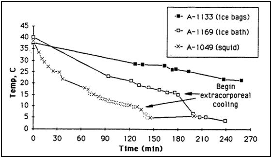
It may also be very useful to estimate body fat and more reliably determine subcutaneous fat cover by using a Skinfold Caliper. A Skinfold Caliper is a device which measures the thickness of a fold of skin with its underlying layer of fat. By measuring skin fold thickness at precise places on the thorax, abdomen and thigh a reasonably accurate measurement of total body fat can be obtained in a healthy individual. In the case of the cryopreservation patient the interest is not primarily in the accuracy of the tool in determining total body fat per se, but rather in the creation of a database of measurements which can serve as an objectifying guide to determining a patient’s degree of fat cover at the time of cardiopulmonary arrest.

Illustrated in the charts and tables below is the procedure for taking Skinfold Caliper measurements in both adult males and females:

Measuring Sites for Males

Site Direction of fold Measurement
Thorax Diagonal Fold is taken ½ the distance between the anterior auxiliary line and nipple.
Abdomen Vertical Fold is taken vertical 2 cm lateral to the umbilicus.
Thigh Vertical Fold is lifted on anterior aspect of thigh midway between inguinal crease and proximal border of patella. Body weight is shifted to left foot.

Table 2: Measuring Sites for Skincaliper Body Fat Determination:


Measuring Sites for Female


Site Direction of fold Measurement
Triceps Vertical Fold is taken midway between the shoulder and elbow joint, on the center of the back of the arm.
Waist Diagonal Fold is taken diagonally above the iliac crest along the anterior auxiliary line.
Thigh Vertical Fold is lifted on anterior aspect of thigh midway between inguinal crease and proximal border of patella. Body weight is shifted to the left foot.


Multiple regression equations exist for calculating the percentage of body fat from the Skinfold Caliper measurements depending upon age and sex (50-52). However, at this time the author believes that the selection of a particular algorithm is not very important. Because there is no typical cryopreservation patient, the consistent collection of reliable data and their reduction to arbitrary numbers which can be compared from case to case is the most important consideration.

Collection of BMI and Skinfold Caliper measurements from multiple patients combined with some objectification of the efficacy of CPS during transport will likely be the most useful tools for comparing various methods of cooling; both external and internal.

Below are the Jackson, et al., equations for calculation of percentage body fat in females and males:

FEMALES:

D = 1.0994921 – (0.0009929 x sum of triceps, suprailiac, thigh) + (0.0000023 x square of the sum of triceps, suprailiac, thigh) – (0.0001392 x age), based on a sample aged 18-55.
Jackson, et al. (1980) Generalized equations for predicting body density of women. Medicine and Science in Sports and Exercise, 12:p175-182.

MALES:

D = 1.10938 – (0.0008267 x sum of chest, abdominal, thigh) + (0.0000016 x square of the sum of chest, abdominal, thigh) – (0.0002574 x age), based on a sample aged 18-61.
Jackson, A.S. & Pollock, M.L. (1978) Generalized equations for predicting body density of men. British J of Nutrition, 40: p497-504.

Figure 22: Actual cooling curves for three adult patients on HLR support, using ice bags, portable ice bath (PIB), and PIB plus SCCD cooling. Patient A-1133 weighed 56.8 kg, patient A-1169 weighed 57.3 kg, and patient A-1049 weighed 36.4 kg. As this data indicates, PIB cooling is approximately twice as effective as ice bag cooling. The spray cooling device (SCCD) increases the rate of cooling by yet another 50% over the PIB (roughly adjusting for differences in patient weights).
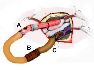
As can be seen in Figure 38, cooling rates with the PIB, even when using stirred water to maximize convective cooling; still result in unacceptably slow rates of cooling. To solve this problem four other methods of cooling, which are amenable to immediate, in-field application have been developed:

1)      Use of chilled parenteral medications; bulk IV fluids given to provide cerebroprotection, such as buffers and hyperosmotic agents are administered (where possible) chilled to 1-2 oC.

2)      Peritoneal lavage with 4 liters of Normosol-R™ chilled to 1-2 oC is used 2x as soon as feasible after pronouncement.

3)      Colonic lavage with1 liter of Normosol-R™ chilled to 1-2 oC is used 4x, as an alternative or prior to peritoneal lavage, as soon as possible during CPS (total of 8 liters).

4)      Liquid assisted pulmonary cooling (LAPC) with 4 liters of 3M FC-75™ perfluorocarbon is used 2x during CPS; the first as soon as possible after placement of an endotracheal tube, and the second 15 minutes following the first.

Detailed instruction on how to perform these adjunct procedures to external cooling are provided later in this Chapter.

References

References are given at the end of Part II of this article.

Footnotes


[1] The other two pillars are the information theoretic criterion of death and the assumed continued advance of technology and medicine.

[2] The Boston Circulatory Arrest Trial was carried out using alpha stat pH management which is no longer used by most centers for pediatric cardiac surgery involving DHCA.

[3] The Q10 rule is much abused in biology and a further exposition of the limitations on its use is presented in Appendix 1.

[4] A few qualifying remark are in order here. This statement holds only when the new therapeutic modality fits well within the existing biomedical paradigms, and social and ethical milieus. Semmelweis and antisepsis in the 18th Century were vigorously resisted whereas the sulfanilamide and penicillin were rapidly embraced 60 years later. Few clinicians question the potential of hypothermia, or the biological basis of its therapeutic action, to the extent it is currently understood. What is questioned is, ‘is it worth it?’

Posted in Cryonics Technology (General), Ischemia-Reperfusion Injury | 2 Comments

Commercial Air Transport of the Cryopreservation Patient

By Mike Darwin

Preparing the Patient for Shipment

Closed chest cardiopulmonary support (CPS) will generally be discontinued either at the start of in-field cardiopulmonary bypass, or when the patient’s frontal sinus or tympanic temperature reaches ~ 15ºC. Due to hypothermia-induced blood sludging, cold agglutination and red blood cell (RBC) aggregation, continuation of CPS below 15ºC is currently deemed to be ineffective and possibly counterproductive.

Many patients will be remote from the facility where cryoprotective perfusion will be carried out and will be transported by common carrier or private carrier over considerable geographical distances. In some cases it will be possible to move the patient using a specialized transport vehicle with on-going extracorporeal support. In other cases the distances will be sufficiently great that the only realistic option is iced-shipment in the absence of perfusion. It is often necessary to use a commercial air freight service to move the patient from one area of the United States to another (or from one country to another).

Preparation Following Extracorporeal Support

Figure 1: The arterial (A) and venous lines (C) should be cross-connected using a 3/8 “x 1/2″ connector (B) with great care being taken to avoid introducing air into the tubing or connector during this operation. A syringe may be used to dribble perfusate or normal saline into the opening between the two lines/connector as they are approximated and connected. The edges of the wound are then approximated around the cannulae and stapled closed.
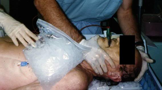
If extracorporeal support and/or blood washout are to follow CPS then a considerable amount of preparation of the patient for shipment will be necessary at the conclusion of extracorporeal support. Once blood washout and cooling to 2-4ºC are achieved, the patient will need to be disconnected from the extracorporeal circuit. This should be done by cutting or “breaking” the arterial and venous lines aseptically and cross-connecting them (avoiding any introduction of air) with a 3/8″-1/2″ sterile connector (Figure 1). The edges of the groin wound(s) are then approximated with the cannulae still in place, the skin at the operative site is toweled dry with a sterile muslin towel or gauze 4x4s, and a waterproof 3-M adhesive drape is placed over the wound and cannulae to prevent water from entering the wound and to stabilize the cannulae against dislodgment.

Figure 2: Combitube in position and capped after arrival of a patient from remote stabilization and blood washout.
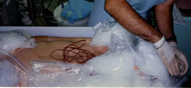
All other lines and catheters should then be capped or clamped using disposable plastic occluding clamps, Kelley clamps, or stainless steel tube occluding forceps, starting with those at the head and working down.

1) If present, the endotracheal tube is plugged or clamped. If either the DEGTA or Combitube are present, the mask is disconnected from the gastric tube/obturator; the gastric tube/obturator is clamped. If the Combitube has been used it should be left in position and plugged, not clamped. If an ET tube or DEGTA were used, and the mouth is packed with 1-2 4×4 gauze (to guard against water entering the airway) with a visible tail of each gauze square left outside the mouth to facilitate easy removal. Do not remove the DEGTA gastric tube/obturator, or let the balloon down, as this may allow seepage of gastric contents into the lungs.

Figure 3: Probe leads on thorax following shipment of patient. The twist ties have just been removed and probe leads are ready for connection to the temperature monitoring equipment in the cryoprotective perfusion facility.
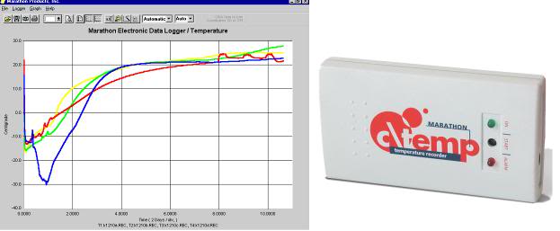

2) Disconnect all temperature probes from the monitor(s) and leave them in place. Probe leads are then coiled up neatly and secured in coils with twist ties. Coiled probe wires are placed on the ventral surface of the body where they may be secured with tape, if desired, to avoid being dislodged during movement of the patient from the PIB to the shipping container. If an auto-logging recording thermometer is being used to monitor the patient while in transit, it should be connected to the probe in closest proximity to the brain (nasal, pharyngeal, etc.). If a DuaLogR or similar recording thermometer is to be used ensure that it is in its watertight case and is placed on the patient’s thorax, well above the probable level of any melt water that may accumulate during shipping.

3) Any IV or central venous lines should be capped using aseptic technique. If caps are not available, the IV set-up must be clamped off and the entire IV set including bag or bottle left connected to the patient (and thus sterile).

4) The Foley catheter is plugged or clamped.  It is not necessary to maintain sterility of the distal end of the catheter.

5) Rectal and peritoneal lavage tubes are left in place and the tubing plugged or clamped. The rectal tube line should be wrapped in several gauze 4x4s to prevent the spread of any feces contaminated drainage.

The patient’s eyes are closed with tape and all visible blood and secretions on the body are cleaned up while the patient still in the PIB. Be certain that blood and other laboratory samples are attached to the right forearm with tape!

The patient’s hands are positioned at his or her side and the legs are fully extended and placed in close approximation to each other. The PIB is then drained of water as completely as possible using the SCCD, or by bailing the water out manually.

Before the patient is removed from the PIB, the air shipment container should be prepared by placing a layer of Zip-Loc bags filled with flaked or small cubed ice over the bottom of the inner container of the shipper. If a heavy-duty body bag is available it should be used to hold the ice bags that will go under the patient. Enough additional ice bags should be prepared in advance to allow for complete and thorough packing of the patient in ice immediately upon transfer to the shipper. Generally, 50 kg (110 lbs) of ice are required to safely refrigerate a patient (who has been pre-cooled to near 5ºC) for 24 to 48 hours in a well insulated[1] container. The nomogram  (Figure 4) will give approximate ice melt values per unit of time for a pre-cooled patient in a metal shipping container insulated with either 2” of expanded polystyrene (EPS) or ½” Rmax R-Matte Plus-3 foil-clad polyurethane foam board, with a stated R value of 3.2.

More ice will be needed if any of the following conditions pertain:

  • The ambient temperature is significantly above 24 ºC.
  • The patient weighs significantly more than 80 kg (176 lb).
  • The patient’s body temperature at the time of packing for shipping is greater than 5ºC.
  • Shipping time is anticipated to longer than 36 hours.

Immediately prior to transferring the patient to the shipment container, excess ice should be removed from the PIB and/or the patient should be elevated in the PIB and the ice should be moved under the patient to raise the patient above any remaining liquid on a bed of ice. The patient is then wiped down with towels while the dorsal surface of the body is allowed to drip free of excess fluid on the bed of ice. A c\temp disposable electronic temperature logger (http://www.temperature-data-logger.com/c_temp.html) should be placed over the patient’s right temple using the accompanying Velcro headband. The c\temp should be set to record temperatures at 58 second intervals (e.g., 5 day setting).The purpose of the temperature data logger is provide quality assurance during patient air shipment that there has been no warming and to provide a record of the patient’s transport temperature for later clinical and scientific evaluation (most patients will still be well above ~ 1ºC when they are placed in the air shipper. The c\temp, along with a representative data stream, is shown in Figure 4, below.

Figure 4: The c\temp dlt temperature logger operates in therange of -81°C to +30°C and may be used for both water ice and dry ice shipment of cryonics patients. The device has a lithium battery with a 2-year shelf life and can acquire data at intervals as brief as 50 seconds for up to 5 days. It measures 9(L) x 5(W) x 1.6(H) cm and 58 grams. Data is downloaded to a PC using proprietary software.

Once the patient is cleaned up and relatively dry, place the patient’s head in a plastic bag (to protect the airway and ophthalmic and nasopharyngeal mucosa from exposure to water) and lightly secure the bag around the patient’s beck with tape. The patient is then moved to the shipment container. If a body bag is being used as a liner to the inner box of the shipment container, it should be filled with Zip-Loc bags containing crushed or small cubed ice.

Figure 5: Nomogram for determining the minimum safe amount of ice required for patient air transport. To determine the amount of ice that will be needed based on the patient’s body weight and temperature place a straight edge between the body weight scale on the right and the patient body weight scale on the left. The quantity of ice that will be consumed (melted) kilograms (kg) is shown on the center scale. Body weight is in kg and temperature is in ºC. Ambient temperature is assumed to be ~24.ºC. Reproduced from: “Ice Melting Time in a Patient Shipment Container by Charles Platt, Aschwin de Wolf, and Jay Wasserlauf; Suspended Animation, Inc, April 2005.”[4]
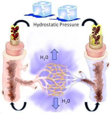
Note: It is very important that the patient’s abdomen and thorax not be heavily loaded with ice as the ice will exert a large hydrostatic force and will displace perfusate (or blood) from the heart and great vessels into the peripheral tissues. This is especially important in the case of patients who are being shipped without the benefit of blood washout, as loading of the chest and abdomen will force blood into the capillaries where ultrafiltration will occur through the capillary membranes. This results in hemoconcentration and plugging of the capillaries with high viscosity sludge of formed elements of the blood. Additionally, in patients where clotting has occurred there is the danger of forcing clots from the central circulation both retrograde and anteretrograde into the arterioles and venules where they may be impossible to dislodge.

Figure 6: Loading the patient’s abdomen and thorax with ice creates hydrostatic pressure in the vasculature. This pressure will result in filtration of water from the blood via the microcirculation into the interstitial space. The result will be massive hemoconcentration of blood components and plasma proteins resulting in a viscous sludge that will be difficult or impossible to displace during cryoprotective perfusion.
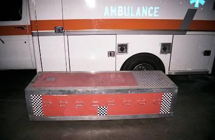
Do not overload the top (ventral surface) of the patient with ice; keep the ice bags on the abdomen and chest to no more than 1″ of ice thickness. Do not place large trash bags or other large plastic bags loaded with ice on top of the patient (See Figure 7)!


Figure 7: Do not load ice atop the patient’s thorax or abdomen (B). Ensure that the c\temp continuous temperature logger is placed next to the patient’s head (A) and that the patient’s laboratory and effluent effluent samples are affixed to his right arm (C).

Special attention should be given to completely packing the patient’s head in ice. Upon completion of ice packing, the body bag is closed (if it is being used) and the inner container is closed and sealed. The outer container is then closed and prepared for shipment.

Ideal Common Carrier Shipment Container

Figure 8: Optimal purpose-built shipping container constructed of welded steel frame and heavy-duty plywood. Note the large number of handles and the special checkerboard marking.
Common carrier transport of the patient requires uncommon preparation and attention to detail if it is to be carried out safely. On two occasions in the past patients have suffered warming or have experienced delays of many hours in transportation as a result of leaking or inadequately insulated shipping containers.[1],[2] Whenever possible a purpose-built shipping container should be used for common carrier transport. A corollary of this recommendation is that a purpose built container should be both kept on hand, and be immediately ready for transport to the site where it will be used.  Shipping containers should always be deployed with the Remote Standby Kit (RSK) when a Standby commences for a clearly terminal patient.

Figure 9: Tamper evident seal placed on patient air shipper after inspection by air freight/TSA security officials prior to shipping. If the lid of the shipper is opened, or an attempt is made to remove the seal, it will be immediately evident.
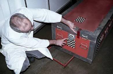
  • It is large enough to accommodate a large adult male (198 cm 110 kg) and still have an adequate volume for 100 kg of ice.
  • Exterior walls are constructed from 3/4″ marine plywood with a welded steel frame making the shipper extremely durable and unlikely to come apart if it is dropped, or to be penetrated by forklift tines during handling.
  • The unit has multiple handles on all sides to facilitate easy lifting by up to 10 people.
  • The exterior lid requires a tool (a heavy-duty Phillips screwdriver) to be opened to discourage casual inspection of the contents, and can be tamper-evident sealed to detect unauthorized entry (Figure 9).
  • The inner container is fabricated from welded, high-impact polypropylene which is fluid-tight, impact resistant, chemically impervious, and can be hermetically sealed. Polypropylene containers of this type also allows the unit to be used for dry ice shipment of patients should straight freezing be necessary.
  • The inner container can be sealed in the field using a self-releasing silicone gasket forming compound which insures that no leakage of refrigerant or body fluids from around the patient can occur.
  • The exterior of the container is marked both by its color (safety orange) and by a black and white checkerboard pattern which allows for easy identification during shipment or if it is delayed or accidentally misrouted. The checkerboard material is vinyl adhesive contact covering available from Kittrich, Corp., (order # 9P23 Con-Tact Brand Covering): http://www.amazon.com/Kittrich-Corp-Con-Tact-Brand-Covering/dp/B001ARHJ24.

Figure 10: Thermometer and alarm assembly at the head-end of the shipment container. The thermometer is a battery-powered unit that is protected by a spring-loaded steel door. The thermometer can be set to sound an audible alarm and stores high and low temperatures in memory. An exterior 110 VAC electrical outlet cover used to protect the remote-sensing battery operated thermometer purchased from a general merchandise store (Wal-Mart).
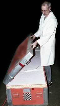

Figure 11: Left: The lid of the shipping container fits flush onto the routed wood of the container box. The insulation for the top of the container is not attached to the lid, thus decreasing wear and tear on the insulation.
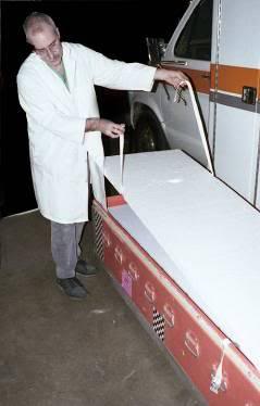
As shown in Figure 10 the container has a battery operated LCD thermometer with alarm and high-low memory capability built into it which is armored against injury. This allows for monitoring of the patient’s temperature during shipment and immediately upon delivery without opening the container. These thermometers operate on a single AA alkaline cell for over a year.

The exterior of the container has a smooth profile with no latches or hinges for anything to catch on. The steel frame is completely smooth, and flush with the walls of the box and has no lip which can hang up on the conveyor belts or become entangled with adjacent freight causing the container to be thrown off the belt, or the frame to bend or deform (both of which happened with previous designs).

Figure 12: Inner top insulating panel of expanded polystyrene foam is lifted to expose the inner liquid-tight box of welded polypropylene.
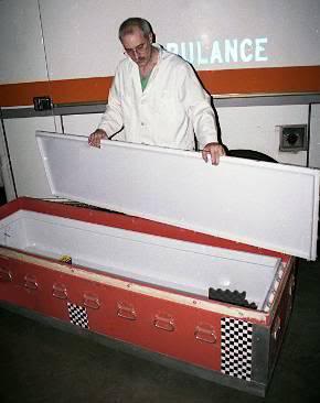
Figure 13: The lid of the inner container has been removed. Note the lip on the lid which allows creation of a non-adhering silicone gasket in the field using Blue RTV Form-A-Gasket, or similar material.
Figure 14: Inner container showing thermometer assembly, sealing lip, spring-loaded catches at 6″ intervals, and boxes of Zip-Loc bags for ice packing inside.
On-Site Fabrication of a Shipment Container

All too often it will not be possible to have a purpose-built container available when needed. The next best alternative is to construct a quality shipper on site if time permits. The following sequence of photographs shows the key elements of such a field-built container. This container has been assembled from three elements which are available from most mortuaries: a sealer casket, a sheet metal air shipping box, and a standard airline casket air shipment tray. The other items necessary for putting such a container together (foil tape, foam or fiberglass building insulation, and a remote sensing (indoor/outdoor) thermometer, etc.) can be obtained from Radio Shack, Wal-Mart or any general purpose hardware or home supply outlet.

Figure 15: An air tray containing a standard metal sealer casket. The air tray consists of a sturdy wooden base with straps to accept a heavy cardboard cover for the casket.
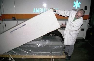
In the past, very reliable water-tight and sealable metal air shipping containers (for routine transfer of human remains between mortuaries) were widely stocked by most mortuaries. Most of these containers were manufactured by Fayette Lock and Tool of Connersville, Indiana (the very best are re-usable shipment cases manufactured by Ziegler Metal Products, and are used almost exclusively by the military, and rarely if ever, by the civilian sector). In the mid-1980′s Fayette Lock and Tool ceased operation and the standard for air shippers dropped dramatically. Today, the typical mortuary air shipment box is crudely made of galvanized metal which is poorly soldered or tack-welded at the joins. Such shippers are not water tight and will leak profusely if filled with ice bags with or without a body bag as a liner.

NOTE: The importance of having a water tight, leak-proof shipment container cannot be overstated. Virtually all common carriers will reject a leaking container, will refuse to ship it if it has been accepted, and will pull it from shipment and notify local health authorities if it begins to leak during shipment.

Sealing the shipment container against leakage thus becomes of paramount importance.  The first step in achieving this is to caulk all the joints of the inner mortuary air shipper with silicone caulk (bathtub or exterior silicone caulk is acceptable) after preparing the surfaces to be caulked by wiping with rubbing alcohol (70% isopropanol in water), acetone (nail polish remover) or other degreasing agents to insure good adhesion of the sealant.

A metal sealer casket is then stripped of its decorative lining and prepared to receive insulation. Foam board (urethane, expanded polystyrene, or other foam board insulation) or fiberglass wool insulation is laid down on the bottom of the sealer casket. The mortuary shipping box is placed atop the insulation and additional foam board is cut to insulate the sides and ends of the box by filling the annulus between the sealer casket and the mortuary shipper.

The lid of the sealer casket is filled with fiberglass wool and/or foam board insulation.  The insulating material can be protected against water and improved in efficiency by using foil backed adhesive tape of the kind used in home gutter repairs to cover the foam board or glass wool.

Figure 16: Sealer casket on an air tray under the cardboard cover.
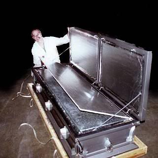
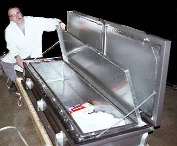
Figure 17: Sealer casket with insulation in the lid, additional foam board insulation between the casket lid and mortuary shipping box, and foam board and fiberglass insulation between the inner and outer containers (casket and mortuary shipper).

In a situation where there is no time to obtain insulation and adhesives it will be necessary to rely on the integrity of the sealer casket to stop water leaks. Blankets, paper towels, wadded up newspaper, bubble wrap, Styrofoam peanuts, or any of a variety of materials may be used for improvised insulation. Whenever possible use absorbent material on the bottom of the sealer casket to absorb any leakage or sweating from the inner container. A multilayered defense against leakage is the best strategy.

Figure 18: Interior of a sealer casket which has been outfitted as an in-field air shipper. A galvanized metal liner has been silicone caulked and a body bag and Zip-Loc bags are present inside to be used when the need arises.
It is important to be aware that actual leakage is not the only potential problem which may cause interruption of shipment and quarantine of a patient in transit. Even if a water-tight container is used, sweating due to water condensation on cold metal may be mistaken for leakage.  Thus, it is very important to have some insulation between the container holding the patient and refrigerant, and the outside of the shipper.

Figure 19: Thermometer/alarm attached to the outside of a sealer casket with self-adhesive Velcro strips. Velcro and thermometer/alarms such as the one above can be purchased at Radio Shack, Wal-Mart or other general merchandise stores.
Sealer Casket Operation

Sealer caskets are made by a number of manufacturers; however, the largest of these is the Batesville Casket Company in Batesville, Indiana. The Batesville sealer is the prototypical sealer casket. A low cost sealer consists of a metal box of 20 gauge steel with a lid which has a rubber gasket and a compression closing mechanism. The metal shell typically has bar-type handles which are attached with penetrating screws (which will leak if the casket is filled to handle height!). Welds are frequently shoddy, are typically backed up with pitch coating, and will leak (usually only slightly) if the container is even partially filled with liquid. The interior consists of fabric, and excelsior or polyester stuffing which is lightly attached to a tack strip of wood, or glued to the metal exterior.

This lining may be ripped away and used (if necessary) as absorbent material for the bottom of shipper. Most sealer caskets will have at least a rudimentary foam mattress and spring and wire tray or bed assembly to receive the body and adjust its height up or down in the casket. This frame should be removed if the casket is to be thoroughly insulated. Low-end sealers cost about $800 US. Mortuary air shippers range in cost from $150 to $175, US.

Figure 20: Gasketed sealing screw-port which covers the lid-locking mechanism of a typical sealer casket.
At the foot end of the sealer on the lip of the box just under the lid is a port capped by a gasketed screw-plug (see Figure 20). The plug is removed to reveal a recessed female hex head bolt which a hex-wrench can be inserted into to lock-down the lid. Typically sealers will come with a hex-handled closure tool (often decorative) called a “casket key.” This tool should be carefully stowed with the casket after it is closed so that the casket can be rapidly and safely opened when it arrives. The casket key hex wrench is of a large non-metric size and is not commonly found in home supply or nonprofessional hardware stores. If the casket key is lost opening the sealer can be very difficult and may require the use of power tools. Additionally, a few manufacturers use different hex-head sizes and, more rarely, unusual key shapes.

It is important to lock the casket with the key prior to shipment in order to get a seal and minimize the chances of leakage. Also, all airlines require that the seal be engaged and some will ask that seal engagement be demonstrated by the shipper prior to their accepting the body for shipment. The casket is sealed by closing the lid and inserting the casket key into the mechanism.  The casket key is then turned clockwise until the lid is pulled tightly down against the bottom of the casket. The key will usually stop turning when the seal is made. Do not lose the casket key!

Figure 21: Casket key being used to close the lid of a sealer casket.
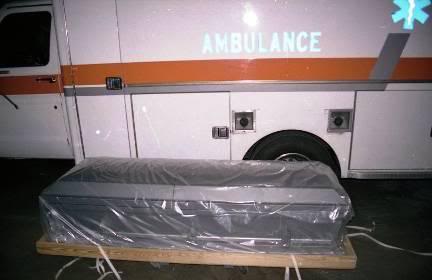

Figure 22: Sealer casket on air tray wrapped in plastic just prior to being covered with a cardboard over-container.
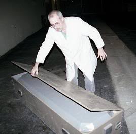
Air trays are as important to facilitating shipment of a patient in a casket as the casket itself. No airline will accept a visible casket as air cargo for public relations reasons. Since freight and baggage can usually be seen being loaded by passengers, all caskets must be covered in a way that makes them unrecognizable as such, i.e., disguises both their surface and unique shape. Airlines will reject a casket which is not covered with an air tray. Most air freight companies will stock air trays but some will not. Especially in smaller communities it is important to acquire an air tray well in advance of need or have a simple plywood box built to cover the casket. The name used by US airlines for a casket air tray is a Jim Wilson tray, an a body being shipped is referred to as a “Jim Wilson.” This is done for public relations reasons as many passengers are unaware that corpses are being transported in the cargo hold and would be distressed to learn that this is the case.

Figure 23: Rudimentary and inexpensive in-field constructed patient air shipper.
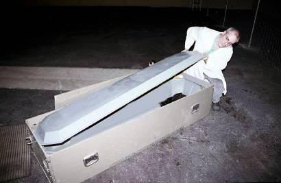
In the event a sealer casket is not available or affordable, many mortuaries can arrange to have a plywood box built on short order, to serve as an outer box for a mortuary shipment box. Some quality mortuary shipping containers are still being manufactured, and, if one of these is available (or a poor quality shipping container can be well-caulked with silicone) it can be used as the inner box. The vertical supports used for adjustable metal shelving can be used as corner bracing and to attach the lid to the box. Trunk handles can be acquired from a hardware store to facilitate lifting, and fiberglass batting (building insulation) can be used to fill the space between the inner and outer box. Figures 23 and 24 show an air shipping containment constructed in just this fashion in the field during the early 1980′s. An exterior electrical outlet cover (for standard 110 VAC service) was used to protect a Radio Shack remote-sensing thermometer.

Figure 24: A mortuary air shipping container positioned inside a plywood outer box and fiberglass building insulation is placed between the metal air shipper and the plywood box.
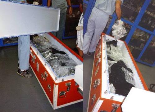
Importance of Insulation

Some human cryopreservation organizations insist that the use of insulated air shippers is unnecessary, and cite firsthand experience with patients transported in a standard metal shipper on an air tray packed in ice during the summer months, arriving with little melting and the patient still “adequately” refrigerated. Such stories are no doubt true, and the author has been forced to ship a patient under such circumstances once in the past. However, such careless handling relies on everything going exactly as planned as well as dumb, blind luck. A patient who is accidentally loaded onto the wrong plane, a patient in an aircraft grounded for mechanical trouble or due to a bomb threat or inclement weather, can experience many hours and in some instances almost a day’s delay beyond anticipated arrival. Failure to plan for these all too commonplace contingencies is grossly negligent.

Figure 25: Patient arrival after air shipment from central Colorado to Southern California during the summer months. The patient was a ~88 kg man whose body temperature was 20.8ºC at the time he was packed in 50 kg of ice inside a custom fabricated cryopatient air shipper insulated with 4” of EPS. Transit time was ~19 hour door to door.[3] Note that most of the ice has been converted to water during transit. The patient’s deep nasopharyngeal arrival temperature was 1.5ºC. This case points up the importance of using ample ice, especially if the patient has not yet cooled to at least 5 ºC before shipment takes place. Complex routing or other factors, such as snowstorms, hurricanes, or other severe weather that might cause the flight(s) carrying the patient to be delayed should be carefully considered. The possibility that the patient may be temporarily lost in shipment or misrouted should also be given consideration as these events can add many hours to transit time.
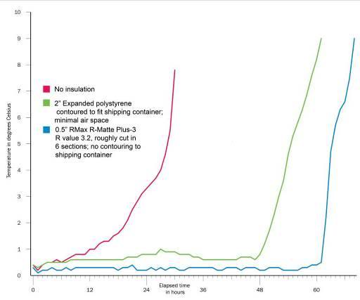
Figure 26: Temperature/time curves measured inside the transport container. Temperature was measured adjacent to the left side of the head of a phantom patient made of thermal neutral expanded polystyrene (EPS) inside a metal air shipping container loaded with 43 kg of water ice. The phantom patient was enclosed in one 3-mil lightweight body bag plus and placed inside another 20-mil heavyweight body bag. Ambient temperature was maintained between 25.5 oC and 24 oC. Data collection for each experiment were synchronized to begin when the first reading on the phantom was between 0 and 0.5oC (earlier values discarded). The air shipper was evaluated un-insulated ___, insulated with 2” of EPS ___, or insulated with ½” of RMax R-MattePlus-3(R value = 3.2) foil backed polyurethane foam insulation ___. Reproduced from: “Ice Melting Time in a Patient Shipment Container by Charles Platt, Aschwin de Wolf, and Jay Wasserlauf; Suspended Animation, Inc, April 2005.”[4]
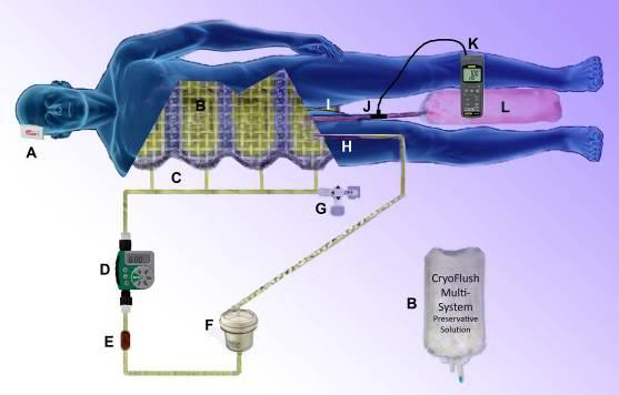
There have already been three cases in the less than 100 patients air shipped by all human cryopreservation organizations so far where a patient’s care was compromised (delays and/or rewarming) due to an inadequately insulated and inappropriately labeled air shipment container. These statistics should serve as ample warning that casual packaging of patients for common carrier transport is unacceptable. Figure 15-23 shows the measured rewarming rates of insulated containers versus an un-insulated container under very conservative conditions with respect to ambient temperature.[4] As can be clearly seen, insulation is absolutely essential for safe air cargo shipment of cryopatients.

Arranging For Air Cargo Transportation

Regulatory Concerns

Removal of human remains from the place of death is regulated to varying degrees in all states and most countries. The complexity and character of these regulations varies enormously, not only from state to state, but from county to county and, often, from day to night in the same location depending upon who is on duty when the call comes in. In most cases, movement of remains after legal death is governed by the following regulatory elements:

1)      A signed death certificate or an agreement to sign a death certificate by the attending physician is required so the patient can be removed from the location of legal death. If the patient is a coroner’s or a medical examiners (ME’s) case, the patient will be transported to the ME’s office for autopsy or further investigation prior to release. Alternatively, a “release number” will be given by the ME or the coroner to waive autopsy or further investigation and release the patient for disposition.

2)      A removal Permit or Transit Permit issued by the County Department of Health is required to transport the patient out of the county where death occurred to another county or another state. Obtaining this permit is usually contingent upon filing a completed death certificate with the Health Department which they certify as acceptable.

3)      A disposition Permit or a Burial Permit is required for intermediate or long-term storage; usually within 2 to 10 days of arriving in the county, or if in the county where legal death occurred, within 2 to 10 days of death. A Disposition Permit is issued only after a certified copy of the Death Certificate and of the Removal or Transit Permit is submitted to the County Health Department where disposition or extended holding is going to occur.

While these elements seem straightforward, their implementation is anything but. Signatures required (funeral directors, physician’s, next of kin, etc.), office hours of the regulatory agencies, local mortuary or public health regulations concerning the shipment of un-embalmed bodies all must be taken into consideration and dealt with professionally and with self-assurance. The ideal person to handle this regulatory burden is a local funeral director who routinely deals with county and state bureaucrats and is thoroughly familiar with the procedures and requirements for inter-county and interstate shipment. The Transport Technician will be fully preoccupied with direct patient care and will have neither the time nor the energy to undertake arrangements for air freight transport of the patient or the associated regulatory burden.

In many jurisdictions (state or county) movement of remains by common carrier without embalming is technically not allowed. This regulation is widely ignored and is sometimes circumvented in the case of cryopatients (where there is no communicable disease posing a public health risk) by having the funeral director certify that blood washout and antibiotic treatment are the equivalent of “disinfection” of the remains. In states or counties where a requirement for embalming is strictly enforced, it may be necessary to move the patient by private carrier (a mortuary vehicle or rented van) to a neighboring jurisdiction, where such rules do not exist, or are not enforced.

The funeral director should also be given the task of making arrangements for the Transport/Standby crew, or critical elements of the crew, to accompany the patient to the cryoprotective perfusion facility. In some cases it will be desirable for key personnel to arrive sooner than it is possible to have the patient arrive. Most funeral directors are skilled at making such arrangements for families of their customers on short notice and many have a good liaison with airlines and local travel agents who can greatly facilitate reservations for staff and cargo space for the patient. Often, the ability to get any staff on any timely flight to the cryoprotective perfusion facility will be solely dependent upon the funeral director making special arrangements with the airline to book them as accompanying the human remains, typically as “family.” Many airlines will “bump” regular passengers to accommodate the next-of-kin of the deceased.

Since the 9/11 attacks on the US the requirements for being able to ship air freight have become onerous. Shippers must be vetted with the freight company and there is a requirement (often changing) for how long they must have been in business before they are acceptable shippers. Most mortuaries will qualify in this regard, and it is critical that shipping arrangements be made through the mortuary, rather than directly by the HCO, unless the HCO has approved status as an air cargo shipper. Neither Federal Express nor UPS will accept human remains or body parts for shipment.

Air Cargo Logistic Considerations

Because of time constraints to get freight loaded rapidly, air freight is often not handled with care by airport personnel. The author has observed a transport container holding a cryopatient nearly drop from a conveyor belt during loading onto an airliner, and has seen caskets containing cadavers (being shipped for conventional disposal) penetrated by forklift tines which were misaligned with the pallet under the casket, and were shoved through the side of the air tray and casket, during hasty loading (resulting in massive damage to the body within). Whenever possible, the Transport Technician should supervise the handling of the patient every step of the way, including on and off the aircraft. Due to recent terrorist acts it has become increasingly difficult for the Transport Technician to do this. Until quite recently it was usually easy for the Technician to get access to air freight facilities and the tarmac to supervise loading of the patient onto the aircraft. This is now all but impossible. However, it is still important to accompany the patient to the air freight depot and to emphasize that extra care should be used in handling the patient, and that every precaution should be taken against misrouting.

Know, in advance, the most convenient and rapidly accessible airport near the HCO’s facilities!

However, the primary consideration must be timely transport of the patient regardless of the airport used. The drive time to other airports accessible to the HCO must be weighed against earlier arrival times. In most cases only one flight will be available at the appropriate time and that will determine which airport is used. Know the alternate airports for the HCO’s location and always choose earlier arrival (and less cold-ischemic time) over convenience, or direct flights. If a direct flight and a flight with stop-overs or plane changes have almost the same arrival time, choose the direct flight, as there is less chance of misrouting of the patient.

In most cases the cooperating mortician or funeral director should be employed to handle airline arrangements since s/he will be most familiar with local shipping requirements, local carriers, and can most reliably arrange transportation to the airport and handle the Department of Health paperwork requirements.

If the weather is subfreezing, it will be critically important to emphasize to airline personnel that the patient must not be allowed to remain in unheated quarters or to be exposed to subfreezing conditions for more than an hour or two, at most.

Pulsed Vascular Rinse Transport

At this time, rigor, and the cold ischemic degradation of the blood brain barrier (BBB) and the peripheral vascular endothelium and basement membrane appear to be avoidable in cryopatients only by the expedient of continuous, asanguineous hypothermic perfusion at 2-4oC.[5-8] An alternative to continuous perfusion is the use of periodic flushes or vascular rinses using a perfusate containing the appropriate substrates.[9] Recently, a perfusate has been developed which is capable of preserving canine hearts for 24 hours in a viable condition.[10] The protocol employed for use of this perfusate involves periodic short intervals of warming the organ to allow metabolic housekeeping to take place during the flush.

Figure 27: PulseFlush vascular rinse extracorporeal perfusion system. The system is pressurized by the body weight of the patient on a nylon mesh restrainer holding four 4 -liter bags containing the pre-chilled perfusate (B). The bags are connected via a manifold (C) to the arterial line. Pulses of flow are delivered every six hours by a modified Orbit 91213 battery operated in-line irrigation controller (D). A flow restrictor in the arterial line (E) before the auto air separating and venting arterial filter (F) establishes the maximum rate of flow from the bags into the patient. Perfusion is very low flow and pressure (~30 mmHg) into the femoral artery (H) and drainage is ‘high’ pressure (10 mm Hg) via a 22 Fr. Biomedicus flat wire femoral venous cannula (I) into a 22 liter flexible plastic effluent collection bag (L). Venous pH and temperature are logged continuously at 5 second intervals using a pH/ORP logger. Extra-cranial temperature is monitored during transport shipment using a c\temp temperature logger: http://www.temperature-data-logger.com/c_temp.html (A).
It is not now technically feasible to follow the transient warming protocol demonstrated to be most be effective in preserving cardiac (and possibly skeletal) muscle viability. However, based on experiments conducted by the author at Critical Care Research, Inc., in 1998-99, periodic vascular rinses are effective at reducing, and in some cases eliminating the onset of cold ischemic rigor in rabbits. To achieve this in human cryopatients a simple system has been developed using a modified Orbit 91213 in-line lawn irrigation controller. Four 4-liter bags of perfusate are pressurized by being placed in a custom made flexible nylon mesh holder and restrainer which the patient is placed atop in the air shipper (Figure 27). The weight of the patient pressurizes the fluid in the bags which is connected via a tubing manifold  to the Orbit 91213 controller. The controller operates on two AA (1.5V) alkaline or (preferably) lithium batteries. The unit is easy to operate and program and should be set to deliver a vascular rinse pulse of 4 minutes duration (4 L) every 6 hours. Instructions for the modified Orbit controller are present as Appendix 1 to this Chapter.

The Orbit 91213 controller can be integrated into the PulseFlush tubing pack and ethylene oxide sterilized with the batteries removed (Batteries must be returned to the device prior to priming of the arterial line). The fluid contact parts of the device are biocompatible (stainless steel, polypropylene, polyethylene and Buna-N). The device performs reliably under harsh conditions (wet environment, subfreezing temperature to -8oC) and, when used in conjunction with the appropriate tubing pack (including a flow restrictor) and perfusate bags holder, it will reliably deliver 4 liters of flow at ~30-40 mm Hg pressure q. 6 hours (1 LPM/min flow rate).

If pulse vascular rinse is being used, it is important to elevate the patient on ice packs as far above the bottom of the inner container as possible (i.e., that still allows for the lid to be closed). The venous effluent collection bag should be placed on the bottom of the inner container with no ice or other material between the bag and the bottom of the inner box. This will provide the maximum amount of gravity ‘fall’ from the venous cannula to the collection bag. The venous line and the venous effluent bag must be primed and completely de-bubbled prior to activation of the system. Failure to do this will result in air-locking of the venous line and failed venous drainage.The venous drainage bag should be placed between the patent’s lower legs with no ice on top of it. Be sure to ensure that there is adequate space for the bag to fill to its 22 L volume.

Once the tubing set has been connected to the patient, the manual override on the Orbit controller should be triggered, and one 4-liter rinse given by gravity before connecting the perfusate bag manifold. This is done in order to establish that the system is functioning properly, assure that venous drainage is adequate, and prime the venous line and the effluent collection bag. Connection of the perfusate manifold must be done with minimal air introduction into the system. The arterial filter must be secured in the filter holder and positioned so that the air separation vent port is ‘upright’ and free from any obstructions. The arterial and venous lines must be carefully positioned to avoid kinking or occlusion of the lines, and be secured in place with Backhaus forceps anchored to the patient’s skin.

The patient air shipper should be clearly labeled with “This Side Up” stickers showing the proper orientation of the container.

References

1.         Jones T: Suspending “Mrs. Stone (Alcor patient #1206). Cryonics 1994, 15(3):9-13.

2.         Quaife A: Personal Communication re: cryopreservation of Mary DeMarr. In. Edited by Darwin M. Berkley, CA; 1973.

3.         Jone T: The cryopreservation of Jim Glennie. Cryonics 1992, 13(9):10-13.

4.         Platt C, de Wolf, A, Wasserlauf, J.: Ice Melting Time in a Patient Shipment Container. In.: Suspended Animation, Inc.

5.         Darwin M, Leaf, JD, Hixon, H.: Case report: neuropreservation of Alcor patient A-1068. 1 of 2. Cryonics 1986, 7(2):17-32.

6.         Darwin M, : Unpublished case report of Alcor Life Extension Foundation patient A-1036. In.; 1988.

7.         Darwin M: Unpublished case report of Alcor Life Extension Foundation patient A-1410, . 1992.

8.         Henson H: The Transport of patient A-1312. . Cryonics 1992, 13(2):18-24.

9.         Fahy G: Polyglycerol compositions for the protection of living systems from states of reduced metabolism. In. Edited by Office USP. USA; 2009.

10.       Fahy G, Wang, TC.: Extended Organ Presrvation. In: World Intellectual Property Organization. Edited by Organization WIP, vol. PCT/US2005/021472. USA; 2004.

Footnotes


[1] A well insulated container is one which has at least 2” of EPS or ½” of of RMax R-MattePlus-3(R value = 3.2) foil backed polyurethane foam insulation

Posted in Cryonics Technology (General) | 2 Comments

The Armories of the Latter Day Laputas, Part 3

By Mike Darwin

“When reason fails, the devil helps!”

— Fyodor Dostoyevsky , Crime and Punishment

The Entropy of Empire

There are, no doubt, many reasons why men aspire to become the chief executive officers (CEOs) of nation-states turned empires, not the least of which is a sincere desire to directly effect the course of these empires’ decision making and thus, history. Rarely is this opportunity granted, because nation-states, and especially imperial nation-states, are driven by an overweening self interest that is nearly perfectly inscrutable. Thus, the course of empires is goverened not so much by the conscious decisions of individual men, as it is by the inevitable collapse of empires; what I call the the “entropy of empire.”

The entropy of empire narrows and constrains the choices any individual actor can  make, and does so most powerfully with respect to actions of the CEO. As a result, the most powerful influence an imperial CEO or emperor is likely to have will occur not as a consequence of the formal or deliberate decisions he makes, but rather, as a consequence of his unintended actions.


Figure 26: As empires enter the arc of decline, their CEOs or emperors become increasingly constrained in their decision making. High stakes, risk aversion, absence of normal feedback (e.g., life in the bubble) and increasing enmity from the larger community all act to reduce the options a leader can take without catastrophically destabilizing the system, or alienating special interests that are perceived as critical to maintaining the status quo.

If you doubt the former, it is only necessary to look to the economic and international policies of the ostensibly liberal Democratic US President, Barac Obama, taken since he assumed the Presidency in 2004, and contrast them with those of George W. Bush, his “ultra-conservative” Republican predecessor. Because the entropy of empire is in play, it is hard to tell where the forigen or economic policy of Bush left off, and that of Obama began.  And so the irony is that it is most often not the ideologically informed policy decisions that mark an imperial CEO out as having effected the course of history, but rather his ancillary, collateral, or wholly unintended actions that influence the turn of events; often none more so than his personal style and temperament. It may be that one (small) reason for the vile arbitrariness and barbarity of emperors such as Nero, Caligula, Stalin or Hitler is that there is the sad realization that the only way they can truly make history is by behaving badly.

There is an unfortunate tendency to think of empires solely in geopolitical terms; as agglomerations of nation-states and territories spanning continents and being possessed of vast wealth and power. In fact, empires come in all sizes, and while all are, relatively speaking, both wealthy and powerful (and ultimately profligate and failed) they can exist whenever conditions allow for the dominance and control of an asset deemed essential by some fraction of the population. The Roman catholic Church prior to the Reformation and the  company owned Appalachian mining town are both examples of empires that can exist apart from the nation-state (and even within it), writ both very large and very small. It is a peculiarity of cryonics under current conditions that, because of its lack of widespread societal aceptance, the absence of meaningful qualitative feedback, and the high threshold of resources and credibility required to capture any of the current microscopic market, new ventures are effectively prohibited, at least within the US. Thus are empires made, and once made, they go on until their time is up; until their entropy collapses them.

The result is that the two extant cryonics organizations can and do operate in market niches which largely remove them from competetion with each other and which allow for the creation of nano-empires which span the globe – empires that are, as are all empires, top-down structured, and which operate on the basis of internally developed plans and goals which are created and executed largely independent of actual market forces and core scientific challenges. In this respect, they bear a striking resemblence in their mode of operation to the planned economies of communist nation-states.

Lessons From the Cold war Arms Race?

Figure 27: US President John Fitzgerald Kennedy (JFK).
John Fitzgerald Kennedy (JFK) stood for the office of President of the United States on 02 January, 1960. At that time he was most widely known as the war hero turned congressman who had authored the Pulitzer Prize-winning book Profiles in Courage, rather than the author of the Harvard senior thesis turned best seller[1] Why England Slept.[2] In fact, Profiles in Courage was ghostwritten,[3] and despite its Pulitzer Prize, it is a mediocre book bordering on bad.  If you want a glimpse into the kind of the mind that JFK possesed, then Why England Slept is the essential read. It is also a haunting read, because Kennedy’s thesis was that the British Empire was already in terminal decline by the early 1930s, and that Britain was in neither the military or economic condition to have opposed Hitler in Munich in 1938. Kennedy’s argument was simple, and in hindsight unassailable: Confront the Nazis in 1938 and rapidly loose the war. Appease them and buy time until the USSR and the US were brought into the conflict, or induced to materially assist Britain, and the war might well be won. When Kennedy finished his thesis in 1940, the verdict on victory was still out, and it would not be confirmed by history until five bloody years later.

Anyone who takes or accepts the credit for the intellectual workproduct of another man, as JFK did when he was complicit in the creation of Profiles in Courage, has shown himself to be a blackguard. It is therefore tempting to look at Why England Slept as merely an apologist tract for the pro-fascist politics of his father, Joseph P. Kennedy, and of pro-fascist-isolationist demographic in America and England in which he had been reared.[4] In the run up to the war, the US had been deeply divided in its sympathies.  While there was a long tradition of Anglophilia stemming from the country’s roots as a British colony, there was also a large German and Irish population which regarded Great Britain with contempt, suspicion, or both.

The economic collapse resulting from the stock market crash in 1929 had led to profound social unrest and the emergence of vigorous socialist and communist political activism throughout the Western world. In Germany, Spain and Italy the advance of communism was seen to have been effectively stopped by Hitler, Franco and Mussolini, and perhaps even more importantly, to have been countered by a movement (Fascism) which offered economic recovery as well as eugenic improvement and a new “scientific system of government.” Mussolini made the Italian trains run on time and Hitler created a vast industrial infrastructure in Germany and pulled the country out of a catastrophic inflationary depression. Both men coupled these acommplishments with a showy ideology that sparkled with glamor and promised to bring order out of chaos. To many, Fascism offered the prospect of lasting peace and prosperity.

While JFK was reared in this ideological milieu, Why England Slept reveals an original thinker who was not just curious about geopolitical history, but able to draw significant and valid conclusions from surprisingly meager data. This ability may well have been the very thing that redeemed him from what, during his campaign for the Presidency, must have seemed a minor indiscretion, and but one of many, with the facts. At the time of JFK’s presidential campaign, elements inside the US Air force were engaged in a major disinformation campaign, principally to convince as many in government as possible, as well asthe American electorate, that there existed a “missile gap” between the USSR and the US. Following the launch of Sputnik in 1957, Air Force general Curtis LeMay and the corps of Air Force intelligence analysts, became convinced that the USSR had perhaps upwards of a hundred ICBMs.[5] By contrast, the US Central Intelligence Agency  CIA (CIA) analysts argued that there were perhaps a dozen. As it turned out, there were only four; all of them liquid fuel SS-6 missiles which required hours of preparation to launch. By contrast, at that time the US had 170 land based Titan and Atlas ICBMs, and was quickly building more. It also had eight George Washington and Ethan Allen class ballistic missile submarines with the ability to launch 16 Polaris missiles; each with a range of 2,200 kilometres (1,400 mi).

“Small” Lie: Big Consequences

Figure 28: US President Dwight David Eisenhower.
Once this debate between the Air Force and CIA analysts was leaked to the press, the Democrats argued that Republican President Dwight D. Eisenhower was not spending enough money on national security and that the US was, as a consequence, open to nuclear annihilation. In his 1960 campaign JFK echoed these charges and used them to considerable effect against his opponent. Later, he alleged he that he took this position because he did not have access to the intelligence data indicating that there was no missile gap, and that in fact the US had vast nuclear superiority in every sphere of nuclear weapons delivery (bomber, ICBM, IRBM, submarine). In fact, JFK had been extensively briefed by the Director of the CIA, Allen Dulles, in July of 1960 – as had his running mate, Senator Lyndon Johnson. Dulles summarized his briefing of the Democratic candidates in a letter to President Eisenhower in August of 1960. Eisenhower, who determined the scope of the briefing, was deeply disturbed by the emergence of what he termed the “military-industrial complex” and was suspicious that the “missile gap” was yet another manifestation of its endless thirst for taxpayer dollars for needless and costly high technology military infrastructure.

JFK and his campaign manager brother, Robert Kennedy, were savvy enough politicians to turn a deaf ear to the truth about the missile gap. Thus, when JFK won the presidency in 1960 he entered office in the position of being unable (and perhaps unwilling) to curtail the explosion in the number US nuclear offensive weapons. Indeed, it was not until USSR premier Nikita Khrushchev began applying pressure in the form of the Berlin Wall to “test” JFK’s mettle as a statesman and leader, that JFK conceded, first to Khrushchev, and later to the world, that not only was there no missile gap, but that the US held an enormous strategic lead. While not news to the Soviets, this public revelation put them in the difficult position of being made acutely and unavoidably aware that they were vulnerable to a “successful” first strike nuclear attack by the US.[6]

The Caribbean Crisis

Figure 29: Soviet Premier Nikita Khrushchev.

Khrushchev and the Politburo had good reason to be concerned about the new American President’s intention’s and aggressiveness following a US sponsored failed attempted invasion of the USSR’s ally, Cuba, in April 1961 (“The Bay of Pigs” invasion).  Kennedy’s perceived “soft” response to the pressure applied from 4 June – 9 November 1961, the second “Berlin Crisis,” which resulted in the erection of the Berlin Wall, encouraged Khrushchev to pursue a redress to the imbalance of nuclear deterrence which both sides now knew and acknowledged existed. The USSR lacked the economic base to maintain ground based ICBM parity with the US, but what it could afford to do was to produce additional inexpensive intermediate range ballistic missiles (IRBMs) and, following the US’ lead in Turkey (with its Jupiter IRBMs), deploy them closer to the US – effectively negating the lack of intercontinental delivery capability. Cleverly, with this action, Khrushchev perceived that he could kill two birds with one stone: redress the missile gap – the real missile gap – and once and for all protect the USSR’s ally Cuba, from invasion by the US.

The Cuban Missile Crisis, the result of the decision taken to place intermediate and short range tactical nuclear ballistic missiles in Cuba, is not known by that name in the Russian speaking world. Rather, it is called the “Caribbean Crisis,” or alternatively, the “October Crisis.” Recently, I sat in Kiev, surrounded by a group of 20 and 30-something Russian and Ukrainian men (and one woman). I was to depart the following morning for the former Soviet ICBM complex in Pervomaysk, and a question had arisen as to why I was interested in going to such a grim place. At which point one of the group leaned forward and said, a bit expectantly, perhaps with irony and perhaps with a little bit of awe, “You know, Mike is the only person here who alive during the Caribbean Crisis!” This was answered by a chorus of “Ahhhs or Umms.” My response was grimmer still: “I was not only alive at the time; I’m old enough to remember it well.”

It is not unreasonable to view the Crisis through the lens of the conflicting ideologies and economic systems of the two nation-sate empires of the time. But to confine the discussion to that plane would be to miss the most important lesson it has to teach, in the same way that the Behaviorists of the first half of the 20th century failed to grasp the criticality of the inner workings of the mind, in giving rise to animal and human behavior. At its most fundamental level the Crisis was about a failure of two adversaries, and ultimately two men, to understand the intent and the fundamental values (moral, ethical and pragmatic) each held. The politics and the world views of the parties involved were important, but not nearly as important as what each side felt, viscerally, about the other.

The Western view of Russia and of communism, is that their economic system (collectivism) was a grotesque and nearly complete failure, which was the direct result of collectivist ideology and practice. This point of view was easily confirmed by examining the USSR, either by econometric criteria, or directly, by visiting.  Russia’s GDP was approximately 10% of the US’s in the early 1960s, and the level of consumer well being and real wealth was a fraction of that in the US at the time. The US science fiction writer Robert Heinlein toured the USSR in 1960, and his travelogue of that visit was published in his book Expanded Universe in 1980. Heinlein argued that any notion that the USSR represented a threat to the US, or to the West, was ludicrous based on his own and his wife’s (Virginia Heinlein’s) firsthand observations of the Soviet standard of living, degree of technological achievement, and population demographics.

Figure 30: In 1960 science fiction author Robert Heinlein, and his third wife, Virginia Gerstenfeld Heinlein (both shown at left on the set of the movie Destination Moon to which Heinlein served as a technical adviser and script writer) toured the Russia where they were, by chance, present during the U-2 crisis when the Soviets shot down a US U-2 spy plane overflying the USSR to gather intelligence (primarily) on Soviet ICBM installations and captured its pilot, Francis Gary Powers (who had failed to use the cyanide ampoule provided to avoid just such a contingency). Virginia Heinlein became fluent in Russian prior to their visit to Russia for the express purpose of being better able to evaluate Soviet society. Both of the Heinlein’s were virulently anti-communist and anti-Soviet. The account of their visit to the USSR was not commercially published until 1980, when it appeared in a collection of Robert Heinlein’s works entitled Expanded Universe.

Heinlein wrote of the medieval state of the country outside of the major cities and he noted, correctly, that Russia was imploding in terms of population, as early as 1960. This latter conclusion was arrived at by the simple expedient of Virginia Heinlein asking every woman she met how old she was, her marital status, how many children she had, and so on; a simple and yet highly reliable way of gathering otherwise impossible to obtain demographic data. This was the kind of spying the CIA should have been doing at the time, and apparently was not. Heinlein went on to question the very possibility that the USSR could have any significant nuclear missile infrastructure in 1960-1. He did this all based on his observations of the general state of the Soviet economy, and of the particulars of that part of their high technology infrastructure he was able to examine as a VIP tourist in 1960. The CIA could have benefitted greatly from his analysis, if not from his rabidly anti-Soviet perspective.

Figure 31: Vladimir in 988, CE. (Holland Park W11, London, UK)

There is no question that this was indeed the state of the Soviet Union in 1960, and so at first glance, it would seem that the analysis of the Soviet Union, and Russia in particular, as a backward and economically blighted state with a contracting population[7],[8] was justified.  However, that was not the whole picture and it fails to account for a number of very material anomalies for which the Russian speaking peoples may be properly credited with being unique. I know of no other example in history where a people have consistently and deliberately reached outside of their culture to select their core social technologies.

Perhaps the first and best documented example of this is the selection of Christianity as the new religion for the Russian people by Vladimir I in 987 CE. The history of Kievan Rus’[9] authored by the Kievan monk Nestor (1056 -1114 CE) during the reigns of Vsevolod I and Svyatopolk II, notes that in consultation with his boyars, Vladimir sent envoys to study the religions of neighboring nations. Nestor reports that Vladimir’s envoys reported back that the Muslim Bulgarians had “no gladness among them; only sorrow and a great stench and that Islam was undesirable due to its taboo against the consumption of alcohol and pork to which Vladimir is said to have remarked that “Drinking is the joy of the Russes. We cannot exist without that pleasure.”

Nestor also describes Vladimir evaluating Judaism and eventually rejecting it, noting that the Jews’ “loss of Jerusalem demonstrated they were no longer in God’s favor.” In the end, Vladimir decided upon Christianity, probably in no small measure because his envoys reported from Constantinople that, “We no longer knew whether we were in heaven or on earth,” describing a majestic High Mass in Cathedral Hagia Sophia, “nor such beauty, and we know not how to tell of it,” and because of the horrid end promised to nonbelievers come the end of the world and the day of last judgment. It should also be noted that substantial power and formidable wealth of the Byzantine Empire at that time was likely also no small disincentive. Forthwith, all of Rus was converted to Christianity by royal ukase.[10]

Figure 32: Vladimir Ilyich Lenin introduces a fundamentally new social and economic system, communism, to the Russian people in 1919.
The adaptation of the Ancient Greek uncial alphabet by Cyril and Methodius, to create Russian Slavonic Cyrillic was a similarly “deliberate” decision, taken in order to facilitate the propagation of Christianity in the Rus via the written word.[11] This pattern of an active and often methodical search and adaptation or adoption of intellectual assets external to the Russian culture has been repeated throughout Russian history. Perhaps the most notable and most recent example of this was the adoption of a radical, untried and very alien economic system in the form of Marxist (-Lennist) communism.[12] The economist and economic historian Jack Hirshleifer has called the opening 5 years of Soviet Communism “the most extreme effort in modern times to do away with the system of private property and voluntary exchange.” It came at an enormous cost in lives and property, which under Stalin, grew to a grotesque and fantastic degree.

Figure 33: Illiteracy in Russia, by sex, between 1856 and 1915.

The idea that the inferiority of the USSR’s  economic performance was solely an artifact of communism infuriated the Soviets, who believed that this judgment was fundamentally unfair and deliberately biased. To some extent, they were justified in this belief. At the time of the Russian Revolution in 1918, Russia was an agrarian monarchy that can only be described as being trapped somewhere between the Middle Ages and the 18th (not the 19th) century. Literacy rates prior to 1900 were in the range of 20%, and while they had risen to ~30% by 1920 (www.marxists.org/archive/lenin/works/1923/jan/02.htm) this should be contrasted with the literacy rate in the US which had been at ~ 90% since the US Revolutionary War in 1776; and a literacy rate in the United Kingdom at the start of the 20th century that was ~ 80%, by comparison.

The US, in addition to having a relatively stable governmental regime from 1800 to 1920, had also benefitted enormously from the abundance of “low hanging resource fruit” available to fuel its economic expansion.[13] At the cost of minimal blood and treasure the US had expanded westward, acquiring enormous assets in the spheres of agriculture, minerals, oil, and lebensraum. An even greater and often overlooked benefit was the vast influx of immigrants, both skilled and unskilled, and all able bodied [14] fleeing various failed and failing economies in Europe, as well as adverse religious, political, and social conditions in their home countries. This vast influx of youthful, often skilled and always able bodied human capital provided vast additional motive force to the engine of US economic expansion. The absence of any significant regulatory burden, near absence of institutionalized corruption, and ready access to vast natural resources drove the US economy forward in an unprecedented way.8

Figure 34: Growth in the USSR’s gross domestic product (GDP) from 1970 to 1990. Ofer, Gur. (1987). “Soviet Economic Growth, 1928 – 1985.” Journal of Economic Literature 25(4):1767 – 1833, and Easterly, William, and Fischer, Stanley. (1995). “The Soviet Economic Decline: Historical and Republican Data.” World Bank Economic Review 9 (3):341 – 371.
 

Figure 35: Growth in the US gross domestic product (GDP) from 1970 to 1990 (blue) compared with the averaged (curve smoothed) growth of the Soviet GDP over the same period of time.
Russia, while gifted with enormous natural resources, lacked the well developed and industrially capable base of human capital that the US enjoyed during the same period, and it was also plagued by a large burden of corruption in the pre-Soviet era. [15] These facts alone would have served as a basis for Soviet antipathy towards the West’s evaluation of Soviet economic performance during the interval from 1918 to 1961.  However, an additional and wholly justified source of Soviet resentment of the Western criticism of “deficient” Soviet economic growth and prosperity, was the effect of WWII on the Soviet economy and the Russian speaking peoples. It is hard for most Westerners who lived through WWII, let alone those alive now, to even begin to understand the devastating impact that the Great Patriotic War (the name by which the Russian speaking peoples refer to WWII, and in particular their prosecution of the war on its eastern front), had on the Soviet economy and on the Soviet peoples.

The Eastern theatre of World War II was the most lethal and costly conflict in human history to date. In excess of 30 million people were killed in this conflict[16] with brutality exercised on the civilian and combatant populations by both sides (i.e., Nazi and Soviet) that was without parallel in scale, if not cruelty, in the history of warfare. As Time magazine noted in a 2008 retrospective on the war in the Eastern theatre: “By measure of manpower, duration, territorial reach and casualties, the Eastern Front was as much as four times the scale of the conflict on the Western Front that opened with the Normandy invasion.”[17]

Figure 36: The blue line shows the probable rate of population growth and the likely absolute numbers that would have been expected if Russia had not experienced the great Patriotic War. Population growth was set back ~ 20 years as a consequence of the war. The relative sharp increases and decreases in population that occurred from 1900 to 1939 were an artifact of the Russian Empire losing territories with ~ 30 million people after the Russian Revolution (Poland 18 mil; Finland 3 mil; Romania 3 mil; the Baltic States 5 million and Kars to Turkey 400 thousands). World War II Losses were estimated between 25-30 million, including an increase in infant mortality of 1.3 million. Total war losses include territories annexed by USSR in 1939-45.

Both Nazi Germany and the Soviet Union utilized “scorched earth” tactics, with the Soviet Union suffering 20 million civilian deaths and total human losses of 26.6 million.[18] The Soviets destroyed as much as possible of their infrastructure and materiel (military and civilian) that they could not evacuate in order to deprive the advancing forces of any benefit. Subsequently, as the Germans retreated from formerly occupied territories, particularly in Ukraine, Russia’s breadbasket, SS Commander Heinrich Himmler ordered SS-Obergruppenfuehrer Prutzmann: “to leave behind in Ukraine not a single person, no cattle, not a ton of grain, not a railroad track … The enemy must find a country totally burned and destroyed.” (September 7, 1943).[19] Acting in concert, the SS and the Wermacht destroyed 18,414 miles of rail lines, flooded mines, razed factories and poisoned countless wells.

In excess of 2 million homes and other structures were burned razed. Nazi Ostland Administrator Erich Koch ordered that “the homes of recalcitrant natives … are to be burned down; relatives are to be arrested as hostages.” The Soviets estimated that the retreating Germans “razed and burned over 28,000 villages and 714 cities and towns, leaving 10,000,000 people without shelter. More than 16,000 industrial enterprises, more than 200,000 industrial production sites, 27,910 collective and 872 state farms, 1,300 machine and tractor stations, and 32,930 general schools, vocational secondary schools and higher educational institutions of Ukraine were destroyed.  Thus, the USSR was subjected to two rounds of comprehensive and devastating destruction of its resources and productive infrastructure.

The direct damage to the Ukrainian national economy caused by the fascist occupation came to 285,000,000,000 rubles.[20] Large numbers of Ukrainians were deported to the Reich for slave labor. The historian Geoffrey A. Hosking has noted that “The full demographic loss to the Soviet peoples was even greater than the raw numbers indicate, since a high proportion of those killed were young men of child-begetting age, the postwar Soviet population was 45 to 50 million smaller than post-1939 projections would have led one to expect.”[21] Considering the economic impact of WWII using total population loss from 1939 to 1945 as a surrogate marker: US losses were 0.32% of the population, UK losses were 0.94% and the USSR‘s losses were a staggering 13.88% of the population, with much of this demographic consisting of young and able bodied individuals at or near their peak period of productivity and reproductive potential.[22]

Figure 37: The mind-numbing human losses suffered by the USSR during the “Great Patriotic War” are put into perspective when compared with the total human losses of the other nation-states involved in the conflict.

What is widely remembered and understood is that the USSR achieved a military victory over Nazi Germany. Too often not considered is that the property damage inflicted by the Axis invasion was estimated to be on the order of 679 billion rubles. The war resulted in the complete or partial destruction of 1,710 cities and towns, 70,000 villages, 2,508 church buildings, 31,850 industrial facilities, 40,000 miles of railroad, 4100 railroad stations, 40,000 hospitals, 84,000 schools, and 43,000 public libraries. And the majority of the USSR’s livestock were slaughtered for food, to prevent exploitation by the enemy or as a result of starvation, disease or escape from captivity.[23]

It is interesting to note that despite its communist/socialist economy, the victory of the USSR over the Axis was in large measure a result of its war industry being able to consistently outperform the Germans, despite the enormous loss of population and land. Stalin’s much ridiculed five-year economic plans, carried out during the 1930s, had resulted in the industrialization of the Urals and central Asia – albeit at great cost in human life and suffering. In 1941, the same trains that transported Soviet troops to the Eastern front were used to evacuate thousands of hastily disassembled factories from Belarus and Ukraine, to areas far removed from the front line. When this industrial capacity was reconstituted east of the Urals, war production could be continued safely out of reach of Luftwaffe bombers. The large increases in the production of war materiel that were necessary to sustain the Soviet war effort were achieved by a large reduction in the civilian standard of living via the application of “total war,” in conjunction with assistance from the US and the UK in the form of the Lend-Lease program, which shipped vast amounts of war materiel via the famed Murmansk convoys.[24]

As German manpower losses mounted and became critical during the last half of the war, they were able to compensate for manpower attrition through the use of slave labor from conquered Eastern European countries, interned Jews and Soviet POWs. Despite the German’s superior production of raw materials they were unable to approach; let alone match Soviet production of war materiel. Much of this disparity was due to a fundamental difference in Soviet manufacturing strategy. In 1943 the Germans made an explicit strategic decision to improve quality over quantity in the production of war materiel, whereas by contrast, the Soviets settled on a strategy to refine and simplify the designs of existing military hardware while steadily increasing the volume of production.

Figure 38: Poster promoting the Marshall Plan (ERP) circa 1950. Note that US flag comprises the wind vane which is the “tail” that helps keep the windmill pointed into the wind and thus on track to generate motive force.
By contrast, the US experienced trivial losses in manpower, capital and systemic infrastructure (i.e., roads, power plants, factories, etc.) and in fact emerged from the war in a better position in terms of global infrastructure and industrialization that when it entered it. The end of WWII also saw the US become a first-class imperial power, replacing Great Britain as the world’s foremost superpower. The US exercised economic and political control over Japan and West Germany via the Marshall Plan (European Recovery Program, ERP) was the $13 billion US aid program to facilitate the rebuilding of European infrastructure and economies in the aftermath of WWII primarily to contain the spread of communism and provide a viable export market for US manufactured goods.[25] The ERP operated from 1948 to 1952 and succeeded in vastly increasing Western European productivity, standard of living and wealth, in no small measure as a result of what has been called the “bonfire of the regulations” wherein international trade barriers were largely eliminated (Europe) and choking internal regulation, excessive taxation and corruption/favoritism were largely swept away. By 1952 as ERP funding ended, the economy of every participating nation-state had surpassed pre-war levels; for all ERP recipients, output in 1951 was at least 35% higher than in 1938.[26], [27]

The ERP was also offered to the USSR and its client states, but they did not accept it. In late September of 1947, the Soviet Union called a meeting of the nine European Communist parties in southwest Poland.[28] The position of the USSR regarding the ERP was that “international politics is dominated by the ruling clique of the American imperialists” who are intent upon the “enslavement of the weakened capitalist countries of Europe.”The communist parties in Europe were instructed initiate a guerrilla struggle, including the use of “sabotage,” against US imperialism in Europe [29] The report further claimed that “reactionary imperialist elements throughout the world, particularly in the U.S.A., in Britain and France, had put particular hope on Germany and Japan, primarily on Hitlerite Germany — first as a force most capable of striking a blow at the Soviet Union.”[30]

The paranoid response to the ERP was likely a result of a complex interaction of factors including Stalin’s psychopathic and paranoid personality, the intense anger and resentment of the USSR to the punishing economic and human losses suffered on the Easter Front in the Great Patriotic War, and finally, deep and not wholly unjustified mistrust of the US’ intentions to profit from and expand its global sphere of influence, as indicated by language in the report to the effect that “the bosses of Wall Street” are “taking the place of Germany, Japan and Italy.” [31]The ERP was also described by the Kremlin as “the American plan for the enslavement of Europe” [32] and it described the global geopolitical situation as being divided “into basically two camps—the imperialist and antidemocratic camp on the one hand, and the anti-imperialist and democratic camp on the other”.[33]

Viewed in this light, the deliberate transformation of Russia into a world class industrial economy (second only to the US) by 1952 in the aftermath of the 1918 Revolution and in the face of the unprecedented devastation and disruption suffered by the Soviet state during WWII, looks considerably more impressive. It is also worth noting that during the early period of nuclear proliferation (1960 to 1970) the Russian economy grew at an impressive rate of ~5% per year – in the absence of capitalism!

Figure 39: US and USSR/Russian Strategic Offensive Nuclear Forces, 1945-1997. Note that the US was solidly ahead in ICBM capability until circa 1968 and that the US  maintained strategic superiority or parity with the USSR throughout the course of the Cold War. Source: Robert S. Norris and Thomas B. Cochran, U.S.-USSR/Russian Strategic Offensive Nuclear Forces, 1945-1996, Nuclear Weapons Databook Working Paper 97-1 (Washington, D.C.: Natural Resources Defense Council, January 1997); Robert S. Norris and William M. Arkin, “NRDC Nuclear Notebook (U.S. Strategic Nuclear Forces, End of 1997),” Bulletin of the Atomic Scientists, January/February 1998, pp. 70-72.; Robert S. Norris and William M. Arkin, “NRDC Nuclear Notebook (Russian Strategic Nuclear Forces, End of 1997),” Bulletin of the Atomic Scientists, March/April 1998, pp. 70-71.
The cost of the Cold War was staggering; for the US the bill was $19.65 trillion (1948-1991) in 2010 dollars, of which $8,731.5 billion (also in 2010 dollars) was expended directly for nuclear arms.[34], [35] Precise data for the dollar cost of the Cold War to the USSR are not available, however it is generally believed that the Soviet Union spent 12-13% of its GDP on military programs in direct support of the Cold War. The cost in hardship to Soviet citizens was vastly greater than this fraction of the GDP might suggest due to the smaller size and lower level of technological sophistication of the Soviet economy.  As the CIA noted in its 1977 A Dollar Cost Comparison of Soviet and US Defense Activities, 1966-1976 (Secret):

“…the dollar cost comparison shows Soviet defense activities to exceed those of the United States by about 40 percent in 1976. If both are measured in terms of estimated ruble costs, the Soviet activities are about 25 percent larger than the US. Thus, the effect of the index number problem is not large enough to alter the basic conclusion that Soviet defense activities overall are currently larger than those of the United States.[36]

The election of JFK, in significant measure on the basis of a bald faced lie about a mythical missile and nuclear arms gap between the US and the USSR, was, in effect, a green light given by the American electorate for the indefinite enfranchisement of  Eisenhower’s darkest nightmare for the post WWII world; the creation of the “military-industrial complex,” and with it the increased likelihood of global thermonuclear annihilation. JFK’s new military strategy, known as “Flexible Response,” constituted a commitment for the US and its allies to remain on a wartime footing and a wartime economy until the Cold War ended in 1991. Beyond the horrendous cost in dollars, the myth of the missile gap further corroded US-Soviet relations, created unnecessary doubt and anxiety in both US citizens and America’s allies. Those in the know in positions of leadership in other nations and in multinational corporations must have had frequent occasion to ask themselves, “If the US, with its massive economy and enormous technological base cannot maintain nuclear parity with the USSR, which is in reality an emerging Third World nation, what kind of leadership and security can we expect from the US?”

If Soviet expenditures for the Cold War were indeed on a par with those of the US, then the approximate dollar value for whole endeavor by both sides would be in the range of $40 trillion 2010 US dollars. To put that into perspective, that is also, give or take, the approximate net worth of the United States of America, at current market value, e.g., $50-60 trillion US! While not the only factor in the collapse of the USSR, the Cold War was very material in its financial implosion. The US and the rest of the West  have been said to have “won” the Cold War, with the tacit assumption being that the US, unlike the USSR, did not spend itself into bankruptcy making weapons of mass destruction and creating and supporting the enormous infrastructure required for their care and feeding. For myself, I doubt very much that the West will escape without paying the same price the USSR did, and possibly considerably more, and with interest.

As peoples, we squandered the funds that would have paid for us to become a space-faring people, to develop self sustaining industrial technology, and above all, to have made vast strides  in the life extension sciences – in particular, with respect to developing suspended animation – and thus, medical time travel. Failure to achieve the latter has condemned billions of human beings to death, making it by far the most expensive war in dollars and lives in the entire history of our species. We cannot continue in this fashion if we are to survive, either as individuals, or as a species. Having said that, there are many lessons we can learn from the Cold War and the effects of its aftermath, which we are now suffering. Some of those lessons will be discussed directly.

End of Part 3

References & Footnotes


[1] The book was made a best seller in large measure by Kennedy’s powerful and influential father, Jospeh P. Kennedy

[2] Kennedy, John F. Why England Slept , Funk, New York, 1940. Reprinted by Greenwood Press, ISBN 0313228744 (1981).

[3] Parmet, Herbert S. Jack: the struggles of John F. Kennedy.  Dial Press, New York (1980).

[4]Swift, Will. The Kennedys Amidst the Gathering Storm: A Thousand Days in London, 1938-1940. Collins/Smithsonian, 352p. ISBN 978-0-06-117356-1 (2008).

[5] Goodman, Melvin A. “Exaggeration Of The Threat: Then And Now“, The Public Record, 14 September 2009. Retrieved 04-19-2011.

[6] Preble, Christopher, John F. Kennedy and the Missile Gap, Northern Illinois University Press , ISBN-10: 0875803326 (2004).

[7]Demography and development in Russia“, UN Development Program, 28 April 2008. Retrieved 04-11-2011.

[8] Global decline in the Russian population did not occur until much later, however, the beginnings of this process were correctly noted by Virginia Heinlein  whose questioning was necessarily confined to large cities in Russia where the Heinlein’s were permitted to visit. Population collapse typically starts in cities where the reproduction rate is lower than in rural and agricultural demographics.

[9] Chadwick, N. G. The Beginnings of Russian History: an Enquiry into Sources, Cambridge University Press, ISBN 0-404-14651-1 (1946).

[10] Moss, Walter G. “A History of Russia Volume I: To 1917,” Anthem Press, London (2002).

[11] The World’s Writing Systems. Oxford University Press. ISBN 0-19-507993-0 (1996).

[12] Richman, Sheldon, “War Communism to NEP: The Road to Serfdom” (PDF). The Journal of Libertarian Studies 5 (1): 89–97, (1981): http://mises.org/journals/jls/5_1/5_1_5.pdf. Retrieved 04-19-2011.

[13] Cowen, Tyler, The Great Stagnation: How America Ate All The Low-Hanging Fruit of Modern History, Got Sick, and Will (Eventually) Feel Better: A Penguin eSpecial from Dutton [Kindle Edition], ASIN: B004H0M8QS (2011).

[14] All immigrants to the US were screened for disease and able-bodied status before being allowed to enter the country; Ellis Island, NY was the principal intake and quarantine facility used for this purpose.

[15] The Great Patriotic War of the Soviet Union, 1941-45: A Documentary Reader, Routledge, pp. 5712-7 ISBN 978-0-7146- (2008).

[16] Krivosheev, G.I. Soviet Casualties and Combat Losses, Greenhill  ISBN 1-85367-pp. 280-7 (1997).

[17] Bonfante, Jordan (23 May 2008). “Remembering a Red Flag Day” Time: http://www.time.com/time/world/article/0,8599,1809018,00.html.

[18] Hosking, Geoffrey A. “Rulers and victims: the Russians in the Soviet Union,” Harvard University Press, Harvard, MA. p. 242. ISBN 0-674-02178-9 (2006).

[19] Dallin, Alexander. German Rule in Russia 1941-1945, Macmillan, London (1957).

[20] Bazhan, M.P. ed. Soviet Ukraine ( Kiev: Editorial Office of the Ukrainian Soviet Encyclopedia, 1969), 569p. Published by the Academy of Sciences of the Ukrainian SSR.

[21] Hosking, Geoffrey A. Rulers and victims: the Russians in the Soviet Union. Harvard University Press, Harvard MA, p.242. ISBN 0-674-02178-9 (2006).

[22] http://en.wikipedia.org/wiki/World_War_II_casualties

[19] Hosking, Geoffrey A. Rulers and victims: the Russians in the Soviet Union. Harvard University Press, Harvard MA, p.242. ISBN 0-674-02178-9 (2006).

[20] http://en.wikipedia.org/wiki/World_War_II_casualties. Retrieved 05-25-2011.

[21] The New York Times, 9 February 1946, Volume 95, Number 32158.

[22] The Lend-Lease program operated in the form a loan. Repayment of that loan to the US proved a significant drain on the economies of its Allies. The UK completed its debt repayment to the US in 2004 and the USSR in 2006: http://www.telegraph.co.uk/finance/2945924/Reborn-Russia-clears-Soviet-debt.html. Retrieved 05-11-2011.

[23] Milward, Alan S. The Reconstruction of Western Europe 1945-51, Berkeley, University of California Press (2006).

[24] Eichengreen, Barry, The European Economy since 1945: Coordinated Capitalism and Beyond, p. 57 (2008).

[25] Mills, Nicolaus, Winning the peace: the Marshall Plan and America’s coming of age as a superpower. Wiley. New York, ISBN 978-0-470-09755-7, p. 195 (2008).

[26] Behrman, Greg. Most noble adventure the Marshall plan and the time when America helped save Europe. New York, Free Press, (2007).

[27] Wettig, Gerhard, Stalin and the Cold War in Europe, Rowman & Littlefield, New York, ISBN 0742555429, p. 146 (2008).

[28] Wettig, Gerhard, Stalin and the Cold War in Europe, Rowman & Littlefield, New York ISBN 0742555429, p.142 (2008).

[29] Behrman, Greg. Most noble adventure the Marshall plan and the time when America helped save Europe. New York, Free Press, 2007.

[30] Wettig, Gerhard, Stalin and the Cold War in Europe, Rowman & Littlefield, ISBN 0742555429, p. 146 (2008).

[31] Wettig, Gerhard, Stalin and the Cold War in Europe, Rowman & Littlefield, ISBN 0742555429, p. 145 (2008).

[32] Rhodes, Richard, Arsenals of Folly: The Making of the Nuclear Arms Race, Knopf, New York (2007).

[33] Stephen I. Schwartz, ed., Atomic Audit: The Costs and Consequences of U.S. Nuclear Weapons Since 1940, (Washington, DC: Brookings Institution Press, 1998). Further information about Atomic Audit can be found at <http://www.brookings.edu/projects/ archive/nucweapons/weapons.aspx>. Retrieved 06-11-2011.

[34] CIA, A Dollar Cost Comparison of Soviet and US Defense Activities, 1966-76 (Secret) SR-77-10140, October 1977, p. 2.


Posted in Culture & Propaganda, Economics, Philosophy | 4 Comments

The Armories of the Latter Day Laputas – Part 2

By Mike Darwin

Figure 11: Linda Chamberlain (and dog Terra) stands next to the emergency entry/egress site of the Titan 1 missile site near Yuba City, CA.
We spent 2 days at Titan 1 Base # 1, which was attached to Beale Air Force Base (Marysville, CA) and located 15 miles northwest of Yuba City and 9 miles west of Live Oak, California. We stayed in a cabover camper perched on a Ford F-450 pick-up truck on the surface, not far from the personnel entrance to the facility, next to which Linda Chamberlain can be seen standing in the photo aboce (Figure 11).

The complex was originally surrounded by both a barbed wire topped chain link and a high voltage electrified fence, and access was via a single paved road, which terminated at a guardhouse. The crew housing and vehicle bays had been demolished by the time we arrived, however the guard post building was still in place. The underground facilities were entered by a hydraulically lifted “personnel door” which can be seen in the photograph above (Figure11). The hydraulic mechanism had been removed by the salvage crew, and the  massive entry door was propped open with wooden beams. Linda Chamberlain is seen standing next to the entrance while Terra (the dog) sniffs the cool, hydrocarbon scented air that was exiting the entrance.

Figure 12: The steel staircase in the entry way was supported by a metal superstructure that was cushioned by large, shock absorbing springs designed to protect the staircase, and any people on it, from damage or injury in the event of a nuclear blast.

A winding flight of steel stairs took us from just below ground level, down perhaps 15-20 meters, to a sublevel that was equipped with air-lock configured blast doors. The sub level floor was supported by large shock absorbers, one of which can be seen at left, in the photo above.

Figure 13: In the foreground and background two of the massive blast doors that guarded the personnel entryway intio the complex.

The blast doors were so massive that it is hard to imagine they could be operated by any less than a couple of men; and yes, they were operated manually. The circular mechanism at the top of the photo above was an overpressure valve which would be closed by the onrush of air in the event of a nuclear  blast. As can be seen below, the main connecting tunnels were quite large.

Figure 14: Junction between three of the connecting passages in the missile site. The red metal framework was originally covered heavy gauge steel-plate flooring. This metal decking was easy to salvage and was removed by salvage contractors after the site was decommissioned in 1965. The structures hanging from the ceiling originally supported plumbing and electrical cables.

The red (corrosion inhibitor) coated metal framework on the “floor” was the support structure for thousands of square feet of heavy steel plate decking. This valuable and easily accessible metal had been removed by the salvage contractor. Walking about in the facility, which was pitch dark, was both hazardous, and psychologically as well as physically draining. This was even more the case in the many areas where seeping rainwater had accumulated – often to near the top of the deck support framework.

The air was permeated with dust, and wads of asbestos hung from the ceilings in many places, or were present as snowy looking mounds of fibers at intervals on the floor. The sweetish odor wafting from the various vents and entrances was the residue of the RP-1which had escaped when the tanks and plumbing that originally contained it were salvaged. RP-1 had leaked into the soil, and where the soil spilled into the tunnels from the holes where the storage tanks once were, the molecules slowly leached into the air. In short, the environment was a cytotoxic and carcinogenic death trap through which we wandered with abandon. Both Fred and I had spelunking experience, so we were not intimidated by the darkness or the tricky footing, but it was difficult, unpredictable and dangerous terrain, and in contrast to caves, there was the added hazard of razor sharp projections of metal, and occasionally, human fecal matter to contend with.

Figure 15:Left to right above, Linda Chamberlain, Fred Chamberlain and Sarge Mc Clintock, in the Control Room of Titan-1 base #1, attached to Beale Air Force Base and located 15 miles NW Yuba City and 9 miles W Live Oak, California.

It is simply impossible to communicate the scale of the place. We humans can build large, and I have been in Grand Central Station, and Madison Square Garden, as well as various super-sized sports arenas. Nevertheless, there is something uniquely impressive about a structure of this size buried 30 stories beneath the earth with a suspended floor cantilevered and spring mounted to withstand a high megaton blast at up to 100 psi of overpressure. Some of our party can be seen in one of the control/command rooms in Figure 15, above. The arch of the concrete dome can be seen in the background.

Figure 16: Human figures are dwarfed by the gigantic proportions of the autonomous, self-maintaining machinery of the of the vanished race of the Krell in the 1956 science fiction film, Forbidden Planet. The Titan-1 site inspired a similar feeling of grandiosity of scale when I visited it in 1974.

If the Powerhouse and Control domes were impressive, they were dwarfed by the overawing massiveness of the missile and antenna silos. There is no experience I have had before, or in the 37 years since, that even remotely approaches the immensity of the scale of the engineering in that Titan site. I sincerely believe the Grand Canyon is nothing more than a big ditch when compared to the view you confront standing in the opening into the Titan silo that you see below. The closest I can come to communicating the experience is to refer to the scene in the classic 1956 science fiction movie Forbidden Planet, where Dr. Morbius shows the 23rd century crew of earth’s United Planets Cruiser C57-D, the machinery of the Krell, a race of beings who had constructed a subterranean, self maintaining technology on a truly fantastic scale which, incidentally, was also the instrument of their annihilation. As the the humans enter the Krell machinery in the film, they appear as ants inside the massive structure which is, we are told, “a cube a mile on a side.”

Figure 17: The illumination present in this photograph (left) is misleading; an artifact of xenon flash photography. The corridors and chambers of the Titan-1 site were illuminated only by carbide lamp and flashlight and it was disturbing, disorienting and dangerous to approach an area where the floor vanished and a vast, black chasm opened up at our feet.

In darkness, we instinctively look down to secure our footing, and when at last we discerned an end to the floor and to our footing, and we shined our lights into the blackness that loomed ahead, what was to be seen was an enormous chasm of space stretching away, seemingly endlessly, terminating in a mirror-black circle of water that gave the appearance of being thousands of meters below our feet (Figure 25). [The deep pool of water had accumulated as a consequence of the ingress of rainwater through the deteriorating cover of the silo].

Everything was on a colossal scale. I can be seen in the photo below, standing in the doorway to a gutted area of the upper level of the control dome. One of the bolts protruding from the concrete at the lower left of the photo above is easily the diameter of a human arm. To move about in such titanic man made spaces is to feel as a citizen of Lillliput transported to Gulliver’s world. I think part of this feeling of being dwarfed and overawed, must have resulted from the absence of any “natural” or terrestrial scales of reference. You are surrounded only by made things, and they are things made on a colossal scale; there are no windows, no trees, no sky; nothing to provide a referential scale.

Figure 18: The author leaning against a doorway into a heavily salvaged area of the Control Room.

Light bulb and battery technology in the mid-1970′s were, by comparison to today’s bright and efficient LED’s and alkaline cells, crude and unsatisfactory. We made our way through the tunnels using the light of carbide lamps of the kind then used by miners and spelunkers. These lamps were fueled by chips of calcium carbide, which were placed in a small metal pressure vessel, to which water was added. The water reacts with the carbide to produce acetylene, which is then burned to produce a flame in front a parabolic reflector. This apparatus, worn on the forehead and secured in place with a head band, was our primary source of light. Short lived, costly, and heavy dry cell batteries were reserved for use when we were “in situ” and wished to explore a large space more completely and at greater leisure. The compact xenon camera flash was a comparatively new invention in 1974, and was what was used to illuminate large spaces sufficiently well to permit photography.

Toxic dust covered every surface, and passageways and machinery were not infrequently defaced with graffiti. The underground warrens of the site attracted not only the restless youth of the area, but also a more serious criminal element, which found the site to be a congenial place for drug fueled partying.

The warren of tunnels seemed endless and it was easily possible to get lost. Fred Chamberlain’s engineering background coupled with his meticulous personality was essential in keeping us on track and out of harm’s way. Unlike the locals, we did not have the luxury of exploring this vast space in increments, being careful to learn our way into the subterranean abyss a little at a time over many visits. The graffiti proved useful in helping us navigate our way, since it provided distinctive landmarks on what was otherwise an alien and thoroughly uniform landscape. The absence of the steel plate floor decking meant that we had to “tightrope” walk along the exposed superstructure of the tunnel floors; an additional source of fatigue and anxiety, since a misstep could mean a potentially seriously injuring fall of a meter or more, often on to sharp or jagged pieces of projecting metal.

Figure 18: A close look at the photo above reveals that there are two openings into the silo; one at top center, and a smaller one to the left it. The smaller of the two tunnel  openings is approximately 2 meters high and is one in which a man can easily stand.

Something of the scale of the silo that housed the Titan missile can perhaps be grasped if you realize that the smaller opening at left in the photo above is a little over 2 meters high. A man standing in that opening and gazing out into the immensity of the silo, decorated as it was with piping the size of a his thigh, and cabling the diameter of his arms, would likely feel as a mouse would, peeping into a concrete grain silo from the vantage of a pipe some good way towards the top of the structure.

Figure 19: It was necessary to tightrope walk on the superstructure that once supported floor decking over large, sometimes deep pools of rainwater seep that had accumulated over the years in the lower areas of the complex. These water filled voids often contained jagged and sharp pieces of metal which made the risk of injury from a misstep substantially greater.

Deep and dangerous voids filled with seep-water would open up at our feet without warning. These were not benign pools of water, but rather were ice cold wells of black water filled with sharp and jagged metal supports covered with rust and corrosion. These hazardous ponds had to be negotiated by “walking the plank” of the girders that once supported the metal decking that comprised the floor.

Tunnels often opened unexpectedly into silos or other large spaces where the floor disappeared without warning and a misstep could end in sudden death, or at very least, in serious injury.

Figure 20: Concrete flooring and graffiti made navigation through the complex a bit easier in some areas.

In a few areas there was concrete flooring, but in most the decking had been salvaged and it was necessary to pick our away perilously along the steel structural members that remained.

Figure 21: Having climbed up one of the few remaining caged ladders in the complex, I stand below two massive shock absorbing springs in one of the antenna silos.

At left and below are a few pictures, including one of me perched on the side of one of the antenna silos. They may provide some additional reference and feeling for the environment, and for the scale of the engineering. We were young then, and perhaps more than a little foolish. I continued to wear the red jacket in these pictures, a promotional giveaway from the Marlborough cigarette company, for a number of years after our adventure into the Titan site, washing it only once before it finally wore out and was discarded. Had I smoked one of Marlboro’s tobacco products, as well as inhaled the asbestos in the Titan site, I might not be here to write these words now.

Figure 22: The massive 125 ton doors of one of the three missile silos and a single remaining tier of the cat walk that once ringed the silo.


Figure 23: Myriads of metal supports project from the walls of one of the missile silos. Scattered remains of electrical conduit and cable trough supports are visible everwhere.


Figure 25: The bottom of the well; a black pool of seep water had collected at the bottom of all three missile silos. The brown stains on the concrete along the side of the silo are a result of rainwater seepage through the doors at the top of the silo. The brown ring of stain at the bottom of the silo shows the degree to which the water level varies from time to time.


End of Part 2


Posted in Cryonics History, Cryonics Philosophy, Culture & Propaganda, Philosophy | 11 Comments

The Armories of the Latter Day Laputas – Part 1

Figure 1: A very special apple orchard in Pervomaysk, Ukraine.

Laputa was the fictional floating city in Jonathan Swift’s 1726 bitingly satirical novel Gulliver’s Travels, whose inhabitants could maneuver about using magnetic levitation. Laptua was populated by a ruling class of technophiles and academics who were scientifically and technologically adept, but who failed to make either moral or practical use of their knowledge. They had mastered magnetic levitation and discovered the two moons of Mars.[1], but at The Grand Academy of Lagado (a send-up of the British Royal Society) vast funds and efforts were expended on research and development of mad schemes, such as “extracting sunbeams from cucumbers, softening marble for use in pillows, learning how to mix paint by smell, and uncovering political conspiracies by examining the excrement of suspicious persons,” (from which the word muckraking is derived). Balnibarbi, the land beneath Laputa, was subjugated by the King of Laputa via the expedient of periodically covering rebellious or dissenting regions with the island’s shadow, blocking sunlight and rain, causing famine, or by aerial bombardment of  Balnibarbi using stones dropped from Laputa; the first instance in which air warfare is known to have been proposed. In extreme cases, the island was simply set down upon refractory cities, literally crushing any rebellion. “La puta” means “whore” in Spanish, a language Swift was fluent in, and there is the understated but very clear message that the Laputians have prostituted their scientific and technical acumen in the pursuit of tyranny and frivolity.

By Mike Darwin

The smell of apple blossoms is light, delicate. and barely detectable any distance from the tree. To catch the scent of the bloom on the air the blossoms must be robust, the trees numerous, and the timing just right. It is a lovely odor, very unlike the strong and exotic odors of citrus or jasmine in bloom; the scents that I’ve grown accustomed to as signaling the arrival of spring these past 30 years that I’ve lived in Southern California.

We left Kiev early in the morning for the 3 hour drive to Pervomaysk, and we arrived just as the moring sun was releasing the scent of the apple blossoms which mixed with the odors of fresh tilled soil and green leafy vegetation –smells that recalled my boyhood in Indiana. May in Pervomaysk might well have been May almost anywhere in Central Indiana, despite the ~ 10 degree difference in latitude. As our driver pulled into the parking lot, the source of the apple blossom scent became apparent. Our destination was awash in carefully tended apple trees, their bark painted white to discourage terrestrial ants and other insects from making the treck up from the ground to terrorize the fruit that would soon be burdening the limbs of these relatively young trees.

In the shade of the trees were various iterations of rocket engines and turbopumps which had been designed to hurl a wide variety of the Union of Soviet Socialist Republic’s (USSR’s) thermonuclear warheads towards targets in Western Europe and the United States, from approximately 1960 to 1988.  For 28 years these engines of destruction had served as the motors to deliver what would have been an absolutely lethal shot to both me and the nation-state I’ve so long inhabited. It is a peculiar feeling to finally, after nearly 30 years, turn, walk around, and actually see and touch the weapons that had been pointed at the back of my head since I first became aware of them on 22 October, 1962.

For the first time in a lifetime it was no longer necessary to imagine the weapon, to guess at its shape, its mass, or its lethality. Impossibly, or so it seemed to me as I stood amidst the apple trees in bloom, I could at last touch it; run my fingers over its elegant countours, marvel at the magnificent engineering, and frankly admire the exquisite craftsmanship of  aerospace grade strainless steel fabrication – workmanship beautifully executed by highly skilled men, whose dedication to their craft was obvious in every line, radius and arc of gleaming silver metal.

Figure 2: The foolish and fawning young courtier Damocles finds himself, per his request, seated on the throne of King Dionysus II of Syracuse, enjoying all the expected benefits of mastery over a kingdom, only to discover the principal hazard; the omnipresent risk of sudden death. King Dionysus thoughtfully made this regrettable accoutrement of power both very real, and very visible, by suspending a sword -  point down, abvove Damcocles’ head – held aloft from it’s pommel by a single horse tail hair.
In his Tuscan Disputations, Cicero relates the story of a fawning young courtier named Damocles, who, in lavishing praise upon the tyrranical King Dionysis the Second, of Syracuse, declaims that he wishes he were so fortunate as to ocuppy the King’s throne, with all its attendant luxuries and bebefits. King Dionysus grants the foolish young man his request, and Damocles reclines upon the throne to begin enoying the luxuries that he percieves are the handmaindens of great power; only ro discover a sword suspended above his head at the pommel by single hair taken from a horse’s tail. We are not told if Damocles had the compsure and the presence of mind to contemplate the craftsmanship of the sword suspended by hair above his head when he took his place on the throne. He should have been fortunate indeed, to have discovered a weapon half so fine as any one of these now inexhorably oxidizing to dust in a newly minited apple orchard outside the village of Pobuzke, in what is, for the moment, the Republic of Ukraine.

Figure 3: Soviet Ambassador Kissoff looks on as US President Merkin Muffley tries to explain the to The Russian Premier that not all of the US aircraft mistakenly attacking the USSR have been shot down: “No. No, Dimitri, there must be some mistake. No, I’m certain of that. I’m perfectly certain of that, Dimitri. Just a second. (puts down phone) You know what he says? He says that one of the planes hasn’t turned back. He says according to information forwarded by our air staff, it’s headed for the missile complex at Laputa...” – President Merkin Muffley, Dr. Strangelove, or How I Learned to quit Worrying and Love the Bomb

The place we had come to was the Intercontinental Ballistic Missile Complex (ICBM) complex at Pervomayask. These facilities were once under the command of the USSR’s Armed Forces Strategic Missile Division’s Regiment 46, which was headquartered there. This facility, and the satellite facilities surrounding it, was in continuous operation from 1961, until the collapse of the USSR in 1988.

For me, there was remarkable symmetry in this visit, for this was not the first time I was an interloper in a thermonuclear ICBM complex. Sometime in the winter of 1974, Fred and Linda Chamberlain and I traveled to Yuba City, California, which is located about 40 miles north of Sacramento. It was a very small town at the time and still has a population of just under 50,000. The purpose of our trip was to investigate a Titan 1  ICBM site that had recently been decommissioned and salvaged, and was up for sale. Alcor was nascent then, and we wanted to explore what we felt would eventually be the kind of  hardened facility that would be absolutely essential both for the long term safety and security of cryonics patients, as well as for credible promotion of cryonics to that critical cross section of the population who knew and understood the real physical risks of such a long-range undertaking in a world controlled by (if not largely populated by) suicidal children at best, and suicidal madmen at worst.

Figure 4: The locatin of  the Titan I missile site outside Yuba City, CA is marked by the red arrow on the map above. It was one of three such sites under the command  of the US Air Force 851st Strategic Missile Squadron which was operational from February of 1961 to March 1965 and was based at Beale Air Force Base in Marysville, California.
The Yuba City site was for sale for an asking price of $250,000[2] and while it was unlikely to be of much use, since it had been gutted by salvage crews, other, comparably priced sites in far better condition (in some cases nearly pristine) were available elsewhere in the country. However, the Yuba City site had two advantages: it was close and it was open and available for inspection to qualified parties. Linda Chamberlain had a real estate license and that provided the necessary qualification.

The Titan I was the first American ICBM based in underground silos, however the Titan 1 missiles were the second generation ICBMs (after the Atlases), and they used as propellant liquid oxygen (LOX) and hughly refined kerosene (RP-1) as fuel – the former of which required truly enormous infrastructure to produce – including a massive power plant and vast quantities of stored petrol to operate it. The Titan site we visited is the black dot closest to Sacramento, CA on the map in Figure 4, above.

I was 18 at the time, and if not exactly a hick kid from Indiana, certainly not a very worldly one. I don’t remember having any idea of what to expect; no anticipation one way or another. I can say that I certainly had no inkling of what I was soon to confront. I’d seen New York City, and summited the Empire State building, so I had some experience of human engineering on a grand scale… But, as it would turn out, nothing in my experience prepared me for what I was to encounter in that cow pasture outside Yuba City. [On second thought, looking at the photo of me then (at right), maybe I was just a hick kid from Indiana, after all.]

In 1961, teams of engineers and construction workers began turning up in rural locations across the Northwestern US, having been preceded by men in black suits who had exercised Federal eminent domain and seized the land of various clueless farmers – often waking them fromtheir sleep in the middle of the night. Literally, the following day, heavy earth moving equipment would arrive and the sides of hills, or the middle of fields, would be transformed into massive construction sites (Figure 6).

Figure 5: Fred Chamberlain’s topographical map from the 1974 expedition showing the general location of the Titan 1 site which near a town called Pennington and in close proximity to Peace Valley which is nestled in the midst of the Sutter Buttes mountains.
The bowels of the earth would be exposed, and in a matter of weeks, 15 story subterranean structures of heavily steel reinforced concrete would start taking shape in a gaping hole in the earth, 30 stories deep, and deeper in some areas. The rolling hills surrounding the Sutter buttes outside of Sacramento were ideal for this kind of construction. The side of a substantial hill could be cut away, the construction proceed, and the hillside be restored to it’s former contour. The area adjacent to the ironically named Peace Valley in the Sutter buttes was ideal in this regard (Figure 5).

Figure 6: Three views of the cconstruction sequence of a Titan 1base underway near Live Oak, California circa 1960. This base was a brother to the Yuba City site, and was also under the command of the 851st Strategic Missile Squadron. In the photo at center, the steel reinforced concrete domes of the Control Room and powerhouse are seen after the concrete has been poured.
So, in the middle of one  night in 1960, a cow pasture on a farm outside of Yuba City, began its transformation into the construction site of what is, in common parlance, called a “Titan missile silo,” although the word silo is wholly inadequate to describe the effort – or the result. In Figures 8 and 9 below, are  schematic renderings of a Titan 1 base which are very close, if not identical to the Titan site near Yuba City. At one end of the complex were the three missile silos; 48.77 meters (160 feet ) deep and 13.41meters (44 feet) in diameter. They were built of steel-reinforced concrete of approximate 1 meter  (3 feet) in thickness.

l6Figure 7: Photographic sequence showing the deployment of the Titan missile for launch. A massive hydraulic elevator raised the missile and its supporting gantry from the underground silo.
Within each silo was a steel gantry that supported the missile, which was perched atop the elevator that carried it to the surface for launch. Each silo was covered by two horizontal doors weighing 125 tons. Adjacent to each silo were the propellant storage and equipment terminal silos, both of which were buried under ~ 6 meters (~ 20 feet) of earth. Several hundred feet away were the Control room and Powerhouse. Both were domed structures built of reinforced concrete and buried ~ 6 meters (~ 20 feet) beneath the surface (Figure 6). The control room was 40 feet high, 100 feet in diameter, and housed the crew, life support stores and the launch control equipment. Nestled between the two buildings was the cylindrical entry portal, 22 meters (72 feet ) deep and 12 meters (38 feet) in diameter, that allowed access to the underground complex.  The entry portal contained a huge elevator capable of moving multi-ton loads into and out of the subterranean facility.

At one end of the complex, approximately 396 meters (1,300 feet) from the farthest silo, were the two radar antennas that were part of the missile’s ground-based guidance system, and whose primary function was to house and deploy the ATHENA guidance radar. The pre-microelectronics (analog) Titan 1 did not have on-board inertial guidance/targeting, and instead relied upon instructions from the ATHENA system to reach its target. The antennas were housed in two silos 20 meters (67 feet) deep and 11.6 meters (38 feet) in diameter. The launch crews raised the antennas above ground as they readied the missile for firing. Before each launch, the guidance radar had to be calibrated by acquiring a special target at a precisely known range and bearing. When the missile was subsequently launched, the ATHENA guidance radar tracked the missile and supplied precise velocity range and azimuth data to it’s onboard guidance system. Because of this, each Titan 1complex could only launch and guide one missile at a time.

More than 762 meters (2,500 feet) of corrugated steel tunnel, 2.75 meters (9 feet) in diameter and buried 12 meters (40 feet) beneath the surface, connected all the structures within the complex.

Figure 8: Layout of  a  typical Titan 1 Missile site.
Figure 9: 3-D schematic rendering of the layout of a typical Titan 1 missile site. The antenna silos housed the two ATHENA guidance radars which had to be operational in order to steer the missiles to their targets. The distance between the antenna silos and the most distant missile silo was between 1,000 and 1,300 feet (400 m).
The Powerhouse dome contained the generating plant for the facility. It’s output was such that it could have supplied the 48,000 residents of Yuba City today with all of the electricity they require. This phenomenal wattage was necessary in large measure to power the the air liquification and oxygen liquefaction machinery required to provide the oxidizer for the RP-1 (Refined Petroleum-1 was a highly refined form of kerosene fuel) in the Titan1 rocket. The base, or support “floors” of both of the Control and Powerhouse domes were made from massively reinforced concrete which and were supported on gigantic shock absorbing springs and cantilevers designed to allow the facility to take an indirect hit from a 40-60 megaton (MT) Soviet thermonuclear warhead (to a maximum terrestrial overpressure of 100 psi), and continue operations uninterrupted, thus preserving the capability for retaliation in the event of a Soviet first strike.

Figure 10: A view of the gutted Powerhouse of a Titan-1 site in Nebraska showing the mammoth scale of its construction.
Soviet warheads during this era were high megatonnage, primarily because their targeting was so poor. If warhead delivery was off by a kilometer (km), or even a few kms, the target would still be destroyed – albeit at the cost of much greater collateral damage – and a greatly increased yield of radioactive fallout.

A total of six squadrons were equipped with the Titan I. Each squadron was deployed in a 3×3 configuration consisting of a deployment of  nine missiles; 3 each in Colorado, Idaho, California, Washington state and South Dakota.  the The close placement of the missiles in 3×3 configuration was necessary because they shared a single ground-based radio guidance system. This had the highly undesirable effect of making them more vulnerable to nuclear destruction – especially with the inevitable improvement in the precision of Soviet missile guidance systems. Thus, a total of  fifteenTitan I ICBM missiles were ready to launch at any given time. What this meant was that in addition to its nuclear equipped B-52 bomber fleet, the US had at least fifteen 4-megaton nuclear weapons in “reserve” in “superhardened” silos to use as in a final retaliatory strike in an endgame scenario.

The Beast in the Belly of the Earth

Figure 11: Titan I missile in situ and ready for launch in its blast-hardened silo.

The Titan I was a two-stage, liquid-fueled rocket manufactured by the Glenn L. Martin Company, the first stage of which delivered 300,000 pounds (1,330 kN) of thrust, with  the second stage providing an additional 80,000 pounds (356 kN) of thrust. As previously noted,  the Titan I utilized RP-1 and LOX as the propellant, the latter of which had to be loaded onto the missile just before launch from an underground cryogenic storage tank, after which the missile was raised above ground on the enormous elevator system, exposing the missile to possible destruction for ~10 minutes before launch. The complexity of the system, combined with its relatively slow reaction time – 15-20 minutes to load, raise and launch the first missile, made it undesirable for rapid retaliatory strikes, and vulnerable to secondary attacks while being readied for launch.

Titan I utilized radio command guidance. An inertial guidance system would have allowed Titan I to proceed to its predetermined target independently without the necessity of using the the ATHENA continuous radio command signals from a ground location, or upon the ability to receive and react to those signals. The inertial guidance system originally intended for it proved un-workable given the state of the art at the time, however, a perfected version  was used in the Atlas E missile. The Atlas series was intended to be the first generation of American ICBMs and Titan II (as opposed to Titan I) was to be the second generation deployed; however, a perceived “missile gap” between the US and the USSR led the Air Force to rush the Titan 1 into early operation.

Figure 11: The payload housing of the Titan 1 Missile containing a ~ 4 megaton thermonuclear warhead.

The Titan I had an effective range of 5,500 nautical miles (10,200 km). The warhead of the Titan I was an AVCO Mk 4 re-entry vehicle containing a W38 thermonuclear bomb with a yield of 3.75 megatons, which was fuzed for either air burst, or ground contact burst. The Mk 4 RV also deployed “penetration aids” in the form of aluminized mylar balloons which replicated the radar signature of the Mk 4 RV, hopefully diverting incoming antimissile artillery away from the warhead. The full specifiications for the Titan 1 rocket are given in Table 1.

Mercifully the Titan 1 was never used for its intended purpose. Instead, a number of the Titan 1’s were used to launch exploratory and manned spacecraft into the solar system and into near earth orbit.


[1] Which would not be discovered for another 150 years.

[2] In 1974 dollars which today equates to $1,092,712.07 in 2010 dollars.


End of Part 1


Posted in Cryonics Biography, Cryonics History, Cryonics Philosophy | 2 Comments

Going, Going, Gone… Part 3

The Urgent Need for a Brain Centered Approach to Geroprotection for Cryonicists

WHAT ABOUT THE SINGULARITY AND OUR IMMINENT RESCUE BY ADVANCES IN GERONTOLOGY?

Figure 19: Putative technological timeline for the development of technologies now thought to be required to rescue patients from cryopreservation. This timeline assumes optimistic but reasonable dates for the development of the required technologies. If this timeline is considered realistic, then maturation of the technologies required to extend lifespan indefinitely for most people now living who are aged 30 or older will not be developed with sufficient rapidity to prevent their being cryopreserved. The times for these events were culled from predictions made within the last by experts in the respective areas as obtained from peer-reviewed articles in a wide range of scientific journals. This graphic compilation and the placement on the timeline are by Mike Darwin, October, 2010.

A common response to this problem in the Cryonicist/Immortalist/Transhumanist community is to invoke the ‘coming of the Singularity,’ a near future time when societal, scientific, and economic change is hypothesized to be so fast we cannot even imagine what will happen from our present perspective, and when humanity will become ‘posthumanity.’118 Unfortunately,  predicting with precision when the Singularity will happen (or even if it will happen at all) is likely to be about as accurate and reliable as predicting when the San Andreas fault will rupture, and as a consequence,  when those of us living in Southern California will experience ‘The Big One.’119 Two generations have come and gone since seismologists first began warning Californians that the Big One was imminent: inevitable yes, but imminent – that’s a vague word when it comes to predicting the future – whether in the case of earthquakes – or technological advances. A careful examination of the time course of the various technological developments required for practical biological immortality, or even for dramatic life extension, as illustrated in Figure 19, gives no grounds for optimism about near-term rescue from senescence.

Beyond analyses like those in Figure 19, there are lots of more mundane reasons why technologies do not develop at anywhere near the rapidity we (or even the experts) think they will. Half a century ago, at the dawn of the ‘Atomic Age,’ there were confident predictions that nuclear power plants would be everywhere, and that ultra-rapid transcontinental atomic-powered trains would be a reality. The fact is that both of these developments were eminently practical – no fundamental technological advances were required. They didn’t happen because there were hidden real or perceived costs in nuclear energy – costs that were deemed so great that the development of these technologies has been stalled for half a century.120,121 The enabling technologies for the definitive treatment of aging, such as genetic engineering, stem cell therapy, and nanotechnology, are vastly more problematic and fraught with hazards than was nuclear power and, as a consequence, it is prudent to anticipate that there will be delays and unexpected costs in their implementation.122-124

Figure 20: Nuclear energy, known as atomic power in the middle of the last century, has been stalled in technological limbo as a result of difficult and largely unanticipated problems associated with it, such as spent fuel disposal, vulnerability to terrorist attacks, and the potential for accidental, uncontrolled release of radioactive materials into the environment.
In addition to the socially obnoxious or technologically hazardous aspects of life extension-enabling technologies, there is also the issue of paying for the enormous cost of their safe and responsible development. 1/2MT, coupled with irresponsible global fiscal policy, has put an enormous and inescapable strain on the economies of the developed world for the foreseeable future. Without doubt, one of the best arguments for radical life extension is the high cost of caring for senescent individuals.

Figure21: Health care costs over the course of human life span are clustered near the end of life, with 2/3rds of all health care dollars being expended in the last two decades of life.
Figure 21 shows how aging rapidly escalates the cost of healthcare near the end of life. As a result of mean life span extension due to 1/2MT, the cost of caring for an increasingly elderly, nonproductive, and ultimately moribund population will rapidly become astronomical, if not altogether unsupportable. The hard reality of this can be seen in Figure 22, which shows the cost of health care for Americans as function of time and a percentage of the Gross Domestic Product (GDP). Thus, the irony is that people who take good care of their health and ‘stay healthy’ actually incur greater health care costs than do those who fail to do so – extension of life span using 1/2MT carries a high cost, indeed.125

Figure 22: US healthcare costs projected to 2015 as a percentage of the GDP.
Currently, health care consumes ~16% of the GDP, and within 5 years healthcare costs will be in the range of 22% of the GDP,126 an amount that is not considered sustainable by economists of any ideological or political stripe. Nor is it conceivable that increases in productivity due to technological advance will serve to bail us out of this fix, or otherwise even partially offset these staggering costs. It is already too late to rescue the individuals who will be generating these expenses (and in fact are generating them now) from senescence. A practical consequence of this will be that money to pay for the research and development of the enabling technologies to slow, prevent, or reverse age associated morbidity will also likely be delayed or altogether absent. These truly unprecedented and frightening costs associated with modest life span extension as a result of 1/2MT will undoubtedly have serious societal implications, as well.127 While it is impossible to predict the future in this regard, it is quite conceivable that there will be a backlash against biomedicine as a result of the hard decisions that will be required, and the shortages both in medical care, and in basic resources that are likely to result.

100 BILLION NEURONS AND COUNTING – DOWN

Figure 23: Left, decline in accommodation (in diopters) as a result of aging: maximal and minimal accommodative amplitudes as a function of age as measured by the ‘push up’ technique in 150 human subjects (Duane 1912) and loss of close accommodation versus decrease in gray matter with age.

As previously noted, the loss of neurons and raw cognitive capability appears to be linear and closely correlated with the age-associated loss of function in other structures/systems that have little or no provision for maintenance during the course of the individual’s life. Loss of close accommodation, also known as presbyopia, occurs in all people who live to age 50 or beyond. Presbyopia results primarily from stiffening of the crystalline lens of the eye due to cross linkage, denaturation, and other damage to the crystallins, the class of proteins that gives the lens its unique optical properties.[1] Degradation of the crystallins, along with other age-associated changes, is the reason people middle-aged and older require reading glasses; the lenses of their eyes are no longer able to focus, or to accommodate sufficiently, to allow for close-up vision.128,129

The nucleus of the lens is compromised of fibers that are derived from cells produced during embryogenesis, which become engorged with crystalline proteins and then experience loss of both the cell nuclei, as well as the other intracellular organelles. These fibers are thus acellular and have no internal machinery for maintaining or replacing the crystallins. Some secondary lens fibers are produced during adult life, but the optical ‘core’ of the lens, the nucleus, consists of fibers that are nonviable and experience little or no protein turnover following infancy.

I’ve plotted the loss of close accommodation against the loss of gray matter in Figure 23. This is a useful comparison, because when close accommodation drops below 3 diopters of true accommodation, it can no longer be ignored, and reading glasses or other corrections to vision become necessary. Because the decay in close accommodation leads the loss of cerebral gray matter, and approximates the beginning of the steepest period of decline in white matter integrity, it may be used as a personal (and near impossible to ignore) indicator of deterioration in cognitive function. It is also almost certain that the progressive, lifelong stiffening of the lens, and the similarly progressive lifelong decline in gray matter volume (beginning at ~ 4-8 years of age) reflect absence of ‘maintenance programming’; in other words, there was insufficient evolutionary pressure to provide for ongoing housekeeping to maintain viability, or to replace damaged cells/structures in these organs. Neurons do not typically divide in mature mammals, and the lens crystallins, which comprise the lens optical fibers, are not replaced after they are laid down during fetal development, or at latest, in infancy or early childhood. The anatomical consideration which underlies this fact is that because the lens capsule completely encloses the lens, the lack of vascularization prevents large scale transport of structural nutrients into the body of the lens to allow for replacement or remodeling of degraded lenticular fibers.130

The crystallins derive their carbon-14 (14C) content from atmospheric carbon dioxide (via ingested vegetation[2]) and in this way share a feature in common with plant life.131 Perhaps one of the few redeeming benefits of the atmospheric testing of atomic weapons during the 1950s and 60s is that it resulted in a large pulse of atmospheric 14C, which subsequently found its way into developing fetuses in gestation at this time. This allows for carbon dating of lens crystallins by comparing the 14C concentration of the lens crystallins to the ‘bomb pulse’ of 14C release that occurred during the era of atmospheric nuclear weapons testing, using tree rings from the same period as a reference.  As it turns out, 14C concentrations fluctuated distinctively year by year with the number of open air tests, and this has allowed for precision dating of lens crystallin proteins. The results of these studies have proved conclusively that almost all lens crystallins are elaborated during fetal development, with only miniscule (and steadily decreasing) additional synthesis over the course of life.131

Far more importantly, this technique has also been applied to the nuclear DNA of human brain neurons, with careful control for local variations in 14C levels, as well as excellent study design to exclude any possible contribution to the 14C of neuronal DNA via DNA methylation. The result of this study has demonstrated conclusively that there is essentially no neuronal cell division in the cerebral cortex of humans after the perinatal period.132 In the case of both neurons and glial cells, the brain cells that we are born with are all that we will have for the remainder of our lives. While neuron components such as cell membranes, organelles, and vesicles undergo dynamic molecular turnover, neuronal and most glial cell DNA, remain atomically unaltered throughout life.  The stunning conclusion to be drawn from this research is that the very atoms that comprise our neuronal DNA at, or shortly after birth, are the same ones that we will die or enter cryopreservation with!133

The implications of this cutting edge research for cryonicists who wish to avoid the neurocognitive devastation inflicted as a consequence of aging are obvious, and need not be belabored here. Unfortunately, mature, clinically available, and FDA-approved therapies to slow or halt brain cell loss are a decade, and likely closer to two decades, away. And when clinical application does come, it will likely be only for the most serious disease states, such as AD, Huntington’s Disease (HD), and Amyotrophic Lateral Sclerosis (ALS). Even in these conditions, access to treatment may be limited by many factors, including high cost and government regulation. Thus, for many of us, even another decade of waiting will be too long.

So what are we to do? There are, encouragingly, many technical approaches to slowing and even to achieving dramatic reversal of cerebral atrophy in aging – none of which call for exotic and invasive procedures such as implantation of stem cells or genetically modified cells into the brain (procedures which also carry great risk of complications, as well as likely unaffordable costs). As hinted at in the previous discussion here of the pleiotropic effects of many of the antidepressants, there has been a very recent explosion of insights that would seem to allow for sophisticated pharmacological manipulations to reduce the loss of brain cell mass and cortical neurons. There is also the less certain prospect of inducing genuine (but limited) CNS regeneration by systemically administered drug treatment. These are topics I will explore in detail in a forthcoming article. The issue at hand here and now is: what are the organizational and strategic approaches that can, and indeed arguably must, be implemented to extend our lives and preserve our cognitive function, NOW.

AN URGENT LESSON FROM THE AIDS EPIDEMIC

Figure 24: The heart of the gay Castro District, much as it looked in 1981 when I was in San Francisco, and AIDS was just beginning.
One of the hardest things for people to understand is that it is possible to do good, without doing good enough; and nowhere is this more the case than in medical research.

In 1981, the carefree and hedonistic place that was the Castro District in San Francisco began to sprout signs in the shop windows along Castro Street showing pictures of peculiar lesions on the skin of young gay men.

Those lesions were Kaposi’s sarcoma (KS) and the puzzle was that this skin cancer, which was almost exclusively a disease of elderly Mediterranean Jewish men, was now appearing on young urban gay men.134 It was a slow growing cancer in the elderly Mediterranean Jewish men – but not in the young gay of men of ‘the Castro’ – in these men it was an aggressive and rapidly fatal disease. The new disease had a tentative name: Gay Related Immunodeficiency (GRID), and it soon became apparent that KS, and an unusual and formerly rare type of pneumonia (Pneumocystis carinii pneumonia (PCP)), were just the two most common presentations of what was soon to become a terrifying and lethal epidemic.135 Within a short time it would become apparent that GRID was not confined to homosexual men, as cases surfaced in hemophiliacs, and shortly thereafter in Haitian immigrants to the US. The disease got a new name: Acquired Immunodeficiency Syndrome (AIDS).136

Figure 25: Photographs of formerly healthy young men began to appear on handmade placards placed in the windows of pharmacies and other businesses on Castro Street, showing the disgusting and terrifying lesions of Kaposi’s sarcoma in mid- to late 1981 and urging immediate medical attention.

Figure 26: The terror sets in: every potential (or actual partner) came to be viewed a potentially lethal encounter.

AIDS was an utterly mysterious disease, and as it would turn out, a surprisingly exotic one. Nothing in the history of infectious disease provided any precedent.  For two years there was not even the barest clue as to what the etiologic agent might be. Speculation raged, with some taking the position it was due to chemically induced damage to the immune system from a combination of recreational drugs, including inhaled nitrates (‘Poppers’).137 In the midst of the uncertainty, one thing was certain – AIDS was a uniformly lethal disease, and a lot of gay men were infected with it. Still, it was not until ~2 years after the epidemic began that the terror began to become more or less universal amongst urban gay men. Universal terror required that most of the target population know at least one person who was dead or dying of AIDS. That point was reached by July of 1982, when a total of 452 cases, from 23 states, had been reported to the Centers for Disease Control in Atlanta, GA (CDC) and certainly by November of 1982 when that number had jumped to 716 cases.138 What happened next was arguably more amazing than the disease itself.

For the first time history, an utterly lethal (and gruesome) disease was targeting a small, relatively affluent minority that was both young and marginalized. These men also comprised a combination of male and female behavioral characteristics that would prove invaluable in shaping their response. Being male, they had the testosterone fueled aggressiveness of straight men, while being gay conferred on them the ‘feminine’ traits of verbal fluency, expertise in the arts, the service industries, and communications. Dying young men with no wife and children to distract them, and with time on their hands as death stalked them, became angry and radicalized. Indeed, the latter two ingredients were already in place as a consequence of their outsider and often vilified status as homosexuals, in a ghettoized and politicized urban environment. The gay rights movement had already set the stage for what was to come.

Figure 27: Radicalization lead to in-your-face and confrontation demonstrations that commanded the attention of the media, the political infrastructure, and most importantly, other gay men with substantial talents and energy, as well financial resources to bring to bear on the problem.
By 1983 demonstrations, peaceful and otherwise, had begun, and those men who found themselves or loved ones dying of AIDS decided to take both research and treatment into their own hands. Broadly, this effort took two forms: intense lobbying and application of pressure within the system to obtain government money at every level to support research and provide care for the dying, and the creation of the ‘AIDS Underground’: a guerrilla effort to find or to develop treatments that would do anything to improve the situation for those ill with or dying of the disease. Those efforts ranged from finding more effective ways to manage symptoms, to a full blown effort to find a definitive cure.  Importantly, any advance in treating the illness and extending the lives of patients suffering from it, was the subject of underground research efforts.

Almost from the start, this effort exhibited a surprising degree of coherence and organization, probably because it sprung from underground newsletters, published by well- organized and detail-oriented personalities, exploring novel treatments and explaining how to manufacture the molecules – or smuggle them into the US. A fascinating and heart-rending account of the AIDS underground is given in the book One Boy at War: My Life in the AIDS Underground by Paul Sergios (ISBN-10: 0679418393).139

Very quickly, gay physicians and scientists began the systematic evaluation of putative compounds to cure, slow the advance of, or even just to treat the various illnesses – from PCP to opportunistic intestinal parasites. From these efforts sprang many useful treatments, including two of the first truly effective antiretrovirals, Dideoxyinosine (DDI) and dideoxycytidine (DDC), inhaled aerosolized pentamadine as a prophylaxis for PCP, and several non-FDA approved antibiotics for the relentless (and lethal) diarrhea caused by cryptosporidium.   Given the small size, marginalized, and minority status of the affected population, truly staggering amounts of money were raised and funneled into precisely targeted research efforts aimed not just at ‘finding a cure’ but at finding effective treatments that would extend the lives of dying men long enough to reach a cure – a completely new idea at the time. In this effort, the gay community was astonishingly effective. They succeeded in redirecting government and private money (completely out of proportion to other healthcare priorities) to mechanistic and empirical drug research. They forced restructuring of Federal (US-FDA) drug approval process for HIV drugs, and they radically altered public perception of and attitude towards the disease by using leverage in the entertainment and  publishing industries, where they had special entre.  As someone who was involved in this AIDS Underground effort, I can attest to its effectiveness, and I personally know of at least two people who were actively dying of AIDS, who are still alive today because they received underground treatments which permitted then to survive until the advent of the protease inhibitor drugs, and the emergence of Highly Active Antiretroviral Therapy (HAART).

AN EXTRAORDINARY AND UNPRECEDENTED RECORD OF ACHIEVEMENT

These men were, by and large, non-scientists, though they had the extraordinary good sense to seek out and find gay men who were scientists, as well as members of the heterosexual scientific establishment who understood their research priorities and who were willing to spend the money raised not as they would have perhaps chosen, but rather how the men who had raised it wanted it to be spent.

While most involved understood the need to undertake complex, long-term mechanistic research in order to understand the pathophysiology and natural history of the etiological agent, that research was in no way to take a back seat to finding effective treatments for the opportunistic infections, AIDS wasting syndrome, and other ancillary diseases that were the proximate cause of the loss of lives for the majority of the men dying from the illness.

Additionally, basic mechanism studies were to be prioritized, and focused not on niche-type research to explore an isolated signaling mechanism, or an observed change, say, in cell morphology, but rather to fund projects that would return insights leading to almost immediate improvement in treatments. A global high priority was to identify and characterize the etiologic agent; and the gay AIDS research community, realizing that the US government was spending tens of millions of dollars on this effort, wisely chose to put their money into the hands of a scientist, Luc Montagnier, with the best track record for this kind of research, who was working at an institution that was sensitive to their priorities and concerns: The Institut Pasteur, in Paris, France. In this approach they were wholly justified, and the Institut quickly isolated the HIV virus as the causative agent of AIDS – leading to the sad spectacle of the US CDC, in the person of Robert Gallo, subsequently trying to usurp credit for this discovery.140

So, let us pause here and do a recap of what happened in the case of AIDS between 1981 and the time the causative organism was identified and a diagnostic test developed:

1981: The Disease is medically identified with fewer than 50 cases reported. This identification took place because a gay physician, Joel Weisman (a DO with a practice of almost exclusively gay men working Los Angeles), noticed unusual symptoms in his patients and began a collaboration with world-renowned UCLA immunologist Michael Gottlieb, leading to publication of the first article on GRID in the New England Journal of Medicine that same year.141

1982: Radicalization, and ultimately ‘militarization,’ of a small, but vocal and increasingly powerful cadre of men who became organized and who used a variety of directly confrontation attacks on the scientific, regulatory, and government infrastructure of the US – including confrontations with the President of the United States and the Catholic Church. Grass roots efforts were also begun to raise money and create NGOs to further a community-determined and community-based approach to research.142,143, 144, 145

1982-1983: Underground experimentation with a wide range of non-FDA approved treatments began, initially disseminated by clandestine publications such as AIDS Treatment News and Notes From the AIDS Underground which was followed almost immediately by the creation of People With AIDS  (PWA) ‘AIDS Buyers Clubs’ to make unapproved medications available to ill and dying gay men.146, 147

The disease was also identified as almost certainly being due to a transmissible infectious agent.

Figure 28: The HIV virus was identified and characterized, and an antibody test developed in an unprecedentedly short period of time.
1983-1985: The Etiologic agent, an exotic and heretofore completely unknown type of retrovirus, was identified and molecularly characterized148,149, (and within less than 4 years of the identification of the epidemic, a highly reliable antibody test was tested, validated, licensed and put into universal clinical application.150, 151, 152, 153 Most of the basic research to achieve these ends was funded by NGOs, industry, and other non-US government sources, such as the Institut Pasteur.

1987: The first (marginally) effective therapeutic drug approved (AZT): zidovudine (AZT),154, 155 was introduced despite very limited effectiveness and  high toxicity over the vociferous objection of the FDA, and other regulatory bodies; as well as a majority of the physicians who comprised the government-funded AIDS research community.156,157 This unprecedented exception in FDA procedures and protocol resulted almost completely from political and financial pressures brought to bear by gay AIDS activists, such as ACTUP.158,159

1987-1995: The AIDS Underground continues aggressive and largely rationally self-experimentation to find treatments and a cure.160 Compound Q is made available through underground Buyer’s’ Clubs161 and subsequently shown to be worthless (and indeed harmful) as are many other putative treatments.162 Two effective antiretrovirals are identified and put into underground use,163,164,165 and a plethora of drugs, of varying degrees of effectiveness, are identified and put into clinical use to treat a wide range of AIDS complications, such as AIDS wasting syndrome,166 Mycoplasm avians intracellularae (MAI), CMV retinopathy, PCP pneumonia, cryptosporidium,167 and a variety of other bacterial and micro-parasite infections refractory to drugs available in the US (then or now).168

Figure 29: By 1994 the molecular mechanics of HIV infection were unraveled allowing the development of the protease inhibitors, converting AIDS into a treatable disease with most patients surviving long-term.
By 1993-1994 the molecular pathophysiology of HIV infection was largely elucidated,169 again due to carefully targeted funding to achieve this end and to not run off on tangents that would have  provided interesting, but not particularly clinically useful insights.

1995: Highly Active Antiretroviral Therapy (HAART) was introduced leading to long-term survival for most patients with HIV/AIDS. This treatment was highly controversial, and was roundly denounced because it employed a ‘cocktail’ of drugs which only worked effectively to control the disease when used in combination.170 This presented fundamentally new problems for the FDA, since they were (and still largely are) geared only to the approval of ‘mono-drug therapy.’ In other words, each drug in the ‘cocktail’ must be shown to be safe and effective, independent of the others!

Thus, effective control of a complex and heretofore unknown type of highly lethal etiologic agent was achieved 14 years after the start of the epidemic. There is simply no precedent for this medical accomplishment, and it is only necessary to look at the sorry pace of progress, and the veritable ocean of tangential and off the mark research, funded by disease-specific NGOs, such as the American Heart Association, the American Cancer Society,171 the Muscular Dystrophy Association and countless others, to understand this.172

These unprecedented advances, in terms of both the absolute progress and the phenomenal rate at which it was achieved, were largely a result of these factors:

  • Outcome (results driven) experimentation focused heavily (indeed almost exclusively) on achieving life extending or lifesaving results in real time.
  • Immediate translation of bench results to bedside application based on the urgency of the affected patients’ needs.173
  • Zero outside regulatory constraints and rational, cost-benefit analyzed, internal, non-governmental regulatory controls.174
  • The understanding that most treatments would prove worthless or harmful (i.e., compound Q) and a studied refusal to become emotionally invested in them: results and only results mattered.
  • High quality local and, ultimately centralized, record keeping and statistical analysis of underground trial results.
  • Rapid feedback and turnaround of results from underground trials: e.g.; ddI, aerosolized pentamidine, ganciclovir were validated.
  • Excellent data sharing, no data hoarding or secrecy, and no overt or covert desire to ‘get rich’ from discoveries that would impede the rapid and complex open and honest flow of data.
  • Ideal interdisciplinary basic, clinician, and bench scientist interaction. The scientists developing treatments were often the same ones employing them (often on themselves) and this provided intimate and detailed feedback about adverse effects, effectiveness, or lack thereof – as well as more nuanced and effective ways of delivering the treatments being developed.175
  • Collaboration with big Pharma companies when required: DDI and DDC became big Pharma drugs, as did aerosolized pentamadine for PCP, and ganciclovir for CMV retinopathy.
  • COURAGE and the ability to understand that human experimentation, preferably by those with the most to lose and the most to gain, is the only path to the development of fast and effective therapies. Animals are not people, just as certainly as people are not animals: and while animal research can provide useful leads, and help to explicate the mechanics of both disease processes and therapies, it is no substitute for human experimentation. The increasing absence of the latter has arguably become one of the most critical elements in slowing medical progress today.

SIMILARITIES BETWEEN THE HIV EPIDEMIC IN GAY MEN AND AGING IN CRYONICISTS

Gerontology is too important to be left to the Gerontologists

-Aschwin de Wolf, February, 2011

Ar first blush, interventive gerontology and AIDS may seem nearly identical in every important way, at least in terms of a subpopulation of the people afflicted: cryonicists. Aging and the cognitive decline associated with it, which includes the progressive and relentless destruction of the very structure, the very fabric of our personal identities, is happening to all of us and it is a 100% fatal disease. What’s more, just as was the case with AIDS, aging facilitates attack of its victims by a veritable deluge of grotesque, debilitating, and dignity-robbing diseases: cancers of almost area of the body, stroke and other cardiovascular diseases, blindness from macular degeneration, and, for everyone, the disfiguring and performance robbing changes that are the core of the aging process itself. It would also be hard to find a group of people in contemporary society more marginalized or vilified than cryonicists are today.

However, the analogy with AIDS cannot be completely sustained, because the median survival rate after diagnosis of AIDS before the advent of HAART typically only ~ 6 months to 2 years. Something which added a sense of urgency most likely not shared by those effected by brain aging, including cryonicists. I suspect that the personal and cultural traits of the male gay community culture are more conducive to pursuing such an aggressive and effective course of action than are the traits of the typical cryonicist, but serious call to arms might still be successful.

Certainly it is disheartening to see projects like this one, Research Project 2010b – Microglia Stem Cells: http://www.imminst.org/Research2010B, being funded by the Immortality Institute. This study is described as aiming to “to replace non-functional microglia with new and young microglia cells derived from adult stem cells. We will inject these young microglia cells into ‘Alzheimer mice’- a model for Alzheimer’s disease.  After giving the cells some time to work, we will sacrifice the mice and measure microglia activity, neurogenesis, proliferation of neuroprogenitors and plaque density in the brain. A reduction in plaque density of Alzheimer mice would be a first proof that the transplanted microglia are performing their expected function.”  It is disheartening not because this isn’t important and worthwhile work, but rather because it is scientific research of the kind that is being well funded by large, well established governmental and NGO research entities, with disease-specific agendas. A quick perusal of Pubmed shows these studies (very similar in design, therapeutic mechanism, and objectives) to have been already undertaken and completed (see Appendix A). These are the very entities who have been trying (unsuccessfully) to cure cancer heart disease, muscular dystrophy and multiple sclerosis, for decades having expended billions of dollars in taxpayers’ and charitable givers’ money with little to show in the way of progress.

THE TAKE-HOME MESSAGE

Figure 30: Survival from age 25 years. Cumulative survival curve for HIV-infected individuals and general-population controls. HIV-infected individuals are divided into three calendar periods of observation. The yellow line indicates the current life expectancy of HIV infected individuals and the red line the life expectancy before HAART was developed.  Lohse N, Hansen A-BE, Pedersen G, et al, Danish HIV Cohort Study. Median survival and age-specific mortality of Danish HIV-infected individuals: a comparison with the general population. In: Program and abstracts of the XVI International AIDS Conference; August 13-18, 2006; Toronto, Canada. Abstract MOPE0310.  View poster: Download PDF

If “Research Project 2010b” were completed successfully in 5 years or even in 3 years, it would still produce no results that will do anything to stop the 1 per second, 85,000 per day, or ~31 million per year brain cells you and I are losing right now – not to mention the concurrent staggering loss in neuronal and glial cell mass and function. What’s more, treatments such as these are invasive, require extraordinarily skilled practitioners to deliver the therapeutic cells to the targeted areas of the brain, carry the risk of serious (surgical) adverse events, and will cost a fortune. They also will require FDA approval and the approval of the medical infrastructure for their application – an infrastructure that does not even recognize aging as a disease and is not going to do so anytime soon. This is wonderful research, but it is research of the kind that was not undertaken by AIDS activists, and that typifies why AIDS was made a controlled and highly manageable condition whilst other, far less complex and equally devastating diseases, remain untreatable.  Project 2010b is Big Medicine, disease-specific research that, at least in my opinion, is one of the last places cryonicists, and others who are trying to stay alive and cognitively healthy now, should put their time, their effort, or their money into (if for no other reason than that others will do that work and do it better).

Rather, we should look to the paradigm that brought HIV/AIDS, a fundamentally new and extraordinarily complex disease, to its knees in a scant 14 years. That paradigm focuses on identifying, funding and executing projects which offer the prospect of immediate intervention that can make material differences in the clinical condition of patients (us!) in the shortest possible time. With the advent of sophisticated brain imaging technology, and indeed even using far simpler, standard clinical MRI, as well as sophisticated psychological neurocognitive testing, such as has been developed by Salthouse and his colleagues,176 it is possible, right now, to begin evaluating the large and rapidly growing pool of molecules, both singly and in combination, that have been demonstrated to slow, and in some cases reverse, both the structural and functional neurocognitive decline associated with so-called ‘healthy aging.’

Instead of the alarmingly reckless, arbitrary, and feedback-free self experimentation that is seen with great frequency on the Immortality Institute Forums, and other life extension-related list-serves, there should be a well designed and well coordinated program of both animal and human experimentation to identify pharmacological interventions that are actually proven to slow, halt, or reverse neurocognitive decline. The imaging technology and the neurocognitive testing (much of which can be administered at a distance using a PC and the Internet) that are now both available and affordable, should be immediately pressed into service to validate the efficacy, or lack thereof, of these potentially powerful therapeutic molecules. If we had nothing more sophisticated or promising than that old standby for the treatment of bipolar disorder, lithium carbonate,[3] such research would be more than justified.  But there are many promising and (so far) largely adverse effect free molecules, that cry out for further animal evaluation, followed by rapid application to those of us humans willing to take the risks – risks that I believe are wholly acceptable within the context of a well designed program of self-experimentation (with careful monitoring for both efficacy, and for the emergence of adverse effects).

Twenty years ago, gay men dying in droves from a bizarre and unprecedented illness made the decision that AIDS research was too important to be left to the mainstream government and big Pharma scientists. Instead, they took personal responsibility for their plight, organized, mobilized, agitated, and struck out on their own in search not just of a cure, but for any treatment that offered the real prospect of longer life, or a reduced disease burden. They achieved amazing things, because they knew they were dying NOW, and because they also well understood that the society they were embedded in fundamentally didn’t give a damn about them – or their priorities.177 If you want graphic proof of just how miraculously effective their efforts were, all you need to do is look at Figure 30, or at mortality data from similar studies.178

We cryonicists are in exactly the same position today. The question is, are we smart enough to realize it, and courageous enough to take the necessary action?

The End


Footnotes


[1] There is currently debate over whether atrophy of the ciliary muscles is contributory to the presbyopia of aging. While atrophy of the ciliary muscles may contribute to presbyopia, there can be no question that the primary causes are changes in lens dimensions with age, age- associated changes in geometry of the zonular attachments, of the lens, and changes in the viscoelastic properties of the lens capsule and the lens crystallins; the latter resulting in increased lenticular stiffness as a result of cross linking and denaturation of lens proteins. Similarly, there has been controversy over whether the continued production of secondary lenticular fibers, resulting in forward displacement of the lens nucleus, is contributory to loss of close accommodation. While it is likely that this is indeed the case, the primary cause appears to be degradation of the lens crystallins. Bron AJ, Vrensen GF, Koretz J, Maraini G, Harding JJ. The ageing lens. Ophthalmologica. 2000 Jan-Feb;214(1):86-104. Review. PubMed PMID: 10657747.

[2] Either directly by consuming plants, or indirectly by eating animals that feed on plants, 14C is acquired from atmospheric CO2.

[3] The doses at which lithium appears to exert a positive effect on cerebral atrophy in both depression and aging appear to be below those required for treatment of frank bipolar disorder. This is important because lithium has a narrow therapeutic margin in the management of bipolar disease with the therapeutic dose being perilously close to the toxic dose. This requires frequent blood work to ensure safe serum lithium levels and thus avoid potentially life threatening complications.

References

119)      Kurzweil, R. Viking Press, The Singularity Is Near: When Humans Transcend Biology 2005, ISBN 0670033847.

120)      Lerner-Lam, A. Waiting for “The Big One”: http://bigthink.com/ideas/19599 Retrieved December 23,2010.

121)      Cohn, Steve.  Too cheap to meter: an economic and philosophical analysis of the nuclear dream. SUNY Press, 1997.  ISBN 9780791433898: http://books.google.com/books?id=qQu_YotSU94C&hl=en ,

122)      Evans, N, Hope, C. Nuclear power: futures, costs and benefits. Cambridge University Energy Research Group, 1984. ISBN 9780521261913. http://books.google.com/?id=CK48AAAAIAAJ .

123)      Maynard, et al., Safe handling of nanotechnology. Nature. 2006;444:267-269; Published online 15 November 2006.

124)      Caruso, D. Intervention: Confronting the Real Risks of Genetic Engineering and Life on a Biotech Planet. The Hybrid Vigor Institute (November 20, 2006) ISBN-10: 0615135536.

125)      Sun, W, Webb, A, Zhivan, N. Does staying healthy reduce your lifetime healthcare costs? Center for Retirement Research at Boston College, May 2010;10-8:1-5. http://crr.bc.edu/images/stories/Briefs/ib_10-8.pdf Retrieved December 23, 2010.

126)      Office of the Actuary in the Centers for Medicare & Medicaid Services annually produces projections of health care spending for categories within the National Health Expenditure Accounts, National Health Expenditure Projections 2009-2019: http://www.cms.gov/NationalHealthExpendData/downloads/NHEProjections2009to2019.pdf .

127)      Chernew, ME, Baicker, K, Hsu, J. The Specter of Financial Armageddon — Health Care and Federal Debt in the United States. NEJM (10.1056/NEJMp1002873) was published on March 17, 2010, at NEJM.org. http://healthpolicyandreform.nejm.org/?p=3170 . Retrieved December 23, 2010.

128)      Heys, KR, Friedrich, MG, Truscott, RJ. Presbyopia and heat: changes associated with aging of the human lens suggest a functional role for the small heat shock protein, alpha-crystallin, in maintaining lens flexibility. Aging Cell. 2007;(6):807-15. Epub 2007 Oct 30. PMID: 17973972.

129)      Boscia, F, Grattagliano, I, Vendemiale, G, Micelli-Ferrari, T, Altomare, E. Protein oxidation and lens opacity in humans. Invest Ophthalmol Vis Sci. 2000;(9):2461-5. PMID: 10937554.

130)      Augusteyn, RC. On the growth and internal structure of the human lens. Exp Eye Res. 2010;(6):643-54. Epub 2010 Feb 18. Review. PMID: 20171212.

131)      De Vries, H. Atomic Bomb Effect: Variation of Radiocarbon in Plants, Shells, and Snails in the Past 4 Years. Science 1958;128: 250–251.

132)      Lynnerup, N, Kjeldsen, H, Heegaard, S, Jacobsen, C, Heinemeier, J. Radiocarbon Dating of the Human Eye lens crystallines reveal proteins without carbon turnover throughout life. PLoS One. 2008;3(1):e1529.

133)      Spalding, KL, Bhardwaj, RD, Buchholz, BA, Druid, H, Frisen, J. Retrospective birth dating of cells in humans. Cell. 2005;122:133-43.

134)      Centers for Disease Control and Prevention (1982). “A Cluster of Kaposi’s Sarcoma and Pneumocystis carinii Pneumonia among Homosexual Male Residents of Los Angeles and Range Counties, California.” Morbidity and Mortality Weekly Report 31 (23): 305–7. PMID 6811844. http://www.cdc.gov/mmwr/preview/mmwrhtml/00001114.htm . Retrieved 2008-05-11.

135)      Gottlieb, MS, Schroff, R, Schanker ,HM, Weisman, JD, Fan, PT, Wolf, RA, Saxon,  A. Pneumocystis carinii pneumonia and mucosal candidiasis in previously healthy homosexual men: evidence of a new acquired cellular immunodeficiency. N Engl J Med. 1981;305(24):1425-31.

136)      Kher, U. A name for the plague. Time. July 27, 1982: http://www.time.com/time/80days/820727.html

137)      Shilts, R. And the Band Played On, St. Martin’s Press, 1987. ISBN 03120099.

138)      “Current Trends Acquired Immunodeficiency Syndrome (AIDS) Update – United States”, MMWR Weekly June 24, 32 1983;(24):309-11.

139)      Sergios, P. One Boy At War: My Life in the AIDS Underground, Knopf; 1st edition, 1993. ISBN-10: 0679418393.

140)       Enserink,M and Cohen, J.  Nobel Prize surprise.  Science Magazine – ScienceNow (online), October, 9, 2008.  http://news.sciencemag.org/sciencenow/2008/10/06-01.html

141)      Gottlieb, MS, Schroff, R, Schanker ,HM, Weisman, JD, Fan, PT, Wolf, RA, Saxon,  A. Pneumocystis carinii pneumonia and mucosal candidiasis in previously healthy homosexual men: evidence of a new acquired cellular immunodeficiency. N Engl J Med. 1981;305(24):1425-31.

142)      ‘Flyer of the first ACT UP action March 24, 1987, Wall Street, New York City’, http://www.actupny.org/documents/1stFlyer.html

143)      Office of Technology Assessment (1985) ‘Review of the Public Health Service’s Response to AIDS’, U.S. Congress, Washington DC., February, p.29

144)      Arno P.S. and Feiden K.L. (1992) ‘Against the odds: the story of AIDS development, politics and profits’, HarperCollins Publishers and Palazzolo J. and Baker R. (1988) ‘Antivirals for HIV infection’, Bulletin of experimental treatments for AIDS (BETA), No.2, November),

145)      Palazzolo J. and Baker R. (1988) ‘Antivirals for HIV infection’, Bulletin of experimental treatments for AIDS (BETA), No.2, November.)

146)      Hodel, D, Wilkinson, SD. “Buyers’ clubs” and the legitimization of the AIDS treatment underground — the PWA (people with AIDS) Health Group as case study.  Int Conf AIDS. 1990 Jun 20-23; 6: 149 (abstract no. Th.D.63): http://gateway.nlm.nih.gov/MeetingAbstracts/ma?f=102181676.html , and http://newroots.drizzlehosting.com/buyclub.html )

147)      The AIDS Epidemic in San Francisco: the medical response, 1981-1984, Volume I, an oral history conducted in 1992-1993, Regional Oral History Office, The Bancroft Library, University of California, Berkeley, 1995

148)      Barre-Sinoussi, F. , Chermann, J-C., Rey, F., Nugeyre, M.T., Chamaret, S., Gruest, J., Dauguet, C., Axler-Blin, C., Brun-Vezinet, F., Rouzioux, C., Rozenbaum, W., and Montagnier, L. Isolation of a T-Lymphotropic retrovirus from a patient at risk for Acquired Immune Deficiency Syndrome (AIDS). Science. 1983;220(4599):868-71.

149)      Ho DD et al. Nature 1995; 373 (6510):123-6 Wei X et al. Nature 1995; 373 (6510):117-22 in NAM (2002) AIDS Treatment Update, Issue 119 November)

150)      Pear, R. AIDS blood test to be available in 2 to 6 weeks. The New York Times, March 3, 1985.

151)      Gunson H. (1986) ‘The blood transfusion service in the UK’, in Proceedings of the AIDS conference 1986, Newcastle upon Tyne, UK’, Edited by Jones P.

152)   Office of Technology Assessment , Review of the Public Health Service’s Response to AIDS, U.S. Congress, Washington DC., February, 1985, p.29

153)    Connor, S. and Kingman, S. The search for the virus, the scientific discovery of AIDS and the quest for a cure. Penguin Books, 1988, p.47.

154)   Approval of AZT, News release, March 20, 1987, Public Health Service, http://fda.gov/bbs/topics/NEWS/NEW00217.html

155)   Fischl M. A., Richman D. D., Grieco M. H., Gottlieb M. S., Volberding P. A., Laskin O. L., Leedom J. M., Groopman J. E., Mildvan D., Schooley R. T., et al. ()”The Efficacy of azidothymidine (AZT) in the treatment of patients with AIDS and AIDS-related complex, a double-blind, placebo-controlled trial’, The New England Journal Of Medicine.  1987(4);317:185-191.

156)   Levine J. (1986) ‘A ray of hope in the fight against AIDS’, Time Magazine, September 29

157)   Food and Drug Administration (FDA) (1988) ‘Making drugs available for life-threatening diseases’, October 19 http://www.fda.gov/bbs/topics/NEWS/NEW00151.html

158)   Act Up (AIDS Coalition to Unleash Power) Capsule History.  http://www.actupny.org/documents/capsule-home.html

159)   Thompson, D. The AIDS Political Machine – Time Magazine. 22 January, 1990: http://www.time.com/time/magazine/article/0,9171,969229,00.html

160)   “The AIDS Quarterly,” Transcript of Frontline: The Trial of Compound Q, produced by WGBH/Boston and hosted by Peter Jennings, May 23, 1989: http://www.pbs.org/wgbh/pages/frontline/aids/docs/compoundq.html

161)   Frey, M, Flynn, R. New News on Q.  Project Inform: PI Perspective #14, June 1994: http://www.projectinform.org/info/pip/14/pip14.pdf

162)   “Compound Q.”  FDA Consumer, Vol. 24, March 1990: http://www.questia.com/googleScholar.qst?docId=5002156398

163)   Mc Vicar, N. AIDS Underground. Sun Sentinel – Fort Lauderdale, Features Lifestyle, 3 March, 1991.

164)   Zonana, VF.  AIDS Underground Drug Tester: Hero or Just a Renegade? Los Angeles Times (LT) – THURSDAY July 6, 1989, Home Edition Page: 1 Pt. 1 Col. 1.

165)   http://aidsinfonyc.org/pwahg/notes/32.html

166)   http://aidsinfonyc.org/pwahg/notes/29.html

167)   http://aidsinfonyc.org/pwahg/notes/31.html

168)   Clumeck, N . Current use of anti-HIV drugs in AIDS.  Journal of Antimicrobial Chemotherapy.  1993;32:133-138

169)   Brown, D. ‘AIDS virus shown by research to be relentless in reproducing’, the Washington Post, January 12, 1995.

170)   Priority press (1995) ‘Update on combination antiretroviral therapy: results of the Delta study’, The Fifth European Conference on clinical aspects and treatment of HIV Infection, Copenhagen Denmark, September 26-29, 1995 www.mednet.ca/html/ppaids07.htm accessed 11/11/02

171)   Kolata, G. Forty Years’ War: Advances Elusive in the Drive to Cure Cancer. The New York Times, Published: April 23, 2009: http://www.nytimes.com/2009/04/24/health/policy/24cancer.html?_r=1

172)   WHO Weekly epidemiological record. 1996;70(50):353-355.

173)   http://aidsinfonyc.org/pwahg/notes/26.html

174)   Ricks, D. Aids Drug Clubs Offer Hope From Overseas. The Orlando Sentinel. July 12, 1992: http://articles.orlandosentinel.com/keyword/buyers-club

175)   Arno P.S. and Feiden K.L. (1992) ‘Against the odds: the story of AIDS development, politics and profits’, HarperCollins Publishers and Palazzolo J. and Baker R. (1988) ‘Antivirals for HIV infection’, Bulletin of experimental treatments for AIDS (BETA), No.2, November

176)  Dennis, NA, Cabeza, R. Neuroimaging of healthy cognitive aging. In F. I. M. Craik & T. A. Salthouse (Eds.), Handbook of aging and cognition: Third edition, Psychology Press 2007. ISBN-10: 080585990X

177)   Andriote, J-M. Victory deferred: how AIDS changed gay life in America, University Of Chicago Press, Chicago and London, 1999, p. 256.

178)   Lohse N, Hansen A-BE, Pedersen G, et al, Danish HIV Cohort Study.  Median survival and age-specific mortality of Danish HIV-infected individuals: a comparison with the general population. In: Program and abstracts of the XVI International AIDS Conference; August 13-18, 2006; Toronto, Canada. Abstract MOPE0310.  View poster: Download PDF
Appendix A

1: Lee HJ, Lee JK, Lee H, Carter JE, Chang JW, Oh W, Yang YS, Suh JG, Lee BH, Jin HK, Bae JS. Human umbilical cord blood-derived mesenchymal stem cells improve neuropathology and cognitive impairment in an Alzheimer’s disease mouse model through modulation of neuroinflammation. Neurobiol Aging. 2010 May 13. [Epub ahead of print] PubMed PMID: 20471717.

2: Heile AM, Wallrapp C, Klinge PM, Samii A, Kassem M, Silverberg G, Brinker T. Cerebral transplantation of encapsulated mesenchymal stem cells improves cellular pathology after experimental traumatic brain injury. Neurosci Lett. 2009 Oct9;463(3):176-81. Epub 2009 Jul 26. PubMed PMID: 19638295.

3: Lee JK, Jin HK, Endo S, Schuchman EH, Carter JE, Bae JS. Intracerebral transplantation of bone marrow-derived mesenchymal stem cells reduces amyloid-beta deposition and rescues memory deficits in Alzheimer’s disease mice by modulation of immune responses. Stem Cells. 2010 Feb;28(2):329-43. PubMed PMID: 20014009.

4: Lee HJ, Lee JK, Lee H, Shin JW, Carter JE, Sakamoto T, Jin HK, Bae JS. The therapeutic potential of human umbilical cord blood-derived mesenchymal stem cells in Alzheimer’s disease. Neurosci Lett. 2010 Aug 30;481(1):30-5. Epub 2010 Jun 19. PubMed PMID: 20600610.

5: Garcia P, Youssef I, Utvik JK, Florent-Béchard S, Barthélémy V, Malaplate-Armand C, Kriem B, Stenger C, Koziel V, Olivier JL, Escanye MC, Hanse M, Allouche A, Desbène C, Yen FT, Bjerkvig R, Oster T, Niclou SP, Pillot T. Ciliary neurotrophic factor cell-based delivery prevents synaptic impairment and  improves memory in mouse models of Alzheimer’s disease. J Neurosci. 2010 Jun2;30(22):7516-27. PubMed PMID: 20519526.

6: Nikolic WV, Hou H, Town T, Zhu Y, Giunta B, Sanberg CD, Zeng J, Luo D, Ehrhart J, Mori T, Sanberg PR, Tan J. Peripherally administered human umbilical cord blood cells reduce parenchymal and vascular beta-amyloid deposits in Alzheimer mice. Stem Cells Dev. 2008 Jun;17(3):423-39. PubMed PMID: 18366296; PubMed Central PMCID: PMC2649688.

7: Lee JK, Jin HK, Bae JS. Bone marrow-derived mesenchymal stem cells reduce brain amyloid-beta deposition and accelerate the activation of microglia in an acutely induced Alzheimer’s disease mouse

8: Spuch C, Antequera D, Portero A, Orive G, Hernández RM, Molina JA, Bermejo-Pareja F, Pedraz JL, Carro E. The effect of encapsulated VEGF-secreting cells on brain amyloid load and behavioral impairment in a mouse model of Alzheimer’s disease. Biomaterials. 2010 Jul;31(21):5608-18. Epub 2010 Apr 28. PubMed PMID: 20430437.

9: Malm TM, Koistinaho M, Pärepalo M, Vatanen T, Ooka A, Karlsson S, Koistinaho J. Bone-marrow-derived cells contribute to the recruitment of microglial cells in response to beta-amyloid deposition in APP/PS1 double transgenic Alzheimer mice.  Neurobiol Dis. 2005 Feb;18(1):134-42. PubMed PMID: 15649704.

Posted in Cryonics Philosophy, Gerontology, Medicine | 21 Comments

Going, Going, Gone… Part 2

The Urgent Need for a Brain Centered Approach to Geroprotection for Cryonicists


By Mike Darwin

SENIOR MOMENTS

Interestingly, and perhaps somewhat hopefully, the decay of PP axons seems confined largely to two of the nine circuits in this system, as shown in Figure11. It is the two circuits that connect layer II of the entorhinal cortex with the dentate gyrus and the CA3 neurons that appear to be degrading with aging, beginning around age 40. Work by Yassa, et al., published in May of last year, has shown that the extent of decay in the PP correlates well with poor performance in delayed recall in cognitive testing of patients who were imaged in their study of PP microstructure deterioration using a word-list learning task sensitive to defects in hippocampal function.55

Figure 11: Schematic of the of hippocampal connectivity circuits showing the perforant pathway (PP), entorhinal cortex layer II (EC2,); entorhinal cortex layer III (EC3); deep, entorhinal cortex deep layers (EC); dentate gyrus (DG); subiculum (Sub). The broken red lines emphasizes that these pathways are decaying as a consequence of ‘normal’ aging.
This work also goes a long way towards explaining the onset of difficulty in recall which occurs in many people starting around early middle age, and which typically becomes progressively more problematic in the decades that follow. For many people, by the time they reach their mid-50s to mid-60s, they are experiencing frustrating ‘word searching’ and the inability to recall well known facts or names – only to have the sought for information ‘come to mind,’  or even days later. This kind of hippocampal output failure is consistent with the deterioration in micromorphology now being discovered in PP axons in middle aged and older humans, and mirrors the same structural and cognitive declines in this pathway that have been observed in rodents.

CORRELATION BETWEEN STRUCTURAL CHANGES AND FUNCTIONAL COGNITIVE DECLINE

The prefrontal cortex and underlying white matter are the last areas of the brain to complete myelination and to reach their maximum volume and, presumably, cell density. They are also the first white matter areas of the cerebral cortex to begin to undergo neurodegeneration. Myelination of the frontal cortex typically isn’t completed until the early to mid 20s, and its relentless degeneration begins essentially upon the completion of its development. This more or less linear degradation of the prefrontal and medial temporal lobe white matter correlates with slowed processing speed initially and, later in life, with declines in all areas of cognition.56

The impact of this structural degeneration on cognitive performance in most areas of intellectual processing is not usually apparent until the mid 50s; many abilities such as verbal fluency continue to increase until the mid 50s or even the early 60s.57 While there is linear brain matter loss with increasing age, specific anatomical areas of the brain degenerate at different rates, with some areas exhibiting volume increases into the mid 50s, after which virtually all areas of the neocortex undergo relentless degeneration (including lesioning, as well as volume loss) until death occurs. There is also considerable variation amongst individuals which appears to occur independent of any discrete pathological processes.58

Paradoxically, the fact that this neurodegeneration is occurring is masked to a large degree by several compensatory mechanisms that preserve overall function, as we shall soon see. This leads to a nearly universal attitude of denial in most aging people who continue to insist, often into their 60s and 70s that they (presumably unlike all the others of their species) are being spared meaningful cognitive decline – and in fact may be intellectually ‘sharper’ as they age, as opposed to actually losing neurocognitive ability.[1] There is a good reason for this omnipresent delusion and that is that even though cognitive performance starts to seriously decline, on average, in the mid 50s, a few cognitive domains increase between age 25 and age 55; thus, the declines in late life often merely bring cognitive performance back down to where it was in the mid 20s. Of course, processing speed in late life is vastly slower than it was when it peaked in the 20s, but verbal memory and abilities, reasoning, and spatial abilities are generally well preserved into late life.59

The devastating impact of the loss of raw processing power is best seen in the absolute decline in mathematical abilities, which decrease relentlessly over the course of a human life span. The graphs in Figure 12 show cognitive performance, as measured by the Seattle Longitudinal Study, a 35-year long longitudinal study (actually a sequential research design – both cross-sectional and longitudinal). As can be seen in Graph A, cross-sectional analysis of data from the study demonstrate that cuts are evident in all cognitive domains in aging, with the exception of preserved verbal and numeric ability. Graph B shows the longitudinal data which demonstrate that declines occur in all cognitive domains after age 55.

Figure 12: Cross-sectional and longitudinal estimates of age-related change in cognition. A) Cross-sectional data from the Seattle Longitudinal Study. Declines are evident in all domains, with the exception of preserved verbal and numeric ability. B) Seven year longitudinal data from the same study. Declines are evident in all domains after age 55, with only processing speed displaying declines before 55. These graphs graph shows cognitive performance as measured by a 35-year longitudinal study (actually a sequential research design – both cross-sectional and longitudinal) [Schaie, K. W. Intellectual Development in Adulthood: The Seattle Longitudinal Study. Cambridge Univ. Press, Cambridge, 1996.]
Figure 13: Compensation for neurodegeneration as a result of life-experiences and learning, as well as physiological compensation within the aging brain itself, as illustrated by the increase in word knowledge over the course of the life span.

At least one reason why these enormous functional cuts are not more apparent, or more devastating to individual functioning, is shown in Graph 13. If word knowledge is used as a marker for life experience and the general accumulation of knowledge, then it becomes apparent that knowing more and having more ‘wisdom’ act to offset some of the losses in processing power. Wisdom, as opposed to knowledge or skill, is likely of particular importance, since wisdom is a complex integration of knowledge with emotional learning and divergent life experiences. The difference between knowledge and wisdom can perhaps best be appreciated by an adult telling a child not to play in traffic, or engage in hazardous behavior, at which point the child has the knowledge, but very likely lacks the wisdom (based on repeated, robust, and emotionally charged experiences) to refrain from such behavior.

There are also compensatory changes in the brain itself in terms of how it utilizes its increasingly diminishing processing resources. For instance, it is well established that there is a generalized, age-related reduction of activity in the occipital cortex as a result of the deterioration of sensory processing. Concurrently, there is also an age-related increase in pre-frontal cortex activity, in an attempt to compensate for these deficits.60-63 This relationship between declining occipital function and sensory deficits is consistent with abundant evidence that perceptual processing declines as a function of aging.64 These declines in occipital cortical function have been documented across many different kinds of visual-spatial tasks and support the view that sensory decline is a major factor in cognitive aging.65

Those with a ‘glass half full’ approach to brain aging will point to the data in Figure 13 and cheerfully exclaim “Brain aging isn’t all that bad – life has it compensations, and whilst your brain is deteriorating with age, you are also becoming wiser, and that offsets some of the losses.” This attitude begs the unasked (and unanswered) question, namely, “What would the cognitive performance of the aging or aged human be if there were no accompanying age-associated neurodegeneration?” That graph has not yet been prepared[2], but the answer should be obvious: people would be vastly smarter and more capable as they aged, as opposed to barely holding their own in just a few domains of cognitive performance, while continuing to deteriorate in most others.

An outstanding and highly accessible paper documenting and integrating the structural and functional deterioration of cognition in aging is, Hedden T, Gabrieli JD. Nat Rev Neurosci. 2004 Feb;5(2):87-96. Insights into the aging mind: a view from cognitive neuroscience. PMID 14735112, which is available as full text from this link: http://brainybehavior.com/blog/wp-content/uploads/2007/11/agingbrain.pdf. I cannot recommend this paper highly enough. Additionally, the Salt Cognitive Aging Laboratory, which oversees the Virginia Cognitive Aging Project (VCAP) at the University of Virginia, is the premier facility in the US (and arguably the world) undertaking active, longitudinal studies of aging. The VCAP study has done comprehensive cognitive assessments in adults ranging from 18 to 98 years of age. Approximately 3,800 adults have participated in their three-session (6-8 hour) assessment at least once, with about 1,600 participating at least twice, and about 450 of them participating three or more times. The data from this project have served as the basis for a veritable cornucopia of scientific publications which are available in the Resources Section of their website http://faculty.virginia.edu/cogage/links/publications/. Nearly 200 papers on the cognitive impact of aging are available free of charge on their website. It is necessary to register with your name and email address to access the papers, but it is well worth it. This reservoir of data would take countless hours of on-line research to gather, and many thousands of dollars to download from the web sites of the respective publishers of these papers.

BRAIN MASS LOSS VERSUS BRAIN CELL LOSS

Of course, MRI has its limits as well, and the most important of these in this context is that this imaging technology still cannot resolve individual brain cells. To do that in living humans we are reliant upon light microscopy of brain tissue taken at autopsy, or during neurosurgical procedures that excise a portion(s) of (or biopsy) the brain. This inability to image brain cells is important, because loss of brain volume does not necessarily equate to loss of brain cells. A passable analogy might be that of the skeletal muscles, which can vary enormously in volume and mass, depending upon an individual’s health and conditioning. A professional bodybuilder has roughly the same number of muscle cells after he has ‘bulked up,’ as he did before he began his regimen of exercise (and typically, performance enhancing drugs, as well). The number of muscle cells hasn’t changed, but their volume and character has, and dramatically so.

CLUES FROM DEPRESSIVE DISORDERS

Figure 14: Loss in hippocampal volume as a function of days of major depression experienced. Jang, SW, Liu, X, Chan, CB, Weinshenker, D, Hall, RA, Xiao, G, Ye K.  Amitriptyline is a TrkA and TrkB receptor agonist that promotes TrkA/TrkB heterdodimerizat heterdodimerization and has potent neurotrophic activity. Chemistry and Biology, 2009;16, x-y.

Over the past decade there has been a revolution in our understanding of depressive disorders (Major Depressive Disorder, Dysthymic Disorder and Bipolar Disorder) and oddly enough, this may be of considerable relevance to the aging brain. Since the discovery of a class of drugs called the tricyclic antidepressants, of which amitryptyline (Elavil) was the prototypical drug, there has been a great deal of puzzlement over exactly how these drugs work.67 This mystery was deepened when the Serotonin Reuptake Inhibitor (SSRI) drugs were introduced in the 1980s, fluoxetine (Prozac) being the first.68 The first and most evident effect of the SSRIs is to raise the level of serotonin in the brain by inhibiting its reuptake after being released by neurons during neurotransmission. Serotonin is a neurotransmitter – a chemical messenger that acts as a signaling molecule between brain cells — and it is critically involved in regulating mood and appetite, among other things.69

The SSRIs rapidly elevate serotonin levels – in fact they do so within a day or two of the initiation of treatment. Paradoxically, depressed patients who genuinely respond to the drugs (rather than to the placebo effect), do not experience improvement until ~2 weeks, and often much longer, after starting SSRI treatment, or treatment with most other antidepressant drugs, for that matter. There is now substantial evidence that the effect of the SSRIs, and of elevated brain serotonin levels, is to stimulate the release of brain cytokines, which promotes neuronal survival and inhibit aopotosis.70

One thing depressed people do with increased frequency that makes understanding the pathophysiology of their illness considerably easier, is to commit suicide. If there is any up-side to this otherwise disastrous act, it has been that the high suicide rate (~9% vs. 1% in the general population)71– across all three types of depression – has provided researchers with the brains of human sufferers of these illnesses to examine, using a variety of increasingly sophisticated techniques, including histochemistry, and advanced methods of microscopy. The findings from these studies have been as startling as they were unexpected. Without exception, the brains of people suffering from long-standing depressive disorders had truly massive losses in gray matter, particularly in the frontal and prefrontal cortex – two areas that are also hit the earliest and hardest by aging. In patients with long-standing Dysthymic and Bipolar disorder, the loss in hippocampal gray matter volume approached 50%, and is strongly correlated with the number of days of depression the patients experience, as can be seen in Figure 14!72

These findings triggered additional investigation in laboratory animal models of depression, as well as in human patients, and it was at that point that another startling discovery was made: antidepressant drugs, from lithium through the tricyclics, and including the SRRIs, cause brain cells to increase their connectivity and volume. In patients who respond well to these drugs, a significant amount of the gray matter volume loss was reversed. It should also be pointed out that there is brain cell loss in depressive disorders as well, and this is not reversed by drug therapy.73

Figure 15: Brain Derived Neurotrophic Growth Factor: space-filling model (left) and ribbon diagram, at right.

The logical next question was, “How are these structurally diverse drugs, with very different effects on brain neurotransmission chemistry, acting to stimulate dendritic growth and interneuronal connections?” The answer seems to be that these very molecularly and pharmacologically distinct drugs all activate a brain cell signaling receptor called Tyrosine kinase B (TrkB). In turn, TrkB receptor activation leads to increased expression/secretion of brain-derived neurotrophic factor (BDNF).74,75 And if you are wondering where I am going with this, the answer is that BDNF and related cytokines that are responsible for maintaining neuronal health and neuronal proliferation decline dramatically with age in humans and show a correlation with cognitive impairments, such as memory loss and decreased problem solving ability. In rats, BDNF promotes the growth and proliferation of cells in the hippocampus (both neurons and glial cells) and it is important in long-term potentiation (LTP) and memory formation. Finally, that old standby from the 1940s for treating refractory depression, Electro-Convulsive Therapy (ECT),76 also appears to work, at least in part, by increasing BDNF and related neuroproliferative cytokines.77

BDNF is not alone in maintaining the health of neurons.  It is but one of a group of small proteins, a beta polypeptide that belong to a family of nerve growth factors classified as the neurotrophins: Nerve Growth Factor, (NGF), Neurotrophin-3 (NT-3), and Neurotrophin 4/5 (NT-4/5) all of which are essential to the health and survival of cerebrocortical neurons. NGF is essential to the survival and dendritic branching of both central and peripheral neurons; and both types of neurons undergo apoptosis in its absence.78 NGF binds to the high-affinity tyrosine kinase receptor (TrkA), phosphorylating it, which in turn leads to the activation of the PI 3 Kinase, ras, and PLC signaling pathways: all of which are pivotal for maintaining neuronal function and viability. There is even evidence that NGF has systemic effects, and may be responsible for maintaining integrated function of the organism as a whole.79

NGF, the first neurotrophin discovered, was identified in 1951 by Levi-Montalcini and Hamburger80 (although this discovery was disbelieved for well over a decade and NGF was not structurally characterized until the early 1970s.)81,82

By the mid-1980s, NGF was shown to be present in the CSF and brain in adulthood, and to be secreted by neurons in both the hippocampus and the neocortex. This discovery raised the possibility that NGF continued to play a role in the adult Central Nervous System (CNS)83 and in 1986  Hefti et al., reported that injections of NGF into the brains of adult rats prevented the degeneration and death of pre-frontal cortex neurons after axotomy; and shortly thereafter it was demonstrated that NGF reversed the atrophy of pre-frontal cortex neurons and ameliorated deficits in learning and memory in aged rats.84 These neurosalvaging, and possibly even neuroproliferative, effects of NGF, were reduplicated in the brains of Rhesus monkeys, where NGF infusions were demonstrated to rescue lesion-induced cholinergic degeneration.85,86 In 1983, the human NGF receptor was cloned,87 and by the early 1990s infusions of human NGF were reported to prevent degeneration of pre-frontal cortex neurons in non-human primates.88 Of course, the catch proved to be that NGF must be present in the right concentration, at the right place, and at the right time, to be both effective and safe.89

ALZHEIMER’S DISEASE: IMPLICATIONS FOR NORMAL BRAIN AGING

While Alzheimer’s disease (AD) is a distinctly pathological process apart from so called normal brain aging, it may nevertheless spur the development of treatments that may slow, or even reverse, some of the structural and cognitive declines that occur in the cerebral atrophy of ‘healthy’ aging. Interestingly, people with depressive disorders (who also have decreased levels of neurotrophins) have about two times the risk of developing AD.90 Although the precise pathogenesis of AD is unknown, certain abnormal histological features accompany the disease. These pathological features include the accumulation of extracellular amyloid, the formation of intraneuronal neurofibrillary tangles, synapse loss, and cellular degeneration.

Figure 16: The ravages of End-Stage AD, as shown at right, can be expected to leave little of the brain structure that comprised the individual’s personal identity.
Cellular degeneration in AD occurs in several neuronal populations in the central nervous system. Among the neuronal populations that are most affected, the loss of basal forebrain cholinergic neurons is particularly severe. Loss of cholinergic neurons in AD correlates with severity of dementia, the density of amyloid plaques in the brain, and the amount of synapse loss. To date, the only FDA-approved therapies for AD focus on augmenting the function of degenerating cholinergic

neurons by increasing their production of acetylcholine, or slowing its degradation after secretion.  The cholinesterase inhibitor donepezil (Aricept) was the prototypical drug in this class.

Preliminary clinical trials that focused on delivering either NGF or BDNF to the brains of Alzheimer’s patients, via delivery systems implanted into the ventricles of their brains, have shown great promise: in some cases slowing or even arresting progression of the disease.91,92

However, the delivery of these neurotrophins, and in particular NGF, was shotgun, and this lack of targeting resulted in unacceptable adverse effects. The choroid plexus within the cerebral ventricles is the source of the cerebrospinal fluid that bathes and supports the brain and spinal cord. Administration of NGF into the ventricles meant that not only was the entire brain being exposed to the molecule; so were the spinal cord and, ultimately, the larger nerve branches of the peripheral nervous system.93 Patients receiving NGF in this fashion developed severe back pain that was thought to result from NGF reaching nociceptive NGF-responsive cell bodies in the dorsal horns and dorsal root ganglia of the spinal cord, as well as sympathetic axon sprouting around cerebral vasculature,94 and migration and expansion of Schwann cells into a thick cellular layer surrounding the medulla of the brain and the spinal cord.95 These patients also experienced inappetance, with marked and dramatic weight loss, probably as a consequence of NGF-mediated activation of satiety centers in the hypothalamus — a phenomenon that is also seen in rats treated in the same fashion with NGF.96

Figure 17: Intraventricular administration of NGF resulted in widespread distribution of the molecule through the central nervous system. This in turn resulted in unacceptable side effects including severe back pain and proliferation of myelin-forming cells.
So far, gene therapy has been very disappointing in the treatment of disease in humans. I believe that this is about to change and change dramatically in the case of AD. And the need could not be more urgent, because not only are we facing an unprecedented number of AD cases as the baby-boomers enter senescence, drug trials for AD have proven, without exception, complete failures.97-99 As I write this, a number of sophisticated clinical trials employing genetically modified fibroblasts (cultured from and then returned to the patient who is to receive the treatment) are underway. These trials aim to halt cholinergic neuronal degeneration and augment the function of remaining cholinergic neurons by directly elevating choline acetyltransferase (ChAT) function in neurons. These two therapeutic interventions are being achieved via the delivery of human NGF to the brain using genetically engineered cells.

Figure 18: Nerve Growth Factor: space-filling model (left) and ribbon diagram, at right.

NGF has been shown to prevent both lesion-induced and spontaneous, age-related degeneration of basal forebrain cholinergic neurons and, even more importantly, NGF infusions reversed both lesion-induced memory loss and spontaneous, age-related memory loss in rodents.100-103

Grafts of primary fibroblasts transduced to express human nerve growth factor have been shown to sustain NGF in vivo gene expression for at least eighteen months in the rodent central nervous system, and these grafts sustain NGF messenger RNA production for at least 14 months in vivo.103 In Rhesus monkeys, ex vivo NGF gene therapy has been demonstrated to sustain NGF protein production in the brain for over a year.[3] In animals from rodents to primates, this procedure has been demonstrated to be safe and well tolerated.104,105

In 2008 an 18 month, open label, prospective Phase I clinical trial of ex vivo gene therapy using the technology developed by Ceregene, Inc., as a treatment for AD patients with a mild degree of cognitive impairment was completed. Participants in the study first underwent skin biopsy to obtain cells for the culture of primary, autologous fibroblasts, which were then genetically modified using an AAV serotype 2-based vector[4] expressing human NGF (CERE-110 ). The patients then underwent carefully targeted intracerebral injections of their own primary fibroblasts delivered to the nucleus basalis of Meynert by stereotactic injection into the region.106,107 This area of the basal forebrain is rich in the cholinergic neurons that are undergoing atrophy and apoptosis as a result of AD. Based upon the positive results from this study, a Phase II randomized, controlled study was initiated by Ceregene in April 2009 (ClinicalTrials.gov identifier: NCT00876863), to evaluate CERE-110 in subjects with mild to moderate AD. The full study results have not yet published; however, the preliminary evidence indicates that the results were positive, with some patients showing marked regression of symptoms and significant cognitive improvement. This was also the case with an earlier study using short-lived fibroblasts.

As is the case in humans, monkeys also experience cerebral atrophy as a consequence of aging. In aged Rhesus monkeys there is atrophy (but not death) of ~40% of basal forebrain cholinergic (BFC) neurons in aging, and ex vivo NGF delivery via genetically modified fibroblasts restored the number of healthy cholinergic neurons to values within 7% of those observed in young monkeys.107 Subsequent experiments in aged primates has also demonstrated that ex vivo gene therapy reversed spontaneous atrophy of BFC neurons.108,109 and reversed cholinergic terminal degeneration through the cerebral hemispheres and midbrain.110,111 Similarly, NGF delivery via ex vivo gene therapy in the aged primate brain also completely restored cholinergic axon density to values observed in young monkeys, and did so without causing any adverse effects. A reason for even greater optimism about the therapeutic promise and long-term utility of this technique is the fact that fibroblasts grafted into the aged primate brain sustained NGF gene expression for at least 18 months, during which time there was marked improvement of both the social and cognitive performance of the animals.112,113

REGULATING NEUROTROPHIN SECRETION

Of course, one problem with engrafting fibroblasts to deliver NGF, or other neurotrophic cytokines, is that they will, necessarily, not be ‘smart’ about when to turn on and off their production of the molecule, and as we have seen in the case of shotgun distribution of NGF in humans, stimulating nerve and glial cell proliferation indiscriminately can have serious adverse effects. Under normal conditions the production of neurotrophic factors is undoubtedly regulated by a feedback loop which prevents both over- and under-secretion of the molecules. It would seem impossible to replicate such a feedback-driven control system absent fully mature genetic engineering technology, but this is not the case. One way it is possible to exercise ‘remote control’ over the implanted fibroblasts, is to genetically engineer them to stop production of the neurotrophin they have been programmed to produce, when minute quantities of a harmless chemical are introduced into their environment.

One system for achieving this end is the ‘tet-off’ viral vector system, which uses tetracycline, or its cousin, doxycycline, to turn on and off neurotrophin production: so called tetracycline regulatable systems.115 The tet-off system has been incorporated into fibroblasts expressing human NGF.  Administration of tetracycline in both in vitro and in vivo experiments has demonstrated that NGF production can be regulated by amounts of doxycycline[5] as low as 1 ng/ml, and that NGF gene expression can be suppressed nearly completely within 24 hours of the start of doxycycline administration.116,117 This has allowed for precision control of NGF-dependent neuronal rescue and axonal sprouting in vivo in rats. Similarly, rescue of cholinergic neurons after development of fimbria-fornix lesions was only evident in rats with NGF-expressing grafts that received no doxycycline in their drinking water, or in other words, only when NGF expression was activated. Animals that received doxycycline (NGF expression turned “off”) failed to show cholinergic neuronal protection after lesioning of the fimbria-fornix.117 Thus, it is now possible to exercise exquisite and rapid control of NGF expression in the mammalian brain by using a tet-off control vector system.

End of Part 2

Footnotes


[1] This same phenomenon is also strikingly present in most patients with Alzheimer’s disease and other cognition eroding dementias. The overwhelming majority of such patients refuse to acknowledge that they are experiencing cognitive cuts even when this is confirmed by several individuals with whom they enjoy a good relationship.

[2] Yet another indication that, as Aschwin de Wolf has pointed out, “Gerontology is too important to leave to the gerontologists,” just as Cryobiology has proved too important to leave to the cryobiologists. We’ll have to do our own work to save our own lives.

[3] MH Tuszynski, the lead investigator in the initial Rhesus monkey studies recently stated during a television interview that they have now demonstrated survival of engrafted fibroblasts with retention of NGF secretion out to 5 years.

[4] Adeno-associated virus vectors (AAV) are genetically engineered versions of the human adenovirus – a virus that causes a common cold- like illness in people.

[5] Doxycycline is used as the controlling molecule as opposed to tetracycline because it more readily crosses the blood brain barrier and also has greater penetration into the myelinated axons of the peripheral nervous systems. The dose of tetracycline needed to operate the tet-off system is so low that it has no other therapeutic effect and is below the threshold required for antimicrobial activity.

References

55)      Yassa, MA, Tugan, T, Muftuler, L, Starka, CEL. Ultrahigh-resolution microstructural diffusion tensor imaging reveals perforant path degradation in aged humans in vivo. PNAS. 2010;107:12687–12691.

56)      Bucur, B, Madden, DJ, Spaniol, J, Provenzale, JM, Cabeza, R, White, LE, Huettel, SA. Age-related slowing of memory retrieval: contributions of perceptual speed and cerebral white matter integrity. Neurobiol Aging. 2008;29:1070–1079.

57)      Schaie, K. W. Intellectual Development in Adulthood: The Seattle Longitudinal Study. Cambridge Univ. Press, 1996.

58)      Gunning-Dixon, FM, Head, D, McQuain, J, Acker, JD, Raz, N. Differential aging of the human striatum: a prospective MR imaging study. AJNR Am J Neuroradiol.  1998;(8):1501-7.

59)      Schaie, K. W. Intellectual Development in Adulthood: The Seattle Longitudinal Study. Cambridge Univ. Press, Cambridge, 1996.

60)      Grady, CL, Maisog, JM, Horwitz, B, Ungerleider, LG, Mentis, MJ, Salerno, JA, Pietrini, P, Wagner, E, Haxby, JV. Age-related changes in cortical blood flow activation during visual processing of faces and location. J Neurosci. 1994;14:1450–1462.

61)      Madden, DJ, Connelly, SL, Pierce, TW. Adult age differences in shifting focused attention. Psychol Aging. 1994;9:528-538.

62)      Cabeza, R, Daselaar SM, Dolcos F, Prince SE, Budde M, Nyberg L. 2004. Task-independent and task-specific age effects on brain activity during working memory, visual attention and episodic retrieval. Cereb Cortex. 14:364–375.

63)   Grady, CL, McIntosh, AR, Craik, FI. Task-related activity in prefrontal cortex and its relation to recognition memory performance in young and old adults. Neuropsychologia. 2005;43:1466–1481.

64)   Schneider, B, Pichora-Fuller, MK. Implications of perceptual deterioration for cognitive aging Research. In: Craik, FI, Salthouse, TA, editors. The handbook of aging and cognition, Psychology Press, 2000. ISBN-10: 080585990X

65)   Lindenberger U, Baltes PB. Sensory functioning and intelligence in old age: a strong connection. Psychol Aging. 1994;9:339-355.

66)   Williamson, RN, Gunning-Dixon, A, Head, F, Acker, JD. Neuroanatomical and cognitive correlates of adult age differences in acquisition of a perceptual-motor skill. Microsc Res Tech. 2000;51(1):85-93.

67)   Jang, SW, Liu, X, Chan, CB, Weinshenker, D, Hall, RA, Xiao, G, Ye, K.  Amitriptyline is a TrkA and TrkB receptor agonist that promotes TrkA/TrkB heterdodimerization and has potent neurotrophic activity. Chemistry and Biology. 2009;16, x-y.

68)   Kelwala, S, Stanley, M, Gershon, S. History of antidepressants: successes and failures. J Clin Psychiatry. 1983;44(5 Pt 2):40-8.

69)   Nutt, DJ, Forshall, S, Bell, C, Rich, A, Sandford, J, Nash, J, Argyropoulos, S. Mechanisms of action of selective serotonin reuptake inhibitors in the treatment of psychiatric disorders. European Neuropsychopharmacology. 1999:9, Supp3, S81-S86.

70)   Martinowich, K, Lu, B. Interaction between BDNF and Serotonin: Role in Mood Disorders. Neuropsychopharmacology. 2008;33:73–83.

71)   Bostwick, JM, Pankratz, VS. Affective Disorders and Suicide Risk: A Reexamination. Am J Psychiatry. 2000;157:1925-1932.

72)   Rantamäki ,T, Hendolin, P, Kankaanpää, A, Mijatovic, J, Piepponen, P, Domenici ,E, Chao, MV, Männistö, PT, Castrén ,E. Pharmacologically diverse antidepressants rapidly activate brain-derived neurotrophic factor receptor TrkB and induce phospholipase-Cgamma signaling pathways in mouse brain. Neuropsychopharmacology.  2007 Oct 32 (10):2152-62. Epub 2007 Feb 21. PMID: 17314919.

73)   Saarelainen, T, Hendolin, P, Lucas, G, Koponen, E, Sairanen ,M, MacDonald, E, Agerman, K, Haapasalo, A, Nawa, H, Aloyz, R, Ernfors, P, Castrén, E. Activation of the TrkB neurotrophin receptor is induced by antidepressant drugs and is required for antidepressant-induced behavioral effects. J Neurosci. 2003 Jan 1 ;23(1):349-57. PMID: 12514234.

74)   Sairanen, M, Lucas, G, Ernfors, P, Castrén, M, Castrén, E. Brain-derived neurotrophic factor and antidepressant drugs have different but coordinated effects on neuronal turnover, proliferation, and survival in the adult dentate gyrus. J Neurosci. 200;25(5):1089-94. PMID: 15689544.

75)   Sheline, YI, Wang, PW, Gado, MH, Csernansky, JG, Vanier, MW. Hippocampal atrophy in recurrent major depression. Proc. Natl Acad Sci Medical Sciences 1996;93:3908-3913.

76)   Groves, JO. Is it time to reassess the BDNF hypothesis of depression? Mol Psychiatry 2007;12: 1079–1088.

77)   Mughal, MR, Baharani, A, Chigurupati, S, Son, TG, Chen, E, Yang, P, Okun, E, Arumugam,T, Chan, SL, Mattson, MP. Electroconvulsive shock ameliorates disease processes and extends survival in Huntington mutant mice. Hum Mol Genet. 2010;Dec 3. [Epub ahead of print] PMID: 21106706.

78)   Freeman, RS, Burch, RL, Crowder, RJ, Lomb, DJ, Schoell, MC, Straub, JA, Xie, L. NGF deprivation-induced gene expression: after ten years, where do we stand?  Prog Brain Res. 2004;146: 111–26. PMID 14699960.

79)   Levi-Montalcini, R.  The nerve growth factor and the neuroscience chess board. Prog. Brain Res. 2004;146: 525–7. PMID 14699984.

80)   Levi-Montalcini, R, Hamburger, V: Selective growth stimulating effects of mouse sarcoma on the sensory and sympathetic nervous system of the chick embryo. J Exp Zool. 1951;116:321-362.

81)   Angeletti, RH, Bradshaw, RA, Wade, RD: Subunit structure and amino acid composition of mouse submaxillary gland nerve growth factor. Biochemistry. 1971;10:463-469.

82)   Bocchini V, Angeletti PU: The nerve growth factor: purification as a 30,000-molecular-weight protein. Proc Natl Acad Sci USA. 1969; 64:787-794.

83)   Korsching, S, Auburger, G, Heumann, R, et al: Levels of nerve growth factor and its mRNA in the central nervous system of the rat correlate with cholinergic innervation.  J. EMBO. 1984;4:1389-1393.

84)   Hefti ,F: Nerve growth factor promotes survival of septal cholinergic neurons after fimbrial transections. J Neurosci. 1986;6:2155-2162.

85)   Fischer, W, Wictorin, K, Bjorklund, A, et al. Amelioration of cholinergic neuron atrophy and spatial memory impairment in aged rats by nerve growth factor. Nature. 1987;329:65-68.

86)   Koliatsos, VE, Nauta, HJ, Clatterbuck, RE, et al. Mouse nerve growth factor prevents degeneration of axotomized basal fore-brain cholinergic neurons in the monkey. J Neurosci. 1990;10:3801-3813.

87)   Fischer, W, Wictorin, K, Bjorklund, A, et al: Amelioration of cholinergic neuron atrophy and spatial memory impairment in aged rats by nerve growth factor. Nature. 1987;329:65-68.

88)   Koliatsos ,VE, Nauta, HJ, Clatterbuck, RE, et al: Mouse nerve growth factor prevents degeneration of axotomized basal fore-brain cholinergic neurons in the monkey. J Neurosci. 1990;10: 3801-3813.

89)   Tuszynski, MH, U, HS, Amaral, DG, et al. Nerve growth factor infusion in the primate brain reduces lesion-induced cholinergic neuronal degeneration. J Neurosci. 1990; 10:3604-3614.

90)   Saczynski, JS, Beiser, A,  Seshadri, S. Auerbach, S, Wolf, PA, Au, R. Depressive symptoms and risk of dementia: The Framingham Heart Study. Neurology. 2010;75:35.

91)   Seiger, A, Nordberg, A, von Holst, H, Bäckman, L, Ebendal, T, Alafuzoffm, I, Amberla, K, Hartvig, P, Herlitz, A, Lilja, A, et al. Intracranial infusion of purified nerve growth factor to an Alzheimer patient: the first attempt of a possible future treatment strategy. Behav Brain Res. 1993;57(2):255-61. PubMed PMID:8117429.

92)   Eriksdotter, Jönhagen, M, Nordberg, A, Amberla, K, Bäckman, L, Ebendal, T, Meyerson,  B, Olson, L, Seiger, Shigeta, M, Theodorsson ,E, Viitanen, M, Winblad, B, Wahlund, LO. Intracerebroventricular infusion of nerve growth factor in three patients with Alzheimer’s disease. Dement Geriatr Cogn Disord. 1998;9(5):246-57. PMID: 9701676.

93)   Emmett, CJ, Stewart, GR, Johnson, RM, et al: Distribution of radioiodinated recombinant human nerve growth factor in primate brain following intracerebroventricular infusion. Exp Neurol. 1996;140:151-160.

94)   Eriksdotter-Jonhagen M, Nordberg, A, Amberla, K, et al: Intracerebroventricular infusion of nerve growth factor in three patients with Alzheimer s disease. Dement Geriatr Cogn Disord. 1998;9:246-257.

95)   Winkler, J, Ramirez, GA, Kuhn, HG, et al: Reversible Schwann cell hyperplasia and sprouting of sensory and sympathetic neurites in vivo after intraventricular administration of nerve growth factor. Ann Neurol. 1997;41:82-93.

96)   Cordeira, JW, Frank, L, Sena-Esteves, M, Pothos, EN, Rios, M. Brain-derived neurotrophic factor regulates hedonic feeding by acting on the mesolimbic dopamine system. J Neurosci. 2010 Feb 17;30(7):2533-41.

97)   Smith, AD. Why are drug trials in Alzheimer’s disease failing? Lancet. 2010 Oct 30;376(9751):1466. PubMed PMID: 21036274.

98)   Extance, A. Alzheimer’s failure raises questions about disease-modifying strategies. Nat Rev Drug Discov. 2010;(10):749-51. PMID: 20885394.

99)   (Disease-modifying drug fails in Alzheimer’s study. Harv Ment Health Lett. 2010;(10):7. PubMed PMID: 20503478.

100)   Francia, N, Cirulli, F, Chiarotti, F, Antonelli, A, Aloe, L, Alleva, E. Spatial memory deficits in middle-aged mice correlate with lower exploratory activity and a subordinate status: role of hippocampal neurotrophins. Eur J Neurosci. 2006;(3):711-28. PMID: 16487153.

101)   Hwang. IK, Yoo, KY, Jung, BK, Cho, JH, Kim, DH, Kang, TC, Kwon, YG, Kim, YS, Won MH. Correlations between neuronal loss, decrease of memory, and decrease expression of brain-derived neurotrophic factor in the gerbil hippocampus during normal aging. Exp Neurol. 2006;201(1):75-83. Epub 2006 May 5. PMID: 16678162.

102)    Pertusa, M, García-Matas, S, Mammeri, H, Adell, A, Rodrigo, T, Mallet, J, Cristòfol,  R, Sarkis, C, Sanfeliu, C. Expression of GDNF transgene in astrocytes improves cognitive deficits in aged rats. Neurobiol Aging. 2008;(9):1366-79. Epub 2007 Mar 30. PMID: 17399854.

103)   Conner, JM, Darracq, MA, Roberts, J, Tuszynski, MH. Nontropic actions of neurotrophins: subcortical nerve growth factor gene delivery reverses age-related degeneration of primate cortical cholinergic innervation. Proc Natl Acad Sci U S A. 2001;98(4):1941-6.

104)   Smith, DE, Roberts, J, Gage, FH, Tuszynski, MH. Age-associated neuronal atrophy occurs in the primate brain and is reversible by growth factor gene therapy. Proc Natl Acad Sci U S A. 1999;96(19):10893-8.

105)   Klein, RL, Hirko, AC, Meyers, CA, Grimes, JR, Muzyczka, N, Meyer, EM. NGF gene transfer to intrinsic basal forebrain neurons increases cholinergic cell size and protects from age-related, spatial memory deficits in middle-aged rats. Brain Res. 2000;875(1-2):144-51. PMID: 10967308.

106)   http://www.redorbit.com/news/video/health/4/gene_therapy_for_alzheimers_disease/22520/ . Retrieved 17 December, 2010.

107)   (Mandel, RJ. CERE-110, an adeno-associated virus-based gene delivery vector expressing human nerve growth factor for the treatment of Alzheimer’s disease. Curr Opin Mol Ther. 2010;(2):240-7. PMID: 20373268.

108)   Schulte-Herbrüggen. O, Jockers-Scherübl, MC, Hellweg R. Neurotrophins: from pathophysiology to treatment in Alzheimer’s disease. Curr Alzheimer Res. 2008;(1):38-44. Review. PMID: 18288930.)

109)   Kordower, JH, Winn SR, Liu, YT, et al. The aged monkey basal forebrain: rescue and sprouting of axotomized basal fore-brain neurons after grafts of encapsulated cells secreting human nerve growth factor. Proc Natl Acad Sci USA. 1991;91:10898-10902. http://www.redorbit.com/news/video/health/4/gene_therapy_for_alzheimers_disease/22520/ Retrieved 17 December, 2011.

110)   Conner, JM, Darracq, MA, Roberts, J, Tuszynski, MH. Nontropic actions of neurotrophins: subcortical nerve growth factor gene delivery reverses age-related degeneration of primate cortical cholinergic innervation. Proc Natl Acad Sci U S A. 2001;98(4):1941-6.

111)   Kordower, JH, Winn, SR, Liu, YT, et al: The aged monkey basal forebrain: rescue and sprouting of axotomized basal fore-brain neurons after grafts of encapsulated cells secreting human nerve growth factor. Proc Natl Acad Sci USA. 1991;91:10898-10902. http://www.redorbit.com/news/video/health/4/gene_therapy_for_alzheimers_disease/22520/ . Retrieved 17 December, 2010.

112)   Smith, DE, Roberts J, Gage, FH, et al: Age-associated neuronal atrophy occurs in the primate brain and is reversible by growth factor gene therapy. Proc Nat Acad Sci USA. 1999;96:10893-10898.

113)   Koliatsos, VE, Clatterbuck, RE, Nauta, HJ, et al: Human nerve growth factor prevents degeneration of basal forebrain cholinergic neurons in primates. Ann Neurol. 1991;30:831-840.)

114)      Tuszynski, MH, Sang, H, Yoshida, K, et al. Recombinant human nerve growth factor infusions prevent cholinergic neuronal degeneration in the adult primate brain. Ann Neuro.l 1991;30:625-636.

115)      Gossen, M, Bonin, AL, Freundlieb, S, et al. Inducible gene expression systems for higher eukaryotic cells. Curr Opin Biotechnol. 1994;5:516-520.

116)       Blesch, A, Conner, JM, Tuszynski, MH: Modulation of neuronal survival and axonal growth in vivo by tetracycline-regulated neurotrophin expression. Gene Ther. 2001;8:954-960.

117)      Blesch, A, Uy, HS, Diergardt, N, et al. Neurite outgrowth can be modulated in vitro using a tetracycline-repressible gene therapy vector expressing human nerve growth factor. J Neurosci Res. 2000;59:402-409.

Posted in Cryonics Philosophy, Gerontology | Leave a comment

Going, Going, Gone…

The Urgent Need for a Brain Centered Approach to Geroprotection for Cryonicists


Part I: A Survey of the Problem & a Proposed New Strategy

By Mike Darwin

1964: Dying Then vs. Dying Now

In 1964, the year The Prospect of Immortality1was published, average life expectancy in the United States (US) was 70.2 years.2 Today the average life expectancy has risen to 78.3 years. Americans are living longer than they ever have before.

When cryonics was conceived, the majority of people dying in the US did so with substantially intact brains; the incidence of dementia in people dying at the age of ~70 in the 1960s was ~1%.3 Currently, the incidence of dementia in Americans dying at the average lifespan (78.3) is ~30%. That presents a formidable problem for today’s cryonicists, many of whom are projected to live into their 90s, where the incidence of dementia, primarily from Alzheimer’s Disease (AD) and cerebrovascular disease, rises to 37.4%.4 If you add to that number those cryonicists who will suffer catastrophic brain damaging injuries from stroke,  trauma, and neurodegenerative diseases other than AD, the number of cryonics patients who will enter cryopreservation with severely compromised Central Nervous Systems (CNSs) rises to somewhere in the vicinity of ~50%! If you add to that number the ‘losses’  suffered by cryonicists from autopsy and long delays to the start of treatment due to medico-legal constraints,  just getting cryopreserved, leaving aside the problems of reanimation, becomes an extremely long-shot proposition.

THE DEAD GERONTOLOGISTS

Figure 1: Some of the dead gerontologists.

Thus, it is pretty apparent that unless medicine makes some truly colossal strides in one hell of hurry, most you now reading this are scheduled for a trip to the freezer – and half of you will make that trip with profound, and likely irreversible damage to your brain. For those of you expecting imminent rescue from Gerontology, I would like to point out that long is the list of names of gerontologists I’ve pinned my hopes on (and in many cases actually known personally), who are now not even footnotes in the contemporary life extension community, but rather, are virtually complete unknowns (Figure1). Nathan Shock,5 Johan Bjorksten, Bernard Strehler,6 Alex Comfort,7 Benjamin Frank, Roy Walford8 and Bill Regelson9 are but a few of the more notable of those names. Most of these men, Alex Comfort10 being an exception, were optimistic about breakthroughs in gerontology occurring in a sufficiently short time frame to save their lives. Unfortunately, they were wrong.

That they were wrong was a tragedy of enormous proportions (and not just for them). But why they were wrong is a matter of profound importance for the medical and economic systems of the entire planet – and especially for us cryonicists, because even with the rapid advances now taking place in the life sciences, medical imaging and computing, the definitive answer to aging is, as we shall see, still almost certainly decades away.

HALFWAY MEDICAL TECHNOLOGY

Figure 2: The Spectrum of current medical technologies practiced today.
The late great physician-philosopher-writer Lewis Thomas first identified the problem in 1974, in his classic book, The Lives of a Cell. Thomas wrote insightfully about four different kinds of medicine we humans are capable of practicing, classifying them as Prevention, No Technology, Low Technology, Halfway Technology and High Technology. I have created a color wheel of Thomas’ medical technologies and added one of my own: Futile Technology – the kind of technology which increasingly characterizes the medicine we practice today (Figure 2).

Prevention, no and low technology medicine are fairly straightforward concepts and do not need our attention here. But High Technology Medicine (HTM), Futile Technology (FT), and especially Halfway Technology (1/2TM), deserve considerably greater scrutiny. For the most part, I will confine in-depth discussion in this article to 1/2TM, however some discussion of  HTM and especially FT are merited.

FT is at the root of the impending meltdown of all Western medical and socioeconomic systems. As can be seen in Figure 6, below, there is global and relentless decline in organ function with aging. An inevitable consequence of this is that the aging individual will require more and more supportive medical technology in order (at first) just to remain functional, and eventually in order to simply remain alive. In theory, the demand for (and the cost of) such medical technology is infinite. As will be discussed later, we are currently expending ~16% of the US Gross Domestic product (GDP) on healthcare, and within 5 years, healthcare costs will be in the range of 22% of the GDP! This is simply not sustainable. Even if cancer, heart disease and Alzheimer’s Disease were cured tomorrow, the problem would not only fail to go away, it would be greatly exacerbated, because as long as neuronal attrition continues in aging, even the best maintained extra-cerebral support system will fail due to brain cell loss.

Currently about 1/3rd of every healthcare dollar in the West is spent on people in the last year of their lives. Arguably most of these expenditures are for technological interventions that offer no realistic prospect of therapeutic benefit to the patients to whom they are applied. They not only fail to to restore health, they also fail to either palliate or to restore any meaningful degree of function, and instead only act to prolong the morbid period and its associated suffering. As such, these technologies are futile – they serve no purpose but to preserve unjustified hope at enormous cost, in both dollars and in human suffering. Improvements in slowing, halting or reversing the age-associated degeneration of extra-cerebral tissues in the absence of brain rejuvenation, can thus justifiably be seen as the ultimate in medical futility.

Contemporary ideas of what constitute HTM typically conjure up images of therapies such as artificial hearts, prosthetic limbs, or homologous organ transplantation. In fact, these are halfway technologies which do not definitively cure, and which may properly be viewed as band aids on the underlying pathology: aging.  Truly high technology medicine is just beginning to emerge, and consists of technologies such as stem cell therapies, tissue engineering and organogenesis; treatments which will definitively, if not durably repair the patient’s injured soma. However, even HTM has its limits in that however effectively extra-cerebral organs and tissues may be repaired or replaced, these technologies would not seem to offer the promise of conferring rejuvenation, let alone indefinite maintenance of  healthy functioning of the brain.

Figure 3: Polio victims on Iron Lung support in a school gymnasium in the mid-1950s.

Thomas elegantly describes Halfway Technology as follows:

“Halfway technology represents the kinds of things that must be done after the fact, in efforts to compensate for the incapacitating effects of certain diseases whose course one is unable to do very much about. By its nature, it is at the same time highly sophisticated and profoundly primitive… It is characteristic of this kind of technology that it costs an enormous amount of money and requires a continuing expansion of hospital facilities… It is when physicians are bogged down by their incomplete technologies, by the innumerable things they are obliged to do in medicine, when they lack a clear understanding of disease mechanisms, that the deficiencies of the health-care system are most conspicuous… The only thing that can move medicine away from this level of technology is new information, and the only imaginable source of this information is research. The real high technology of medicine comes as the result of a genuine understanding of disease mechanisms and when it becomes available, it is relatively inexpensive, relatively simple, and relatively easy to deliver.” —Lewis Thomas11

To understand the difference between 1/2TM and HTM, Thomas used the paradigm of the Polio epidemics of the mid-20th century as an example.12 Today, very few people understand either what the Polio epidemics of the 1950s were like, or the divergent ways that both researchers and clinicians sought to address the scourge. On the one hand, hundreds of thousands of people were contracting polio, with many suffering irreversible bulbar paralysis; which meant that they were unable to breathe. They were conscious and very much alive, but they were unable to use their respiratory muscles to ventilate themselves.

Figure 4: Jonas Salk, discoverer of the first clinically deployed Polio vaccine.

For many of such paralyzed patients, a relatively new medical device in the form of the Iron Lung represented an opportunity to go on living. In some patients the paralysis retreated, or vigorous physical therapy allowed them to recover sufficiently that they could once again breathe on their own.13 But for many, the Iron Lung was a life sentence of paralyzed immobility inside a cylindrical ‘steel coffin’ as seen in Figure 3.

A minority of scientists at that time believed that it might be possible to defeat Polio by the expedient of a vaccine,14 and so an intense competition for funds began between those who sought to secure more Iron Lungs to support the ever growing legion of patients with respiratory paralysis, and those who sought to understand the fundamental basis of the disease (in the context of their technological era) and treat it by eliminating it.15 In other words, these researchers wanted to get to the root cause of the illness and stop it there, rather than to develop ever more sophisticated Iron Lungs, and other prostheses, to pinch-hit for the muscles rendered useless and atrophied by Polio.

It is, as they say, all history, now. In one of the most rapid translations of bench research to bedside application, Jonas Salk and his colleagues developed a workable Polio vaccine16 which was rolled out for public use in 1955 – the year I was born – just in time to ensure that yours truly would not end up in an Iron Lung, or be ‘lucky’ enough to escape a brush with Polio confined to wheelchair, or using walking braces with a case of ‘simple paralysis,’ as did US President Franklin Delano Roosevelt. 1/2TM is Iron Lungs, and the Salk and later Sabin vaccines, were HTM. Insulin treatment for diabetes, artificial hearts/ left ventricular assist devices, total hip and knee replacements, and drugs for hypertension are also all halfway medicine. They treat the clinical manifestations of disease with varying degrees of efficiency and cost effectiveness, but they do not ever affect a cure.

CONSIDER JANE FONDA

But let’s be clear, 1/2TM is not to be despised, or even ridiculed. Just a few days ago, the movie actress and one-time leftist political activist Jane Fonda, was making the rounds on television to promote her new exercise videos. Fonda is now 74 years old and has a new boyfriend. She also has been successfully treated for breast cancer (with reconstructive surgery) and has had a total hip and knee replacement, as well as replacement of the crystalline lenses of her eyes.17 In Figure 5, at left, we see Fonda she appears today.

Figure 5: Jane Fonda as she looks today and at right, how she might well look without Halfway Medical Technology.
If you believe the hair is hers, or that her face has been spared the usual ravages of aging (perhaps from so much Hollywood clean living), well, you’re entitled to your illusions. At right in Figure 5, is what a typical 74 year old looks like in much of the world – including much of the developed Western world where lens implants are not affordable in cataract surgery, joint replacement is not available or affordable, and the bisphosphonates are not an option for preventing osteoporosis. Indeed, Fonda would be lucky to be hobbling about on a cane given her degree of joint degeneration. It is more likely that she would be a customer of the oft advertised ‘Scooter Store’ if it weren’t for her considerable personal wealth and 1/2TM.

Instead, she is doing exercises from her new video on TV, and talking about her sex life, enhanced by that knee replacement (as she commented recently on the Ellen DeGeneres program)18 and that, in no small measure, is because those prosthetics, crude as they are, have allowed her to stay mobile and active. And we can be reasonably sure that another halfway therapy, the bisphosphonate class of drugs, has held osteoporosis in check for her, as well. Without the conditioning made possible by functioning hip and knee joints, and the good eyesight required to use them effectively, these drugs would have only limited benefits. And one more thing, that Ms. Fonda is not sporting a pair of coke bottle eye glasses is the result of the very recent development of implantable replacement lenses for the treatment of cataracts.  So, 1/2TM can do some pretty impressive, and some pretty important things, including keeping old people mobile, sighted, cosmetically younger appearing (by decades), and yes, even alive a lot longer, as is the case for Ms. Fonda.  All of which is reassuring for those of us rapidly skittering down the rabbit hole into the dark and twisted ‘wonderland’ of old age.

Figure 6: Left, physiological decay as a consequence of aging (data and Graph by Benjamin Shock) and at bottom, the current fraction of medical resource consumption by type of medical technology.

But, we need to be careful, because no technology comes without a price, and as it turns out, 1/2TM carries the ‘Mother of All Price Tags.’ While Jane Fonda looks ~50 on the outside, and singer and actress Cher, at age 64, looks even better, it is critical to understand that this is not the case on the inside. [Here, I must pause to give Cher great credit when she said, just a few weeks ago on an ABC Nightline segment aptly entitled, ‘If I Could Turn Back Time,3 that “aging sucks” and that “for me, old age gets in the way.”19 ]

The real physiological condition of Jane Fonda (and Cher), as well as that of you or me, can be inferred from the graph in Figure 6, which shows the decay of physiological reserves in multiple organ systems that occur as a result of aging. This data and graph were, by the way, compiled by one of the dead Gerontologists I mentioned at the start of this article, Nathan Shock.20

Figure 7: Singer-actress Cher at age 64.

Fifty years ago, when I was a boy, a common aphorism was that everyone loses 10,000 brain cells a day – and triple that number for every time they got really drunk (the latter was a very material additional fact in my neighborhood). Since I was ~7 years old at the time, and didn’t drink, I filed this information away, and largely forgot about it. And when I began to seriously study the neurobiology of aging in my 30s, the issue of brain cell loss in aging was something I again considered, but mostly in the context of pathological states – and advanced age – people in their 70s, 80s and 90s had cerebral atrophy; not healthy young men in their 30s. Then there was the reassuring fact that there are ~100 billion neurons in the typical human brain,21,22 and that number seemed comfortably vast, even at a spend-down rate of 10K neurons per day.[1] My erroneous assumption at that time was that brain cell loss must mirror the sharp declines in function seen in other organ systems – most which begin in middle age. It was thus a problem for old people – and I was not old at age 30 (or so I thought).

CEREBRAL ATROPHY IS THE CENTRAL PROBLEM OF AGING FOR CRYONICISTS

It turns out that the old adage about losing 10K of brain cells a day was regrettably much further from the mark than I thought, either when I was 7, or when I was 37. Cerebral atrophy is a big problem in aging, and it turns out the process begins not in middle age, but at approximately 2 years of age – at least for the neurons that comprise the gray matter of the cerebral cortex.23 Brain cell loss and degeneration become morphologically apparent in the brain’s white matter by the time we are in our early 20’s, although there is evidence that more subtle changes have been afoot for much longer.24 Losses in gray matter volume proceed approximately linearly with age in normal aging, and the average gray matter volume decreases from ~390 mL at age 22, to ~300 ml at age 82.25 Total loss in brain mass between age 20 and age 80 is, on average, ~450 g, or roughly 1/3rd of our youthful brain volume. If you are not on the metric system, all you need to know is that an average human brain weighs ~3 pounds when you are age 20, and by the time you are 80, your brain will weigh a pound less. And that is absent disease – if you have Alzheimer’s, hypertension, or atherosclerosis (cardiovascular disease) your losses will be greater – a lot greater.26

Figure 8: Gray matter loss with aging.

Top: Voxel Based Morphometry (VBM) analysis of gray matter changes in aging. (A) Colored voxels show regions demonstrating significant negative correlations between gray matter volume and age (p < 0.05, fully corrected for multiple comparisons across space). Clusters are overlaid on the MNI152 template brain. Images are shown in radiological convention. (B) Plot to illustrate relationship between age and mean gray matter volume across all significant voxels. The pink triangles represent female subjects. [From: Giorgio, A, Santelli, L, Tomassini, V, Bosnell, R, Smith, S, De Stefano, N, Johansen-Berg, H. Age-related changes in grey and white matter structure throughout adulthood. Neuroimage. 2010;51(3):943-51.Epub 2010 Mar 6.]

Bottom: Growth and aging changes in gray matter for 116 living healthy individuals. Gray matter volume reached maximum by 6 to 9 years of age and thereafter declined linearly. [From: Courchesne E, Chisum HJ, Townsend J, et al.: Normal brain development and aging: quantitative analysis at in vivo MR imaging in healthy volunteers. Radiology. 2000;216:672.]

“A good functional analogy might be to consider the brain as an hourglass, with cell loss proceeding at slightly different rates for different individuals, but nevertheless being inexorable, and continuing until the last grain of sand, or in this case neuron, has passed from top to bottom – or that enough have that the integrated functioning of the organism is no longer possible.”
The take-home message in the paragraph above, in case you missed it, is that, “losses in gray matter volume proceed approximately linearly with age in normal aging,” and that’s a real problem, because if you look at the decline in the function of most organ systems in aging in Figure 6, you’ll notice that virtually all of the decay starts at around age 30[2].27 That’s hopeful, in a way, because it suggests that it is a developmental, and therefore likely genetically orchestrated process, and that if we can understand the alterations in gene expression that accompany these declines, we can potentially reverse them. Unfortunately, in the case of brain cells, especially those of the gray matter where we do most of our processing, and where we arguably ‘reside’ as thinking beings, the losses start before puberty, and are quite advanced by the time we are in our in 30s.28-30 The most likely implication of this pattern of life-long and continuous cell loss, is that the brain has no intrinsic capability for robust cellular repair, or replacement. A good functional analogy might be to consider the brain as an hourglass, with cell loss proceeding at slightly different rates for different individuals, but nevertheless being inexorable, and continuing until the last grain of sand, or neuron in this case, has passed from top to bottom – or that enough have that the integrated functioning of the organism is no longer possible.

The near linear loss of gray matter volume and the accompanying heavy losses in gray matter neurons poses a severe problem for the aging cryonicist because they imply that ever more sophisticated advances in 1/2TM, and even HTM, exclusive of true brain rejuvenation, will lead to our becoming neurological struldbrugs,[3] and that is a condition from which not even cryonics can resurrect us.

As is true in much of cutting edge medicine, there is controversy over the clinical significance of these losses in gray and white matter. For instance, it is known that part of procedural learning (how to drive a car, play the piano, or recite your ABCs) is a result of the paring down of connections between some brain cells, and other poorly understood changes in the structure of the white matter. Similarly, selective apoptosis of both gray and white matter neurons seem associated with developmental milestones, including sexual maturation. There has even been dispute over whether or not the ‘normal’ cerebral atrophy of aging is as widespread as previously thought,31 or whether it affects the critical cognitive areas of the brain as badly as it does others. One recent study reported a relative sparing of the hippocampus, the part of the brain critical to memory storage and retrieval. Unfortunately, the dissenters are almost certainly wrong, and cerebral atrophy appears to be global, relentless, and largely pathological.

The controversy, such as it is, stems from the fact that, until very recently with the advent of quantitative brain imaging techniques based on analyses of Magnetic Resonance Imaging (MRI) derived structural data, such as Voxel-Based Morphometry (VBM) and volumetric analyses, it has not been possible to image the regional patterns of grey matter (GM) and white matter (WM) volume loss.32,33

It is surprisingly difficult to get quantitative data of this kind from cadaver brains, and especially difficult to obtain it from the brains of healthy, living subjects when the only unequivocally reliably means of measurement involve removal, fixation and dissection of the subjects’ brains!

Figure 9: Group-averaged diffusion tensor images of anisotropy of white matter in young and normal elderly. Parallel movement of water molecules through white matter results in anisotropic diffusion, with greater anisotropy (and so greater white matter density) indicated by brighter areas. Older adults tend to show decreased white matter integrity compared with younger adults, with the greatest age-related declines occurring in anterior cortex. (Head, D. et al. Differential vulnerability of anterior white matter in non-demented aging with minimal acceleration in dementia of the Alzheimer type: evidence from diffusion tensor imaging. Cereb. Cortex (in press). This paper offers a comprehensive DTI study of white matter changes in normal and demented aging and demonstrates the loss of fiber tracts, gliosis and scarring that occur in the so called ‘healthy’ aging brain.
Just as importantly, it has been virtually impossible to image the structural changes in long nerve processes in the brain before the even more recent advent of a technique called diffusion tensor imaging (DTI).34 DTI allows for quantification of alterations in white matter microstructure during aging, and that is something that is otherwise almost impossible to do, even with serial sections of brain tissue examined by light microscopy.  Thus, for the first time, literally within the past 2-3 years, we are getting a clearer picture of the neuropathology of ‘normal’ aging, and it isn’t a pretty one.35-40

The development of DTI has been especially useful in documenting age-related changes in white matter, and there is now solid evidence that one of first areas of the brain to undergo age-related white matter decay is the medial temporal lobe (MTL),41 which is the area of the brain that is central to the formation of new memories, and in particular, to the acquisition of new factual information and to remembering events.42-44 Changes in the MTL are first observed (and remain most pronounced in) the perforant path (PP). The PP is so called because it perforates the subiculum[4] and carries input from the entorhinal cortex to the hippocampus, where memory consolidation and encoding are thought to be moderated.45,46

Lesioning of the PP results in defects in memory and learning which are broadly similar to those seen when the hippocampus itself is injured or ablated.47-51 In rats and nonhuman primates there is typically a loss of upwards of 25% of PP axons with aging, and studies of the brains of AD patients, as well as those of patients with mild cognitive impairment (MCI), have revealed widespread synaptic loss, and markedly reduced axon density of PP.53,54

Figure 10: VBM-style analysis of WM changes with age. (A) Colored voxels show regions where WM volume shows a significant linear (blue) or non-linear (green) relationship with age (p < 0.05, fully corrected for multiple comparisons across space). Clusters are overlaid on the MNI152 template brain. Images are shown in radiological convention. (B, C) Plots to illustrate relationship between age and mean WM volume across all voxels showing a significant linear (B) or nonlinear (C) relationship with age. The pink triangles represent female subjects. Giorgio et al. The graph in the green bordered box below shows white matter volume as evaluated by conventional MRI using T1 weighted imaging. This data shows a steady increase in WM volume until age ~40, followed by a modest decline in advanced old age. However, using more sophisticated directional Voxel Based Morphometric imaging, as shown in the purple bordered box at the top of this page, WM changes are revealed to be complex, inhomogeneous between brain hemispheres, and begin in the early 20’s. As can be seen in the VBM white matter graph (purple box) there are, in fact, extensive loses in WM, however they are regional in nature as opposed to the global losses experienced by gray matter as a function of ‘normal’ aging. Growth and aging changes in white matter for 116 living healthy individuals. White matter volume rapidly increased until 12 to 15 years of age, and thereafter increased at a slower rate, plateauing at approximately the fourth decade of life. [From Courchesne E, Chisum HJ, Townsend J, et al.: Normal brain development and aging: quantitative analysis at in vivo MR imaging in healthy volunteers. Radiology. 2000;216:672.]
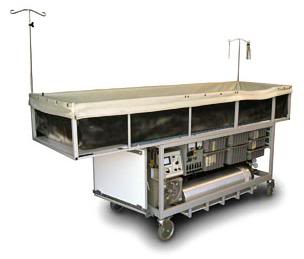
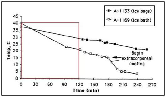
Footnotes

[1] Actually, it turn out this seat-of the-pants estimate was excessively optimistic, since the average loss of neocortical neurons is far higher, proceeding at a rate of about 1 per second, 85,000 per day, or ~31 million per year: Pakkenberg, B., Pelvig, D., Marner,L., Bundgaard, M.J., Gundersen, H.J.G., Nyengaard, J.R. and Regeur, L. Aging and the human neocortex. Exp. Gerontology, 38:95-99, 2003 and Pakkenberg, B. and Gundersen, H.J.G. Neocortical neuron number in humans: effect of sex and age. J. Comp. Neurology, 384:312-320, 1997.


[2] The start and the rate of decline are variable, with some organ systems showing deterioration starting in youth. However, for the organ systems shown Figure 6, deterioration does not begin until around age 30.

[3] In Jonathan Swift’s savagely satirical novel Gulliver’s Travels, the name struldbrug is given to those humans in the country of Luggnagg who are born normal, but are in fact immortal. Although the struldbrugs do not die, they do nonetheless continue aging. Swift describes the plight of the struldbrugs in terms almost any resident in an nursing home today (who is still compos mentis) would immediately understand: “when they have completed the term of eighty years, they are looked on as dead in law; their heirs immediately succeed to their estates; only a small pittance is reserved for their support; and the poor ones are maintained at the public charge. After that period, they are held incapable of any employment of trust or profit; they cannot purchase lands, or take leases; neither are they allowed to be witnesses in any cause, either civil or criminal, not even for the decision of meers and bounds.”

[4] The subiculum receives input from CA1 and entorhinal cortical layer III pyramidal neurons and is the main output of the hippocampus. The pyramidal neurons send projections to the nucleus accumbens, septal nuclei, prefrontal cortex, lateral hypothalamus, nucleus reuniens, mammillary nuclei, entorhinal cortex and amygdala and as such, is the principal routing network for information from the hippocampus. The subiculum is also critically involved in the formation of procedural memories.

End of Part 1


References

1)      Ettinger, RCW. The Prospect of Immortality, Doubleday, 1964: http://www.cryonics.org/book1.html Retrieved 29 December, 2010.

2)      World Development Indicators database: http://www.nationmaster.com/graph/hea_lif_exp_at_bir_tot_yea-life-expectancy-birth-total-years&date=1964#source Retrieved 17 December, 2010.

3)      http://aging.senate.gov/crs/aging1.pdf Retrieved 02 February, 2011.

4)       Plassman, B, Langa, K, Fisher, G. Wallace, R. Prevalence of Dementia in the United States: The Aging, Demographics, and Memory Study. Neuroepidemiology. 2007;29(1–2):125–132. Retrieved 04 February, 2011.

5)      Cook, J. Nathan Shock, Pioneer on Aging, New York Times, Obituaries, Published: November 15, 1989: http://query.nytimes.com/gst/fullpage.html?res=950DE1D61E31F936A25752C1A96F948260 ) Retrieved 04 February, 2011.

6)      Coles, L. Stephen, “The Life and Contributions of Professor Bernard L. Strehler, Founding Editor-in-Chief of  Mechanisms of Aging and Development, Professor of Biology at the University of Southern California [February 21, 1925 May 13, 2001],” Mechanisms of Aging and Development, Vol. 123, pp. 821-5 (2002): Retrieved 03 February, 2011.

7)      Rayner, E. Alex Comfort, Obituary, The Guardian, 28 March, 2000: http://www.guardian.co.uk/news/2000/mar/28/guardianobituaries . Retrieved 04 February, 2011.

8)      Maugh, II, TH. Obituary: Roy Walford, 79, Obituary; Eccentric UCLA Scientist Touted Food Restriction, Los Angeles Free Press: http://losangelesfreepress.com/obituary-roy-walford-79-eccentric-ucla-scientist-touted-food-restriction/ ). Retrieved 04 February, 2011.

9)      Epps, R. Dr. William Regelson, Obituary, Style, Richmond Virginia, 04 February, 2011: http://styleweekly.com/ME2/dirmod.asp?sid=&nm=Articles%2FNews&type=Publishing&mod=Publications%3A%3AArticle&mid=8F3A7027421841978F18BE895F87F791&tier=4&id=A24A05558531439EA6A4A43D345B7CBF . Retrieved 04 February, 2011.

10)   Alex Comfort, Wikipedia: http://en.wikipedia.org/wiki/Alex_Comfort . Retrieved 04 February, 2011.

11)   Thomas L. The technology of medicine. In: The Lives of a Cell. New York, NY: Viking Press; 1974:31–36.

12)   Silver, JK, Wilson, DJ. Polio Voices. Santa Barbara: Praeger Publishers. 2007 p. 141.

13)   Wilson DJ. And they shall walk: ideal versus reality in polio rehabilitation in the United States.  Asclepio. 2009;61(1):175-92.

14)    Smith, JS. Patenting the Sun: Polio and the Salk Vaccine. William Morrow & Co; 1st edition. 1990. ISBN-10: 0688094945.

15)   Bookchin, D, Schumacher, J. The Virus and the Vaccine, Macmillan, 2004. ISBN 0312342721.

16)   Juskewitch JE, Tapia CJ, Windebank AJ. Lessons from the Salk polio vaccine: methods for and risks of rapid translation. Clin Transl Sci. 2010;3(4):182-5.

17)   Fonda, J. My Life So Far, Random House, New York, 2005. ISBN 978-0-375-50710-6 (0-375-50710-8).

18)    http://blog.zap2it.com/pop2it/2010/12/jane-fonda-on-breast-cancer-hip-replacement-on-ellen.html Retrieved 23 December, 2010.

19)   http://www.hulu.com/watch/194332/abc-nightline-just-cher-if-she-could-turn-back-time Retrieved 04 February, 2011.

20)   Shock NW. Age changes in some physiologic processes. Geriatrics. 12;40-48:1957.

21)   Pakkenberg, B, Pelvig, D, Marner,L, Bundgaard, MJ, Gundersen, HJG, Nyengaard, JR, Regeur, L.  Aging and the human neocortex. Exp. Gerontology. 2003;38:95-99.

22)   Pakkenberg, B. Gundersen, HJG. Neocortical neuron number in humans: effect of sex and age. J. Comp. Neurology, 1997;384:312-320.

23)   Giorgio, A, Santelli, L, Tomassini, V, Bosnell, R, Smith, S, De Stefano, N, Johansen-Berg, H. Age-related changes in grey and white matter structure throughout adulthood. Neuroimage. 2010;51(3):943-51.Epub 2010 Mar 6.

24)   Hedden T, Gabrieli, JD.. Insights into the aging mind: a view from cognitive neuroscience. Nat Rev Neurosci. 2004;5(2):87-96. PMID: 14735112.

25)   Courchesne, E, Chisum, HJ, Townsend, J, et al. Normal brain development and aging: quantitative analysis at in vivo MR imaging in healthy volunteers. Radiology. 2000;216:672.

26)   Franke. K, Ziegler, G, Klöppel, S, Gaser, C. Alzheimer’s Disease Neuroimaging Initiative. Estimating the age of healthy subjects from T1-weighted MRI scans using kernel methods: exploring the influence of various parameters. Neuroimage.2010;15;50(3):883-92. Epub 2010 Jan 11. PMID: 20070949.

27)   Shock NW. Age changes in some physiologic processes. Geriatrics. 1957;12:40-48.

28)   Giorgio, A, Santelli, L, Tomassini, V, Bosnell, R, Smith, S, De Stefano, N, Johansen-Berg, H. Age-related changes in grey and white matter structure throughout adulthood. Neuroimage. 2010;51(3):943-51. Epub 2010 Mar 6.

29)   Franke K, Ziegler, G, Klöppel, S, Gaser C; Alzheimer’s Disease Neuroimaging Initiative. Estimating the age of healthy subjects from T1-weighted MRI scans using kernel methods: exploring the influence of various parameters. Neuroimage. 2010;50(3):883-92. Epub 2010 Jan 11. PMID: 20070949.

30)   Madden, DJ, Whiting, WL, Huettel, SA, White, E, MacFall, JR, Provenzale, J.M. Diffusion tensor imaging of adult age differences in cerebral white matter: Relation to response time. Neuroimage, 2004;21:1174–1181.

31)   Burgmans, S, van Boxtel, MP, Vuurman, EF, Smeets, F, Gronenschild, EH, Uylings, HB, Jolles, J. The prevalence of cortical gray matter atrophy may be overestimated in the healthy aging brain. Neuropsychology. 2009;(5):541-50.

32)   Basser, PJ, Pierpaoli, CJ. Microstructural and physiological features of tissues elucidated by quantitative-diffusion-tensor MRI. Magn Reson B. 1996;(3):209-19.

33)   Basser, PJ, Mattiello, J, Le Bihan, D. MR diffusion tensor spectroscopy and imaging. Biophys. J. 1994;66:259.

34)   Dennis, NA, Cabeza, R. Neuroimaging of healthy cognitive aging. In F. I. M. Craik & T. A. Salthouse (Eds.), Handbook of aging and cognition: Third edition, Psychology Press 2007. ISBN-10: 080585990X

35)   Augustinack, JC, Helmer, K, Huber, KE, Kakunoori, S, Zöllei, L, Fischl, B. Direct visualization of the perforant pathway in the human brain with ex vivo diffusion tensor imaging. Front Hum Neurosci. 2010;4:42. PMID: 20577631

36)   Yassa, MA, Tugan Muftuler, L, Starka, CEL. Ultrahigh-resolution microstructural diffusion tensor imaging reveals perforant path degradation in aged humans in vivo. PNAS. 2010;107:12687–12691.

37)    Abe, O., Aoki, S., Hayashi, N., Yamada, H., Kunimatsu, A., Mori, H., et al. Normal aging in the central nervous system: Quantitative MR diffusion-tensor analysis. Neurobiology of Aging. 2002;23(3):433–441.

38)   Antonini, A, Leenders, KL. Dopamine D2 receptors in normal human brain: Effect of age measured by positron emission tomography (PET) and [11C]-raclopride. Annals of the New York Academy of Sciences. 1993;695, 81–85.

39)   de Leon, M.J, George, AE, Tomanelli, J., Christman, D, Kluger, A, Miller, J. et al. Positron emission tomography studies of normal aging: A replication of PET III and 18-FDG using PET VI and 11-CDG. Neurobiology of Aging. 1987;8(4):319–323.

40)   Hedden, T, Riddle, DR, editor. Chapter 11 –in Imaging Cognition in the Aging Human Brain in Brain Aging: Models, Methods, and Mechanisms. CRC Press, 2007.

41)   Ranganath C. A unified framework for the functional organization of the medial temporal lobes and the phenomenology of episodic memory. Hippocampus. 2010 Nov ;20 (11):1263-90. Review. PubMed PMID: 20928833.)   http://dml.ucdavis.edu/articles/ranganath_2010_UnifiedFramework.pdf

42)   Wang, WC, Lazzara, MM, Ranganath, C, Knight, RT, Yonelinas, AP. The medial temporal lobe supports conceptual implicit memory. Neuron. 2010;68(5):835-42. PMID: 21144998; PubMed.

43)   Sauvage, MM, Beer, Z, Ekovich, M, Ho, L, Eichenbaum, H. The caudal medial entorhinal cortex: a selective role in recollection-based recognition memory. J Neurosci. 2010;30(46):15695-9. PMID: 21084625.

44)   Bjørnebekk, A, Westlye, LT, Walhovd, KB, Fjell, AM. Everyday memory: self-perception and structural brain correlates in a healthy elderly population. J Int Neuropsychol Soc. 2010;16(6):1115-26. Epub 2010 Oct 15. PMID:20946708.

45)    Yassa, MA, Tugan Muftuler, L, Starka, CEL. Ultrahigh-resolution microstructural diffusion tensor imaging reveals perforant path degradation in aged humans in vivo. PNAS. 2010;107:12687–12691.

46)   Burke, SN, Barnes, CA. Neural plasticity in the aging brain. Nat Rev Neurosci. 2006;7:30–40.

47)   Wilson, IA, Gallagher, M, Eichenbaum, H, Tanila, H. Neurocognitive aging: prior memories hinder new hippocampal encoding. Trends Neurosci. 2006;29:662–670.

48)    Milner, B, Squire, LR, Kandel, ER. Cognitive neuroscience and the study of memory. Neuron. 1998;20:445–468.

49)   Squire, LR, Stark, CEL, Clark, RE. The medial temporal lobe. Annu Rev Neurosci 2004;27:279–306.

50)   Witte, MP, Van Hoesen, GW, Amaral, DG. Topographical organization of the entorhinal projection to the dentate gyrus of the monkey. J Neurosci. 1989;9:216–228.

51)   Witter, MP, Amaral, DG. Entorhinal cortex of the monkey: V. Projections to the dentate gyrus, hippocampus, and subicular complex. J Comp Neurol. 1991;307:437–459.

52)   Witter, MP. The perforant path: Projections from the entorhinal cortex to the dentate gyrus. Prog Brain Res. 2007;163:43–61.

53)   Hyman, BT, Van Hoesen, GW, Kromer, LJ, Damasio, AR.  Perforant pathway changes and the memory impairment of Alzheimer’s disease. Ann Neurol. 1986;20:472–481.)

54)   Scheff, SW, Price, DA, Schmitt, FA, Mufson, EJ. Hippocampal synaptic loss in early Alzheimer’s disease and mild cognitive impairment. Neurobiol Aging. 2006;27:1372–1384.

Posted in Gerontology, Medicine | 4 Comments

A Visit to Alcor


“Are you interested in the whole body or the neuro? And would you like fries and a Coke with that?”


By Mike Darwin


Introduction


A short while ago, I hosted 3 visitors from the Russian cryonics organization KrioRus. During my visit with them in Moscow two and a half years previously, I had exhorted them to visit the US and see first-hand any hub of cryonics activity there, so that that they could see and learn for themselves what cryonics was really like, and how it was actually being pursued in the US. When I wrote to Alcor President and CEO Max More to try and schedule a visit on short notice, I jokingly included the aside that if I was not permitted to be on premises, I would go elsewhere… It was intended to be a humorous reference to a time in the not so distant past when I was not allowed into Alcor because my homosexuality was “unacceptable” to a former Alcor President (apparently being incompatible with his religious beliefs).

To my considerable surprise, Max took this remark seriously, and polled the Alcor Board on the matter. Surprise was replaced with shock when I subsequently learned that Alcor Director Saul Kent had expressed concerns that my visit to Alcor accompanying the Russians would be used as tool to “attack Alcor.” I still find this shocking, but depending upon how you define attack, maybe Saul was closer to the truth than I imagined possible. I’ve since been told that apparently there were speculations that “touring Russians” might be just an excuse of some kind to gain entry to Alcor.[1] Why this should be necessary is still a mystery to me, since as far as I knew I could visit by making by an appointment, presumably like anyone else.


Figure 1: The Alcor Mobile Advanced Rescue Cart (MARC), a portable extracorporeal support platform.

I saw quite a few unexpected things at Alcor, such as that the MARC is no longer in use and lies covered in dust (not even tarped) and laden with ‘junk’ in the ambulance bay; it has been replaced by waist high PIB fabricated from stainless steel. All of which begs the point of why a hugely complex and costly technological platform, in the form of a mobile operating room was developed, if it isn’t going to be used for extracorporeal support?


Figure 2: Left to right, Danila Medvedev, President of the Russian Transhumanist Association, Max More, Ph.D., President and CEO of Alcor and Valerija Pride, President of KrioRus. Photo by Stanislaw Lipin; courtesy of KrioRus.

A Long History of Serious Deficiencies in Patient Care

Shortly after he took office, I sent Max the 100+ plus page “Report of the Committee to Evaluate Alcor Procedures” which was presented to the Alcor Board of Directors on 04 April, 2002. This report came about because Saul Kent invited me over to his home in Woodcrest, California to view videotapes of two Alcor cases which troubled him – but he couldn’t quite put his finger on why this was so. What I saw shocked and disgusted me. Patients were being stabilized at a nearby hospice, transported to Alcor (~20 min away) and then CPS was discontinued, the patients were placed on the OR table and, without any ice on their heads, they were allowed to sit there at temperatures a little below normal body temperature for 1 to 1.5 hours, while burr holes were drilled, and the neck vessels were cannulated. Distilled water was being used to irrigate the brain and burr holes were done with a wood burr and electric drill with no cooling of the bone under the burr – smoke could be seen coming from the burr wound! Since the patient had no circulation to provide blood to carry away the enormous heat generated by the action of the burr on the bone, the temperature of the underlying bone (and brain) must have been high enough to literally cook an egg.

In one case, a patient’s head was removed in the field and, because they had failed to use a rectal plug, the patient had defecated in the PIB. The result was that feces had contaminated the neck wound, and Alcor personnel were seen pouring saline over the stump of the neck whilst holding the patient’s severed head over a bucket trying to wash the fecal matter off the stump. These are just a few of the grotesque problems I observed.

As a consequence, a “blue ribbon” Committee was put together to comprehensively review Alcor patient videos and records, and to make a field inspection of the Alcor facility. This Committee produced a comprehensive report and presented it to the Alcor Board. [Darwin, M., et al., Report of the Committee to Evaluate Alcor Procedures, Presented to the Alcor Board of Directors on April 4th, 2002.] Max More knew nothing of this until I sent him a copy after he was President for about one and a half months.

A Profound and Fundamental Error Unendingly Repeated

A short while later, Alcor published the first detailed case report since the A-1097 case report was published in 2006. A careful reading of this case report which is available at http://www.alcor.org/Library/pdfs/casereportA2435.pdf discloses that there is something terribly wrong that happens during this case, but unless you really understand cryonics as medicine, you are not likely to see it. The patient was pronounced at 6:58 PM and was transported to Alcor packed in ice while receiving CPS with the LUCAS. At ~ 8:42 PM, 1 hour and 42 minutes later, a median sternotomy is begun (excerpt from the case report, emphasis mine):

Continuation of CPS

Move to surgical table


7. Surgical Procedures


Manual CPS was continued on the patient while fresh ice was brought in from Alcor’s ice machine. The existing ice was removed from on top of the patient and she was pulled out of the ice bath and placed on the OR surgical table at 8:11 pm.


Testing of the sternal saw prior to the surgery revealed that the flexible shaft had frozen up. The problem was handed to Richard to deal with, and he had it unfrozen in a few minutes. The shaft was subsequently disassembled and lubricated.


The patient’s head was shaved to prep for the burr holes to be drilled. These are used to monitor the temperature of the patient’s brain as well as a way to visually watch for swelling. While the burr holes were being drilled using a craniotome perforator, Aaron prepped the patient’s sternum with Betadine, an antiseptic agent used topically to destroy microbes. Dr. McEachern cleaned each of the burr holes as they were completed. At 8:42 PM, she stood on a step stool to gain a higher position above the patient in order to perform a median sternotomy. This is a procedure in which a vertical inline incision is made along the sternum, after which the sternum itself is divided to provide access to the heart for cannulation. Dr. McEachern cut through the skin of the chest with a scalpel. The guide of the saw blade was placed below the sternum. Richard operated the foot pedal on the floor as she guided the saw up the sternum. She talked through the procedure, step by step, to Aaron so he could gain additional training in its operation and application. After the sternum separation was completed, the chest was opened with Finochietto spreaders and the pericardial sac was exposed. Access to the heart was accomplished by cutting through the pericardium.


Dr. McEachern performed an arterial cannulation of the heart by sewing a purse-string suture into the wall of the aortic arch, puncturing the vessel within the purse-string, and advancing and securing the catheter. She then repeated this process for venous cannulation of the heart, going into the right atrium and advancing the cannula into the inferior vena cava. This process took approximately one hour and was completed at 9:48 PM. A thermocouple was placed under the dura through the burr hole at 3.0 hours post-arrest. Brain temperatures were taken under the dura and beneath the brain (nasopharyngeal).


Now the washout could begin. This process is used to replace the patient’s blood with cryoprotectant. The extracorporeal perfusion circuit had been primed with B1 base perfusate solution prior to the surgery and was being circulated through the bypass loop. When cannulation was complete, the circulation was transferred from the bypass loop into the cannulas. The circuit was switched from closed to open circulation and the blood was washed out of the patient’s vascular system, the visual blood concentration going from opaque to light pink.

The graph of below shows a typical patient’s cooling rate in the PIB.

Figure 3: The red-boxed areas of the two cooling curves above show the likely temperature range a patient would have cooled to during 1 hour and 44 minutes of closed chest cardiopulmonary supported external cooling using ■ ice bags or □ Portable Ice Bath (PIB) cooling.

Assuming the patient cooled at the rate that A-1169 did above, then the best that could be expected is that the patient was at ~ 22-24 deg C at 1 hour and 44 min post arrest. Examination of the graph below (Figure 4) shows that the “safe” circulatory arrest times for a “healthy” patient who has had no prior ischemic insult and who is fully oxygenated at various temperatures during cardiac or neurosurgery:

Figure 4: Probable safe circulatory arrest time vs. temperature for humans, as calculated using the Hypothermic Metabolic Index (HMI). [Ungerleider R, Gaynor,  JW.: The Boston Circulatory Arrest Study: An analysis. J Thorac Cardiovasc Surg 2004, 127:1256-1261.]

The reason these graphs are of relevance is that in order to do a median sternotomy you must, necessarily, stop CPR. So, yet again, Alcor took a patient with essentially uninterrupted CPS, brought them into the OR and then exposed her 1 hour and 42 minutes of ischemia! A few days before we went to Alcor, I had sent Max a letter pointing out that this had happened yet again, and that Aaron Drake’s report was badly flawed and that it was clear he was wholly inadequately trained to be doing cases unsupervised. I received no reply.

Our visit to Alcor was surreal – absolutely surreal. The first thing Max did when we entered was to show Danila Medvedev and Valerija Pride swatches of wall paper and explain that the office was he was occupying was “not his” and that it was to be redecorated. He then proceeded to do the same with me. Danila and I looked at each other as if to say, “Are you kidding us? Can this even be real?”

An Unkempt Kitchen in a 2-Star Hotel?

When we arrived at Alcor we were informed that a member was in need of Standby and very likely cryopreservation, and shortly after we entered the facility, the Remote Standby Team left with their equipment. I have been at Alcor only at brief intervals over the past 20 years, with the exception a stint for a month or so as subcontractor working for Suspended Animation, Inc. (SA) in 2002. I thus have mental “snapshots” of the state of the facilities separated by considerable intervals of time. This was the most disturbing snapshot I’ve seen so far. The operating room was unkempt. The floors were scuffed, stained, dirty, and had obviously not been waxed in a long time (Figure 5).

Figure 5: Scuffed and dirty floors, dusty shelves and a disorganized appearance are fair descriptors of the Alcor operating room. I wouldn’t consider medical treatment in a facility with this appearance – nor for that matter would I like to dine in a restaurant with a kitchen in such a state. Photo by Stanislaw Lipin; courtesy of KrioRus.

I inquired when preparation of the operating room would start for the patient who was to be perfused, presumably the next day, and I was told that they were “done.” When we entered the OR I observed tubing strung on the pumps of the heart lung machine console, and had assumed it was training tubing, since some of it was hanging down to the floor in front of the console inside a large, open ZipLoc bag (Figure 5).

Figure 5: Cryoprotective tubing dangling from pump console in a Ziploc bag; proper gas permeable sterilization wrapping (such as Kimlon™) was nowhere in evidence. Photo by Stanislaw Lipin; courtesy of KrioRus.

There were discarded tubing (ethylene oxide) sterilization caps lying around, and refuse, some of it apparently in place for some time, such as twist ties and paper backing from discarded sterilization pouches, lying covered in dust on the computer/instrumentation cart adjacent to the pump console (Figures 7 & 8).

Figure 6: Alcor operating room perfusion and data acquisition equipment. The refractometer heads used to continuously acquire cryoprotectant agent concentration to the left of the computer. Photo by Stanislaw Lipin; courtesy of KrioRus.

Figure 7: Dust covered lower shelf of the stainless steel data acquisition cart. The piece of sterilization packaging and the twist tie appeared to be covered in a film of dust, as well. Photo by Stanislaw Lipin; courtesy of KrioRus.

Figure 8: The recirculating reservoir perched precariously atop the magnetic stirring table used to mix cryoprotectant concentrate into the recirculating perfusate. Photo by Stanislaw Lipin; courtesy of KrioRus.

On the monitor shelf atop the pump console there was a spray bottle that had apparently been labeled “alcohol” (what kind? one wonders) with a Sharpie marker, and a laboratory wash bottle sitting next to it containing a liquid – but with no label (Figure 8).

Figure 9: At top, long view of the neuroperfusion enclosure and at bottom close up showing area of apparent blood contamination. Photo by Stanislaw Lipin; courtesy of KrioRus.

Disturbingly, the neuroperfusion enclosure had what appeared to be a residue of dried blood/perfusate in what appeared to be a defect in the adhesive seal where the waste diversion plate is cemented to the side wall of the enclosure (Figure 9). The drain line from the neuroperfusion enclosure (where biohazardous fluid will collect to be disposed of) was sitting unsecured in a ~20 L Costco Kirkland laundry detergent pail (Figure 10).

Figure 10: Costco laundry soap pail containing the unsecured biohazardous waste line from the neuroperfusion enclosure. Photo by Stanislaw Lipin; courtesy of KrioRus.

Figure 11: Alcor Operating room tableaux “fully readied” for a human neuropatient cryoprotective perfusion. Photo by Stanislaw Lipin; courtesy of KrioRus.

The recirculating reservoir was dangerously small making microbubble embolization of the patient during cryoprotective perfusion all but inevitable, since the cold, polymer-rich, viscous perfusate develops stable foam as a consequence of vortex formation and air entrapment from the action of the mixing magnetic stir-bar.

Figure 12: One possible scheme for achieving uniform mixing of cryoprotectant concentrate with the perfusate recirculating through the patient using a static mixer. A concentrate mixing pump continuously removes a large fraction of the recirculating perfusate from a cardiotomy or venous reservoir. This perfusate is then passed through a combination static mixer-heat exchanger where turbulent flow from inertia reversal and radial mixing uniformly blend the added cryoprotectant concentrate with the recirculating perfusate. The static mixer also serves as a heat exchanger. The blended and chilled perfusate then return to the venous reservoir.

Figure 13: At (A), a typical static mixer array. Static mixers allow thorough mixing of almost any kind of liquid or slurry without the introduction of air or the use of moving parts with the attendant seals. Because static mixers are of necessity a mixing element housed within a tube or cylinder, they make ideal heat exchangers since the fluid flowing inside the mixing tube elements is repeatedly thin-filmed and passed over the tube surface. It is thus possible to very efficiently combine mixing with heat exchange, as can be seen in the combination mixer-heat exchanger seen in B, above.

This has been a repeated problem in previous cases, and has been the subject of numerous advisory communications between Alcor Director Brian Wowk and me, among others. In fact, despite these repeated warnings (increase depth and volume of the recirculating reservoir, use a floating lid or replace the stir bar assembly with an in-line mixer: Figures 12 & 13), this same phenomenon was noted on during the cryoprotective perfusion of patient A-1097 in January of 2006. A detailed paper documenting this effect and demonstrating a simple way to eliminate it was first published in 1994. ( see: http://www.cryocare.org/index.cgi?subdir=bpi&url=tech5.txt)

I quote from this 1994 paper:

“A consequence of the stirring of the recirculating reservoir by the rapidly spinning magnetic stir bar is the generation of an air vortex in the recirculating perfusate. While this vortex is very effective at both rapidly and completely mixing the concentrate with the perfusate in the recirculating reservoir, it is also very effective at introducing air into the recirculating perfusate as well. At rates of rotation fast enough to achieve good mixing; the bottom of the vortex of air reaches the rapidly rotating stir bar. Air is thus turbulently mixed into the perfusate where it forms bubbles of widely varying size; the smallest of which are very stable. As the concentration of cryoprotectant rises, and the viscosity of the solution correspondingly increases, air bubbles generated by stirring in the recirculating reservoir become more and more stable and begin to saturate the recirculating perfusate creating large amounts of foam.”

And from A-1097’s case report:

“A differential vascular resistance check was done by clamping off and then unclamping the left and right carotid artery respectively. After clamping off the left carotid artery the pressure rose to 72 mm. After clamping off the right carotid artery the pressure rose to 140 mm. At 11:55 foaming was identified in the mixing reservoir, with worse foaming observed at 12:17.”

Figure 14: The recirculating reservoir sitting unsecured atop the mixing stir table. Note that the PVC tubing has not been adanced the requisite 3 barbs over the connector at the bottom (right) of the reservoir and that there is no cable tie in place to prevent accidental disconnection of the tubing. Photo by Stanislaw Lipin; courtesy of KrioRus.

It was also sitting akimbo on the magnetic stir table and the connections to and from it, including the critical withdrawal connection at the bottom was neither cable tied, nor pushed over the third barb of the tubing connector at the bottom of the reservoir (these are standard minimum practices for securing tubing against disconnection in extracorporeal medicine).

When I inquired as to how the recirculating reservoir would be secured during perfusion, I was told, “It’ll sit still when it has liquid in it.” If you can look at the picture above and concur with that answer, especially considering that Alcor has a history of pumping this small reservoir dry during perfusion and introducing air in the extracorporeal circuit, then you are a more courageous soul than me. Consider the recommendation made to Alcor by the formal Committee commissioned to evaluate and suggest corrective actions when Alcor’s cryopreservation procedures were found to be severely deficient in 2002:

4.13: Enlarge and alarm the recirculating reservoir.

The recirculating perfusate reservoir ran dry more than once during a recent case. The reservoir should be larger and should have an alarm system that is triggered by a low level of perfusate. At the least one specific person should be assigned the task of monitoring the reservoir.

Cargo Cult Security for Patients?

Then there was the lunacy of the “armored” patient care bay, which Max proudly showed us (Figure 15).

Figure 15: Blast resistant bullet proof window looking into the Alcor patient care bay. The hardened transparent window is backed up with a retractable steel curtain window cover. Photo by Stanislaw Lipin; courtesy of KrioRus.

I can’t even begin to imagine what all this cost (including reinforcing the perimeter walls). It looks very impressive, and no doubt has considerable “sales” value to the naive, or the foolish.

However, if anyone actually looks past the looking glass (which happens to be blast resistant, in this case) what they will see is the following reality.

Figure 16: View through the window into the patient care bay (PCB). A loose piece of the foil faced cardboard sheeting which covers the plywood roof decking and structural supports that comprise the roof of the PCB are highlighted by the red arrows. Photo by Stanislaw Lipin; courtesy of KrioRus.

The red arrows point to the foil-faced cardboard reflective “insulation” that covers the space between the perlins in the patient care bay. One piece has been left (un-anesthetically) loose adjacent to what appears to be a run of sprinkler pipe. What this told me was that roof of the PCB bay is a paper bag. It almost certainly consists of sheets of plywood or particle board decking covered with a layer of roofing felt, and finally the roofing material itself. Typically, this kind of construction can barely withstand the weight of a 250 lb man. Go up on the roof and walk around yourself, and you’ll immediately get a feel for what I’m talking about – the roof will give and spring back as you walk on it. It is minimally engineered for load bearing, and this fine, and a damn good thing in earthquake country, where tilt-up concrete industrial building construction was first developed.

I can then go to Google Earth and quickly verify that, as of 11-2009, there were no structural or other evident reinforcements to the outside of the roof over the PCB. Maybe there are now (doubtful), but this is very easy to determine, either directly, for the cost of an aerial photograph ($300), or by checking with building and code enforcement to see if any structural permits were issued, and inspections subsequently done. Of course, the easiest way is just to climb up on the building and take a look. The single most important and most elementary security precaution any institution can take to increase the safety of its physical plant is to protect its perimeter. This is why sensitive and vulnerable government and corporate installations are surrounded by fences, patrolled by guards (and often dogs) and where feasible, protected by bollards against bomb bearing vehicular attack. Alcor’s perimeter is unsecured.

Figure 17: Google Earth view of the Alcor facility in 2009 shows no evidence of external (surface) reinforcing and no evidence of razor wire or other perimeter defenses on the roof of Alcor building in general, or the patient care bay in particular.

Figure 18: Metal lid covering bigfoot dewar at Alcor. Photo by Stanislaw Lipin; courtesy of KrioRus.

It is also evident that the dewars have no cladding, and that the softest spot is the top of the units where, in order to save both weight and money (again perfectly reasonable), the tops of the foam neck-plugs, as is the industry standard for cryogenic dewars, are fabricated from aluminum, or perhaps a tough plastic, such as ABS. However, in this case, no guessing is required; it is evident that the cover is metal and it is, judging from its thickness, aluminum.

Figure 19: Unsecured facility perimeter and roof of the patient care bay at the Alcor facility in Scottsdale, AZ. The blast and fire resistant neurovaults sit abandoned in the parking lot (red hash mark).

Google even shows me the long abandoned blast resistant neuro-vaults sitting in the parking lot (red hash mark), and confirms that the PCB roof is a standard wooden deck and asphalt configuration which will look structurally just about like this:

Figure 20: Type of roof construction used in the Alcor facility. The large composite wooden beams running from left to right are gluelams – machined pieces of wood glued together under high pressure. The gluelams are the primary load bearing elements of the roof. The single beam structural elements that connect the gluelams are the perlins. The perlins provide most of the structural support for the plywood or chipboard decking of the roof.

What is more, very few, if any people who want to do the patients harm will walk into Alcor on a tour. That’s almost ludicrous, especially when there are much more attractive alternatives. And the most attractive alternative is simply walk up to or drive by the building, and hurl a fragmentation-type explosive device, such as a pipe bomb, on the roof. A more serious and targeted approach would be to climb up on the roof and position heavy explosive charges exactly where they are deemed to do the most damage.

Figure 21: Historically, bollards made of wood, metal or concrete have been used to prevent accidental or deliberate vehicular intrusion into areas where unacceptable damage would result (above, top right).  More recently, concrete barriers such as the ones seen at top left have been used for this purpose. (Photos by Mike Darwin.) The use of concrete barriers, or K rails is, however, not only unaesthetic, it does not provide  protection against the intrusion of armed pedestrians, or 2-wheel vehicles such as bicycles and motorcycles. An effective alternative is the use of high impact and climb-over resistant fencing such as Ameristar Fence Products, Inc., (Tulsa, OK), Impasse™ high-security fencing system. With the company’s integrated cabling system, this product provides anti-ram defense against forced entry and ballistic attack, and is able to stop one 15,000 lb vehicle traveling 40 mph. This product meets Department of State’s K8/L1 and K8/L2 ratings.

So, in effect, the Alcor PCB is the equivalent of a Bugatti Verynon Sports car, which has excellent door locks and a great alarm system – all of which are of little utility in the event you leave the roof off!

Figure 22: The Bugatti Verynon features an excellent alarm system and a sporty, removable roof. The value of the alarm system is considerably diminished if the car is left parked with the roof off.

It is, of course, possible to really protect the patients against these kinds of threats, as well as radiation damage by placing the patient dewars in below ground silos as seen in Figure 23. But it isn’t pretty, although I guarantee you that this, or some variation of it, will be a whole lot more effective and less costly.

Figure 23: Truly effective blast, earthquake and radiation protection were achieved by CryoSpan in the late 1990s by the expedient of constructing in-ground steel reinforced concrete silos. At top, engineer Mark Connaughton works on the wooden support framing used to maintain the shape of the mold prior to pouring the concrete. While not offering “sexy” photo opportunities, such silos provide robust and affordable protection to cryonics patients. Photos by Charles Platt.


Subsequently, Max and I corresponded about these issues until it became clear that he was becoming angry with me. He denied that there was any current wastefulness at Alcor (including their 10 paid employees), challenging me to come up with line item examples; except, of course, I couldn’t do this because Alcor has published no financial reports in four years. In fact, such reports don’t exist (not yet, anyway). He did not respond to my query about why the MARC is no longer in use and lies covered in dust (not even tarped) and laden with ‘junk’ in the ambulance bay; it has been replaced by a waist high PIB fabricated from stainless steel. All of which begs the question of why a complex and costly technological platform, in the form of a mobile operating room was developed, if it isn’t going to be used for extracorporeal support?

Our correspondence pretty much ended with me telling him, “You may not agree with what my vision for Alcor was (in 1987) at this point in time, but the really unfortunate thing, for all involved, is that you probably have no idea what it was, nor why I am unhappy at the waste of millions of dollars of contributed member money[1] in the intervening decades. And Max, those millions were wasted.”

Every “criticism” I made he took as a personal challenge to his competence. Danila described it aptly as, “Horrible.” It now seems clear to me that nothing I can do from behind the scenes will change Alcor. I’ve been working quietly for over a decade now, and things just keep getting worse. I had planned for this contingency, and now I think it is time to proceed in creating an alternative organization and to providing some of the nocioception that Alcor has been spared these past 20 years. I like Max a great deal – we have been good friends for over 20 years.But this is business, serious life or death business.

I have been through at least 4 iterations of what amounted to effectively rebooting, or trying to be reboot cryonics organizations. It takes a long time to do that: ~5 years just to get some equilibrium and the core resources in place. Surprisingly, even throwing vast amounts of money into such efforts does nothing to accelerate the pace, and may even slow it. I’m old, and I am sickened at the thought of having to go through this exercise, even as a participant, let alone as a leader, yet again. Having said that, it is becoming clear with each passing year that this kind of effort was probably inevitable given our nearly complete lack of understanding of what was (and arguably still is) really required to do cryonics in a sustainable fashion. Certainly, there is no escape from this in most other endeavors – and especially not in fundamentally new ones. I rarely meet innovators or entrepreneurs on the “cutting edge” (an expression I loathe) who don’t have sad tales to tell about how many corporate entities they created and cycled through before they found a stable and durable platform (if they ever succeeded in doing so at all).

As to Alcor’s status and prospects, I don’t think Alcor is likely to fail in any kind of immediate or catastrophic way. It’s current and past deficiencies are primarily of a kind that, given cryonics’ fundamental lack of normal market feedback, will not be evident to cryonicists, let alone to the public, even if pointed out to them. The legacy of the Cryonics Society of California (CSC) is proof of that reality. So, that’s not what I’m saying.

The Stench of Impending Failure?

What I am saying is that there are certain infallible signs that an enterprise is in deep trouble, and while perhaps not in immediate danger of going under, is only going to continue to exist under highly favorable conditions. I’ll be quite specific in a moment, but I want to a spend few moments more on what is the really the more important point I have to make.

I’ve traveled the world and visited just about every kind of enterprise imaginable. Whether it is a restaurant Florence, a medical clinic in Hyderabad, or an ICU in Mumbai or Moscow, there is often this unmistakable gestalt (stench) of a profoundly dysfunctional business which is evident within minutes of being on the premises. Please note that I am not saying that all failing enterprises exhibit this aura, because I’m sure they don’t. Enron probably seemed in fine fettle until just before the end. But I am saying that when that ambience is present, the enterprise is in extremis. Under normal market conditions that would mean that you could reasonably (soon) expect the doors to be shuttered (or the floor to be removed, in the Middle East). The exceptions are small town businesses that constitute micro-monopolies, government operated facilities in the undeveloped world, and occasionally, religious orders or other institutions in terminal decline, but who have a trust fund or other stipend to sustain them.

Cryonics: A Product or an Unproven Experimental Procedure?

While in many important ways Alcor is the descendant of Cryonics Society of New York (CSNY) and the no-nonsense, tell like it is approach to cryonics of Curtis Henderson, this is not by any means the full picture. CSNY had perhaps half the paradigm right. The other half that came to constitute the “mature” Alcor approach of the 1980s was pioneered by Fred and Linda Chamberlain, and was one of the Alcor assets already in place, if dormant, when IABS and I arrived on the scene in 1981. That approach was a clear acknowledgement and understanding that cryonics was an experimental procedure, that Alcor was a mutual aid organization, and that recruiting members who were also experimental subjects, was not the same as selling cryonics to customers.

There was, in fact, a strong aversion to marketing cryonics as an ordinary product, or even as a “regular” medical treatment. A consequence of this attitude was that everyone who interfaced with the public, until shortly before I left, had an almost fanatical attitude about how signing up should be communicated to the member. The interesting, and indeed remarkable thing about this is that I do not ever recall doing any training or any scripting of how to handle callers who were prospective members, or who were gathering information for same. It was just something that was “organic” and a part of Alcor’s small, but very well defined corporate culture at that time.

A corollary or a logical extension of this “first contact” strategy was that we spent a lot of time and expended a lot of effort not only continuously educating already signed up members, but actively ensuring that their informed consent was maintained over time. The Cryonics Institute (CI) never had that paradigm and Alcor lost it, and as result, members got translated into customers. I believe this is a critical failure mode for a cryonics organization – any cryonics organization – because whatever else cryonics patients are, they are not customers, and neither are members customers before they become patients. The definition of customer is innocuous enough: A party that receives or consumes products (goods or services) and has the ability to choose between different products and suppliers. Superficially it would seem to fit the bill for cryonics. Unfortunately, it is not what is present in that definition that is problematic, it is what is missing.

By contrast, consider the definition of the word patient:

1. Bearing or enduring pain, difficulty, provocation, or annoyance with calmness.

2. Marked by or exhibiting calm endurance of pain, difficulty, provocation, or annoyance.

3. Tolerant; understanding: an unfailingly patient leader and guide.

4. Persevering; constant: With patient industry, she revived the failing business and made it thrive.

5. Capable of calmly awaiting an outcome or result; not hasty or impulsive.

6. Capable of bearing or enduring pain, difficulty, provocation, or annoyance: “My uncle Toby was a man patient of injuries” (Laurence Sterne).


noun:


1. One who receives medical attention, care, or treatment.

2. Linguistics A noun or noun phrase identifying one that is acted upon or undergoes an action. Also called goal.

3. Archaic One who suffers.


The core of the definition of patient, which was the basis of the adoption of the word in medicine, is “one who demonstrates calm endurance of pain, difficulty, provocation, or annoyance.” Anyone involved in cryonics for very long will quickly come to know, viscerally, which of these terms best applies.


No law yet prohibits marketing cryonics as a consumer product, though some do exist prohibiting its marketing at all. One of the few advantages to age (especially if accompanied by global travel) is that you get to see how diverse societies handle certain classes of problematic services or undertakings – ones that are destabilizing to the social matrix, or which carry a high potential for fraud or exploitation of the individual, especially vulnerable individuals. While there is considerable variation in the details, the general approaches used are remarkably similar within societies that share the same basic values. So, while there is the sale of vital organs in places like India (something that is fast being regionally legislated against), you see either a complete absence of this practice, or the same limited workarounds, in virtually all of the West and Near East. As a relevant aside, it was fascinating to watch the arc of gambling in the Russian Republic, which went from ubiquitous, to exactly where it was in the US 20 years ago; mostly prohibited, except in special zones that are problematic to gain access to and which focus the predation on the wealthy (there is essentially no middle class in Russia). This change occurred in ~2 years!


The point here is that any enterprise operating as a low temperature mausoleum with the added benefit of prospective resurrection, and doing so on a razor thin margin of costs, is very likely headed for trouble. And precisely because they are primarily a customer service organization engaged in selling only the first half of the “product” (i.e., cryopreservation and storage) they will simply not “get it” when the second half of the product is neglected or threatened (i.e., stewardship and resuscitation). Reanimation is a matter for our friends in the future, and an inevitable consequence of this is that day-to-day things that impact its likelihood today are likely to be viewed as of little or no consequence. Indeed, they are not even likely to be perceived at all. I gained enormous respect for Melody Maxim’s ability to inflict harm[2] when she correctly perceived that CI (and thus ACS) had surrendered all practical control over their patients to the Michigan Cemetery Board by the “simple” act of submitting to state regulation.


That and (many) other insights aside, the one of most relevance to this discussion happened when I was dozing on the couch in the reception area (the Russians proved equally exhausting and rewarding to travel with). As I went in and out of consciousness, I could hear the phone being answered at Alcor, and the first thing that caught my attention and increased my level of alertness for the next call was that most of the incoming calls were from people who were apparently inquiring about cryonics services. Maybe I was just there on an unusually busy day, but I heard several calls – more than two and less than five, in the hour plus interval I intermittently slumbered. The side of the calls I could hear went very much like this: “Alcor Foundation,” silence “yes we offer that.” Silence “Are you interested in the whole body or the neuro. The whole body is $200,000 and the neuro is $80,000. Silence. “The whole body is your entire body, and the neuro is just your head.” Silence. “We’re located in the Scottsdale Airpark, which is just outside of Phoenix, and if you would like to arrange a tour we would be glad to show you through our facilities.” Silence. “Most people pay for it with life insurance, and if you like, I can refer to our life insurance representative.” Silence. “Well, if you are ever in the Phoenix metro area, please consider making arrangements to see our facilities.” Silence. “Thank you.”


As I sat there, semiconscious, I had two recurring thoughts that played tag with each other in my fogged brain:


1) Are you interested in the whole body or the neuro? And would like fries and a coke with that?


2) Please, please, let me be dreaming.


I told Max about this and he expressed some concern, informed me that the person handling the inquiries was not a signed up cryonicist, but was nevertheless “a very good employee and very loyal to cryonics.” He told me he would look into the matter (after all, I was dozing) and that he would see about scripting such encounters should it prove necessary. As I sit here in Dulwhich, London, and watch the heavy gray North Sea clouds parade across the fast darkening sky whilst spitting rain, I shudder at the thought of the theatre d’absurb that scipt will be written and performed in.


There is a large and very material difference between continuing to advertise for and accept guests if you are a hotel with fire code deficiencies (that is aware of this and working to fix them), versus being a hotel that is actually on fire. It is a peculiarity of certain kinds of institutions that they will, in fact, continue to solicit and escort new guests to their rooms, even as fire from an already engulfed kitchen, barrels up stairwells with no fire doors. Such behavior happens not infrequently in enterprises where there is any material separation of “operations” from “feedback” (as any number of the recent real estate and financial debacles demonstrate).


What Next?


In my opinion, Alcor is a hotel on fire, already seriously engulfed, and with no plan of any kind, let alone a detailed one, for extinguishing the blaze and rebuilding the infrastructure. A better analogy might be a sinking ship – because the passengers can’t just stroll over to the venue across the street – they are stuck where they are. I expect that most reading this will tell me that this is not so, that I am mistaken, or that I am excessively pessimistic, or even malicious…and that’s fine. I’m not about insisting that everyone share my opinions. I may in fact be wrong. However, what I am not wrong about is that Alcor is profoundly dysfunctional, and that many discrete and general problems can be objectively identified that will likely lead to its eventual failure or replacement. I can also say with a high degree of confidence that those members who are not customers, are either aware of this situation, or are very dissatisfied with Alcor’s performance (particularly its wastefulness and low quality of service).


Opinions aside, what I believe is necessary, which in this case is both an opinion and a fact, is some sort of detailed, credible acknowledgement and understanding of the problems, coupled with a realistic plan for fixing them. Preferably a plan that does not involve asking members for yet more money to hire yet more staff.


Footnotes


[1] Ironically, approximately $4 million of that money came from the Dick Jones estate; a bequest which I was instrumental in directing to the operating fund. For this advice I feel a great deal of humiliation and regret and will sincerely apologize to Dick if I ever get the chance.

[2] And conversely to do good by pointing out soft spots and likely failure modes.


A Visit to Alcor

“Are you interested in the whole body or the neuro? And would like fries and a coke with that?”

By Mike Darwin

A short while ago, I hosted 3 visitors from the Russian cryonics organization KrioRus. During my visit with them in Moscow two and a half years previously, I had exhorted them to visit the US and see first-hand any hub of cryonics activity there, so that that they could see and learn for themselves what cryonics was really like, and how it was actually being pursued in the US. When I wrote to Alcor President and CEO Max More to try and schedule a visit on short notice, I jokingly included the aside that if I was not permitted to be on premises, I would go elsewhere… It was intended to be a humorous reference to a time in the not so distant past when I was not allowed into Alcor because my homosexuality was “unacceptable” to a former Alcor President (apparently being incompatible with his religious beliefs).

To my considerable surprise, Max took this remark seriously, and polled the Alcor Board on the matter. Surprise was replaced with shock when I subsequently learned that Alcor Director Saul Kent had expressed concerns that my visit to Alcor accompanying the Russians would be used as tool to “attack Alcor.” I still find this shocking, but depending upon how you define attack, maybe Saul was closer to the truth than I imagined possible. I’ve since been told that apparently there were speculations that “touring Russians” might be just an excuse of some kind to gain entry to Alcor.[1] Why this should be necessary is still a mystery to me, since as far as I knew I could visit by making by an appointment, presumably like anyone else.

Figure 1: The Alcor Mobile Advanced Rescue Cart (MARC), a portable extracorporeal support platform.

I saw quite a few unexpected things at Alcor, such as that the MARC is no longer in use and lies covered in dust (not even tarped) and laden with ‘junk’ in the ambulance bay; it has been replaced by waist high PIB fabricated from stainless steel. All of which begs the point of why a hugely complex and costly technological platform, in the form of a mobile operating room was developed, if it isn’t going to be used for extracorporeal support?

Figure 2: Left to right, Danila, Medvedev, President of the Russian Transhumanist Association, Max More, Ph.D., President and CEO of Alcor and Valerija Pride, President of KrioRus.

Shortly after he took office, I sent Max the 100+ plus page “Report of the Committee to Evaluate Alcor Procedures” which was presented to the Alcor Board of Directors on 04 April, 2002. This report came about because Saul Kent invited me over to view videotapes of two Alcor cases which troubled him – but he couldn’t quite put his finger on why this was so. What I saw shocked and disguste. Patients were being stabilized at a nearby hospice, transported to Alcor (~20 min away) and then CPS was discontinued, the patients were placed on the OR table and, without any ice on their heads, they were allowed to sit there at a temperatures a little below normal body temperature for 1 to 1.5 hours, while burr holes were drilled and the neck vessels were cannulated. Distilled water was being used to irrigate the brain and burr holes were done with a wood burr and electric drill with no cooling of the bone under the burr – smoke could be seen coming from the burr wound! Since the patient had no circulation to provide blood to carry away the enormous heat generated by the action of the burr on the bone, the temperature of the underlying bone must have been high enough to literally cook an egg.

In one case, a patient’s head was cut off in the field and, because they had failed to use a rectal plug, he had defecated in the PIB. The result was that feces had contaminated the neck wound, and Alcor personnel were seen pouring saline over the stump of the neck whilst holding the patient’s severed head over a bucket trying to wash the fecal matter off the stump! These are just a few of the grotesque problems I saw.

As a consequence, a “blue ribbon” Committee was put together to comprehensively review Alcor patient videos and records, and to make a field inspection of the Alcor facility. This Committee produced a comprehensive report and presented it to the Alcor Board. Max More knew nothing of this until I sent him a copy after he was President for about 1.5 months. A short while later, Alcor published the first detailed case report since the A-1169 case report was published in 2006. A careful reading of this case report which is available at http://www.alcor.org/Library/pdfs/casereportA2435.pdf and discloses that there is something terribly wrong that happens during this case, but unless you really understand cryonics as medicine, you are not likely to see it. The patient was pronounced at 6:58 PM and was transported to Alcor packed in ice while receiving CPS with the LUCAS. At ~ 8:42 PM, 1 hour and 42 minutes later, a median sternotomy is begun (excerpt from the case report, emphasis mine):

Continuation of CPS

Move to surgical table

7. Surgical Procedures

Manual CPS was continued on the patient while fresh ice was brought in from Alcor’s ice machine. The existing ice was removed from on top of the patient and she was pulled out of the ice bath and placed on the OR surgical table at 8:11 pm.

Testing of the sternal saw prior to the surgery revealed that the flexible shaft had frozen up. The problem was handed to Richard to deal with, and he had it unfrozen in a few minutes. The shaft was subsequently disassembled and lubricated. 15

The patient’s head was shaved to prep for the burr holes to be drilled. These are used to monitor the temperature of the patient’s brain as well as a way to visually watch for swelling. While the burr holes were being drilled using a craniotome perforator, Aaron prepped the patient’s sternum with Betadine, an antiseptic agent used topically to destroy microbes. Dr. McEachern cleaned each of the burr holes as they were completed. At 8:42 PM, she stood on a step stool to gain a higher position above the patient in order to perform a median sternotomy. This is a procedure in which a vertical inline incision is made along the sternum, after which the sternum itself is divided to provide access to the heart for cannulation. Dr. McEachern cut through the skin of the chest with a scalpel. The guide of the saw blade was placed below the sternum. Richard operated the foot pedal on the floor as she guided the saw up the sternum. She talked through the procedure, step by step, to Aaron so he could gain additional training in its operation and application. After the sternum separation was completed, the chest was opened with Finochietto spreaders and the pericardial sac was exposed. Access to the heart was accomplished by cutting through the pericardium.

Dr. McEachern performed an arterial cannulation of the heart by sewing a purse-string suture into the wall of the aortic arch, puncturing the vessel within the purse-string, and advancing and securing the catheter. She then repeated this process for venous cannulation of the heart, going into the right atrium and advancing the cannula into the inferior vena cava. This process took approximately one hour and was completed at 9:48 PM. A thermocouple was placed under the dura through the burr hole at 3.0 hours post-arrest. Brain temperatures were taken under the dura and beneath the brain (nasopharyngeal).

Now the washout could begin. This process is used to replace the patient’s blood with cryoprotectant. The extracorporeal perfusion circuit had been primed with B1 base perfusate solution prior to the surgery and was being circulated through the bypass loop. When cannulation was complete, the circulation was transferred from the bypass loop into the cannulas. The circuit was switched from closed to open circulation and the blood was washed out of the patient’s vascular system, the visual blood concentration going from opaque to light pink.

The graph of below shows a typical patient’s cooling rate in the PIB:

Figure 3: The red-boxed areas of the two cooling curves above show the likely temperature range a patient would have cooled to during 1 hour and 44 minutes of closed chest cardiopulmonary supported external cooling using ■ ice bags or □ Portable Ice Bath (PIB) cooling.

Assuming the patient cooled at the rate that A-1169 did above, then the best that could be expected is that the patient was at ~ 22-24 deg C at 1 hour and 44 min post arrest. If you look at the graph below you see the “safe” circulatory arrest times for a “healthy” patient who has had no prior ischemic insult and who is fully oxygenated at various temperatures during cardiac or neurosurgery:

Figure 4: Probable safe circulatory arrest time vs. temperature for humans, as calculated using the Hypothermic Metabolic Index (HMI).

The reason these graphs are of relevance is that in order to do a median sternotomy you must, necessarily, stop CPR. So, yet again, Alcor took a patient with essentially uninterrupted CPS, brought them into the OR and then exposed her 1 hour and 42 minutes of ischemia! A few days before we went to Alcor, I had sent Max a letter pointing out that this had happened yet again, and that Aaron Drake’s report was badly flawed and that it was clear he was wholly inadequately trained to be doing cases unsupervised. I received no reply. Our visit to Alcor was surreal – absolutely surreal. The first thing Max did when we entered was to show Danila Medvedev and Valerija Pride swatches of wall paper and explain that the office was he was occupying was “not his” and that it was to be redecorated. He then proceeded to do the same with me. Danila and I looked at each other as if to say, “Are you kidding us? Can this even be real?”

When we arrived at Alcor we were informed that member was in need of Standby and very likely cryopreservation, and shortly after we entered the facility, the Remote Standby Team left with their equipment. I have been at Alcor only at brief intervals over the past 20 years, with the exception a stint for a month or so as subcontractor working for Suspended Animation, Inc. (SA) in 2002. I thus have mental “snapshots” of the state of the facilities separated by considerable intervals of time. This was the most disturbing snapshot I’ve seen so far. The operating room was unkempt. The floors were scuffed, stained, dirty, and had obviously not been waxed in a long time.

Figure 5: Scuffed and dirty floors, dusty shelves and a disorganized appearance are fair descriptors of the Alcor operating room. I wouldn’t consider medical treatment in a facility with this appearance – nor for that matter would I like to dine in a restaurant with a kitchen in such a state.

I inquired when preparation of the operating room would start for the patient who was to be perfused, presumably the next day, and I was told that they were “done.” When we entered the OR I observed tubing strung on the pumps of the heat lung machine console, and had assumed it was training tubing, since some of it was hanging down to the floor in front of the console inside a large, open ZipLoc bag.

Figure 5: Cryoprotective tubing dangling from pump console in a Ziploc bag; proper gas permeable sterilization wrapping (such as Kimlon™) was nowhere in evidence.

There were discarded tubing (ethylene oxide) sterilization caps lying around, and refuse, some of it apparently in place for some time, such as twist ties and paper backing from discarded sterilization pouches, lying covered in dust on the computer/instrumentation cart adjacent to the pump console.

Figure 6: Alcor operating room perfusion and data acquisition equipment. The refractometer heads used to continuously acquire cryoprotectant agent concentration to the left of the computer.

Figure 7: Dust covered lower shelf of the stainless steel data acquisition cart. The piece of sterilization packaging and the twist tie appeared to be covered in a film of dust, as well.

Figure 8: The recirculating reservoir perched precariously atop the magnetic stirring table used to mix cryoprotectant concentrate into the recirculating perfusate.

On the monitor shelf atop the pump console there was a spray bottle that had apparently been labeled “alcohol” (what kind? one wonders) with a Sharpie marker, and a laboratory wash bottle sitting next to it containing a liquid – but with no label.

Figure 9: At top, long view of the neuroperfusion enclosure and at bottom close up showing area of apparent blood contamination.

Disturbingly, the neuroperfusion enclosure had what appeared to be a residue of dried blood/perfusate in what appeared a defect in the adhesive seal where the waste diversion plate is cemented to the side wall of the enclosure. The drain line from the neuroperfusion enclosure (where biohazardous fluid will collect to be disposed of) was sitting unsecured in a ~20 L Costco Kirkland laundry detergent pail.

Figure 10: Costco laundry soap pail containing the unsecured biohazardous waste line from the neuroperfusion enclosure.

Figure 11: Alcor Operating room tableaux “fully readied” for a human neuropatient cryoprotective perfusion.

The recirculating reservoir was dangerously small making microbubble embolization of the patient during cryoprotective perfusion all but inevitable, since the cold, polymer-rich, viscous perfusate develops stable foam as a consequence of vortex formation and air entrapment from the action of the mixing magnetic stir-bar.

Figure 12: One possible scheme for achieving uniform mixing of cryoprotectant concentrate with the perfusate recirculating through the patient using a static mixer. A concentrate mixing pump continuously removes a large fraction of the recirculating perfusate from a cardiotomy or venous reservoir. This perfusate is then passed through a combination static mixer-heat exchanger where turbulent flow from inertia reversal and radial mixing uniformly blend the added cryoprotectant concentrate with the recirculating perfusate. The static mixer also serves as a heat exchanger. The blended and chilled perfusate then return to the venous reservoir.

Figure 13: At (A), a typical static mixer array. Static mixers allow thorough mixing of almost any kind of liquid or slurry without the introduction of air or the use of moving parts with the attendant seals. Because static mixers are of necessity a mixing element housed within a tube or cylinder, they make ideal heat exchangers since the fluid flowing inside the mixing tube elements is repeatedly thin-filmed and passed over the tube surface. It is thus possible to very efficiently combine mixing with heat exchange, as can be seen in the combination mixer-heat exchanger seen in B, above.

This has been a repeated problem in previous cases, and has been the subject of numerous advisory communications between Alcor Director Brian Wowk and me, among others. In fact, despite these repeated warnings (increase depth and volume of the recirculating reservoir, use a floating lid or replace the stir bar assembly with an in-line mixer), this same phenomenon was noted on during the cryoprotective perfusion of patient A-1097 in January of 2006. A detailed paper documenting this effect and demonstrating a simple way to eliminate it was first published in 1994. ( see: http://www.cryocare.org/index.cgi?subdir=bpi&url=tech5.txt)

I quote from our 1994 paper:

“A consequence of the stirring of the recirculating reservoir by the rapidly spinning magnetic stir bar is the generation of an air vortex in the recirculating perfusate. While this vortex is very effective at both rapidly and completely mixing the concentrate with the perfusate in the recirculating reservoir, it is also very effective at introducing air into the recirculating perfusate as well. At rates of rotation fast enough to achieve good mixing; the bottom of the vortex of air reaches the rapidly rotating stir bar. Air is thus turbulently mixed into the perfusate where it forms bubbles of widely varying size; the smallest of which are very stable. As the concentration of cryoprotectant rises, and the viscosity of the solution correspondingly increases, air bubbles generated by stirring in the recirculating reservoir become more and more stable and begin to saturate the recirculating perfusate creating large amounts of foam.”

And from A-1097’s case report:

“A differential vascular resistance check was done by clamping off and then unclamping the left and right carotid artery respectively. After clamping off the left carotid artery the pressure rose to 72 mm. After clamping off the right carotid artery the pressure rose to 140 mm. At 11:55 foaming was identified in the mixing reservoir, with worse foaming observed at 12:17.”

Figure 14: The recirculating reservoir sitting unsecured atop the mixing stir table. Note that the PVC tubing has not been advanced the requisite 3 barbs over the connector at the bottom (right) of the reservoir and that there is no cable tie in place to prevent accidental disconnection of the tubing.

It was also sitting akimbo on the magnetic stir table and the connections to and from it, including the critical withdrawal connection at the bottom was neither cable tied, nor pushed over the third barb of the tubing connector at the bottom of the reservoir (these are standard minimum practices for securing tubing against disconnection in extracorporeal medicine).

When I inquired as to how the recirculating reservoir would be secured during perfusion, I was told, “It’ll sit still when it has liquid in it.” If you can look at the picture above and concur with that answer, especially considering that Alcor has a history of pumping this small reservoir dry during perfusion and introducing air in the extracorporeal circuit, then you are more courageous soul than me. Consider the recommendation made to Alcor by a formal Committee commissioned to evaluate and suggest corrective actions when Alcor’s cryopreservation procedures were found to be severely deficient in 2002:

4.13: Enlarge and alarm the recirculating reservoir.

The recirculating perfusate reservoir ran dry more than once during a recent case. The reservoir should be larger and should have an alarm system that is triggered by a low level of perfusate. At the least one specific person should be assigned the task of monitoring the reservoir.

Then there was the lunacy of the “armored” patient care bay, which Max proudly showed off.:

Figure 15: Blast resistant bullet proof window looking into the Alcor patient care bay. The hardened transparent window is backed up with a retractable steel curtain window cover.

I can’t even begin to imagine what all this cost (including reinforcing the perimeter walls). It looks very impressive, and no doubt has considerable “sales” value to the naive, or the foolish.

However, if anyone actually looks past the looking glass (which happens to be blast resistant, in this case) what they will see is the following reality.

Figure 16: View through the window into the patient care bay (PCB). A loose piece of the foil faced cardboard sheeting which covers the plywood roof decking and structural supports that comprise the roof of the PCB are highlighted by the red arrows.

The red arrows point to the foil-faced cardboard reflective “insulation” that covers the space between the perlins in the patient care bay. One piece has been left (un-anesthetically) loose adjacent to what appears to a run of sprinkler pipe. What this told me was that roof of the PCB bay is a paper bag. It almost certainly consists of a sheet of plywood or particle board decking covered with a layer of roofing felt, and finally the roofing material itself. Typically, this kind of construction can barely withstand the weight of a 250 lb man. Go up on the roof and walk around yourself, and you’ll immediately get a feel for what I’m talking about – the roof will give and spring back as you walk on it. It is minimally engineered for load bearing, and this fine, and a damn good thing in earthquake country, where tilt-up construction was first developed.

I can then go to Google Earth and quickly verify that, as of 11-2009, there were no structural or other evident reinforcements to the outside of the roof over the PCB. Maybe there are now (doubtful), but this is very easy to determine, either directly, for the cost of an aerial photograph ($300), or by checking with building and code enforcement to see if any structural permits were issued, and inspections subsequently done. Of course, the easiest way is just to climb up on the building and take a look. The single most important and most elementary security precaution any institution can take to increase the safety of its physical plant is to protect its perimeter. This why sensitive and vulnerable government and corporate installations are surrounded by fences, patrolled by guards (and often dogs) and where feasible, protected by bollards against bomb bearing vehicular attack. Alcor’s perimeter is unsecured.

Figure 17: Google Earth view of the Alcor facility in 2009 shows no evidence of external (surface) reinforcing and no evidence of razor wire or other perimeter defenses on the roof of Alcor building in general, or the patient care bay in particular.

Figure 18: Metal lid covering bigfoot dewar at Alcor.

It is also evident that the dewars have no cladding, and that the softest spot is the top of the units where, in order to save both weight and money (again perfectly reasonable), the tops of the foam neck-plugs, as is the industry standard for cryogenic dewars, are fabricated from aluminum, or perhaps a tough plastic, such as ABS. However, in this case, no guessing is required; it is evident that the cover is metal and it judging from its thickness, aluminum.

Figure 19: Unsecured facility perimeter and roof of the patient care bay at the Alcor facility in Scottsdale, AZ. The blast and fire resistant neurovaults sit abandoned in the parking lot (red hash mark).

Google even shows me the neuro-vaults sitting in the parking lot (red hash mark), and confirms that the PCB roof is a standard wooden deck and asphalt configuration which will look structurally just about like this:

Figure 20: Type of roof construction used in the Alcor facility. The large composite wooden beams running from left to right are gluelams – machined pieces of wood glued together under high pressure. The gluelams are the primary load bearing elements of the roof. The single beam structural elements that connect the gluelams are the perlins. The perlins provide most of the structural support for the plywood or chipboard decking of the roof.

What is more, very few, if any people who want to do the patients harm will walk into Alcor on a tour. That’s almost ludicrous, especially when there are much more attractive alternatives. And the most attractive alternative is simply walk up to or drive by the building, and hurl a fragmentation-type explosive device, such as a pipe bomb, on the roof. A more serious and targeted approach would be to climb up on the roof and position a heavy explosive charge exactly where they are deemed to do the most damage.

So, in effect, the Alcor PCB is the equivalent of a Bugatti Verynon Sports car, which has excellent door locks and a great alarm system – all of which are of little utility in the event you leave the roof off!

Figure 21: The Bugatti Verynon features an excellent alarm system and a sporty, removable roof. The value of the alarm system is considerably diminished if the car is left parked with the roof off.

It is, of course, possible to really protect the patients against these kinds of threats, as well as radiation damage. But it isn’t pretty, although I guarantee you that this, or some variation of it, will be a whole lot more effective and less costly.

Figure 22: Truly effective blast, earthquake and radiation protection were achieved by CryoSpan in the late 1990s by the expedient of constructing in-ground steel reinforced concrete silos. At top, engineer Mark Connaughton works on the wooden support framing used to maintain the shape of the mold prior to pouring the concrete. While not offering “sexy” photo opportunities, such silos provide robust and affordable protection to cryonics patients.

Subsequently, Max and I corresponded about these issues until it became clear that he was becoming angry with me. He denied that there was any current wastefulness at Alcor (including their 10 paid employees), challenging me to come up with line item examples; except, of course I couldn’t do this because Alcor has published no financial reports in four years. In fact, they don’t exist (not yet, anyway). He did not respond to my query about why the MARC is no longer in use and lies covered in dust (not even tarped) and laden with ‘junk’ in the ambulance bay; it has been replaced by a waist high PIB fabricated from stainless steel. All of which begs the question of why a complex and costly technological platform, in the form of a mobile operating room was developed, if it isn’t going to be used for extracorporeal support?

Our correspondence pretty much ended with me telling him, “You may not agree with what my vision for Alcor was (in 1987) at this point in time, but the really unfortunate thing, for all involved, is that you probably have no idea what it was, nor why I am unhappy at the waste of millions of dollars of contributed member money[2] in the intervening decades. And Max, those millions were wasted.”

Every “criticism” I made he took as a personal challenge to his competence. Danila described it aptly as, “Horrible.” It now seems clear to me that nothing I can do from behind the scenes will change Alcor. I’ve been working quietly for over a decade now, and things just keep getting worse. I had planned for this contingency, and now I think it is time to proceed in creating an alternative organization and to providing some of the nocioception that Alcor has been spared these past 20 years. I like a Max a great deal – we have been good friends for over 20 years.

I have been through at least 4 iterations of what amounted to effectively rebooting, or trying to be reboot cryonics organizations. It takes a long time to do that: ~5 years just to get some equilibrium and the basic resources in place. Surprisingly, even throwing vast amounts of money into such efforts does nothing to accelerate the pace, and may even slow it. I’m old, and I am sickened at the thought of having to go through this exercise, even as a participant, let alone a leader, yet again. Having said that, it is becoming clear with each passing year that this kind of effort was probably inevitable given our nearly complete lack of understanding of what was (and arguably still is) really required to do cryonics in a sustainable fashion. Certainly, there is no escape from this in most other endeavors – and especially not in fundamentally new ones. I rarely meet innovators or entrepreneurs on the “cutting edge” (an expression I loathe) who don’t have sad tales to tell about how many corporate entities they created and cycled through, before they found a stable and durable platform (if they ever succeeded in doing so at all).

As to Alcor’s status and prospects, I don’t think Alcor is likely to fail in any kind of immediate or catastrophic way. It’s current and past deficiencies are primarily of a kind that, given cryonics’ fundamental lack of normal market feedback, will not be evident to cryonicists, let alone to the public, even if pointed out to them. The legacy of the Cryonics Society of California (CSC) is proof of that reality. So, that’s not what I’m saying.

What I am saying is that there are certain infallible signs that an enterprise is in deep trouble, and while perhaps not in immediate danger of going under, is only going to continue to exist under highly favorable conditions. I’ll be quite specific in a moment, but I want to a spend few moments more on what is the really the more important point I have to make.

I’ve traveled the world and visited just about every kind of enterprise imaginable. Whether it is a restaurant Florence, a medical clinic in Hyderabad, or an ICU in Mumbai or Moscow, there is often this unmistakable gestalt (stench) of a profoundly dysfunctional business which is evident within minutes of being on the premises. Please note that I am not saying that all failing enterprises exhibit this aura, because I’m sure they don’t. Enron probably seemed in fine fettle until just before the end. But I am saying that when that ambience is present, the enterprise is in extremis. Under normal market conditions that would mean that you could reasonably (soon) expect the doors to be shuttered (or the floor to be removed, in the Middle East). The exceptions are small town businesses that constitute micro-monopolies, government operated facilities in the undeveloped world, and occasionally, religious orders or other institutions in terminal decline, but who have a trust fund or other stipend to sustain them.

While in many important ways Alcor is the descendant of Cryonics Society of New York (CSNY) and the no-nonsense, tell like it is approach to cryonics of Curtis Henderson, this is not by any means the full picture. CSNY had perhaps half the paradigm right. The other half that came to constitute the “mature” Alcor approach of the 1980s was pioneered by Fred and Linda Chamberlain, and was one of the Alcor assets already in place, if dormant, when IABS and I arrived on the scene in 1981. That approach was a clear acknowledgement and understanding that cryonics was an experimental procedure, that Alcor was a mutual aid organization, and that recruiting members who were also experimental subjects, was not the same as selling cryonics to customers.

There was, in fact, a strong aversion to marketing cryonics as an ordinary product, or even as a “regular” medical treatment. A consequence of this attitude was that everyone who interfaced with the public, until shortly before I left, had an almost fanatical attitude about how signing up should be communicated to the member. The interesting, and indeed remarkable thing about this is that I do not ever recall doing any training or any scripting of how to handle callers who were prospective members, or who were gathering information for same. It was just something that was “organic” and a part of Alcor’s small, but very well defined corporate culture.

A corollary or a logical extension of this “first contact” strategy was that we spent a lot of time and expended a lot of effort not only continuously educating already signed up members, but actively ensuring that their informed consent was maintained over time. The Cryonics Institute (CI) never had that paradigm and Alcor lost it, and as result, members got translated into customers. I believe this is a critical failure mode for a cryonics organization – any cryonics organization – because whatever else cryonics patients are, they are not customers, and neither are members customers before they become patients. The definition of customer is innocuous enough: A party that receives or consumes products (goods or services) and has the ability to choose between different products and suppliers. Superficially it would seem to fit the bill for cryonics. Unfortunately, it is not what is present in that definition that is problematic, it is what is missing.

By contrast, consider the definition of the word patient:

1. Bearing or enduring pain, difficulty, provocation, or annoyance with calmness.

2. Marked by or exhibiting calm endurance of pain, difficulty, provocation, or annoyance.

3. Tolerant; understanding: an unfailingly patient leader and guide.

4. Persevering; constant: With patient industry, she revived the failing business and made it thrive.

5. Capable of calmly awaiting an outcome or result; not hasty or impulsive.

6. Capable of bearing or enduring pain, difficulty, provocation, or annoyance: “My uncle Toby was a man patient of injuries” (Laurence Sterne).

n.

1. One who receives medical attention, care, or treatment.

2. Linguistics A noun or noun phrase identifying one that is acted upon or undergoes an action. Also called goal.

3. Archaic One who suffers.

The core of the definition of patient, which was the basis of the adoption of the word in medicine, is “one who demonstrates calm endurance of pain, difficulty, provocation, or annoyance Anyone involved in cryonics for very long will quickly come to know, viscerally, which of these terms best applies.

No law yet prohibits marketing cryonics as a consumer product, though some do exist prohibiting its marketing at all. One of the few advantages to age (especially if accompanied by global travel) is that you get to see how diverse societies handle certain classes of problematic services or undertakings – ones that are destabilizing to the social matrix, or which carry a high potential for fraud or exploitation of the individual. While there is considerable variation in the details, the general approaches used are remarkably similar within societies with the same basic values. So, while there is the sale of vital organs in places like India (something that is fast being regionally legislated against), you see either a complete absence of this practice, or the same limited workarounds, in virtually all of the West and Near East. As a relevant aside, it was fascinating to watch the arc of gambling in the Russian Republic, which went from ubiquitous, to exactly where it was in the US 20 years ago; mostly prohibited, except in special zones that are problematic to gain access to and which focus the predation on the wealthy (there is essentially no middle class in Russia). This change occurred in ~2 years!

The point here is that any enterprise operating as a low temperature mausoleum with the added benefit of prospective resurrection, and doing so on a razor thin margin of costs, is very likely headed for trouble. And precisely because they are primarily a customer service organization engaged in selling only the first half of the “product” (i.e., cryopreservation and storage) they will simply not “get it” when the second half of the product is neglected or threatened (i.e., stewardship and resuscitation). Reanimation is a matter for our friends in the future, and an inevitable consequence of this is that day-to-day things that impact its likelihood now are likely to be viewed as of little or no consequence. Indeed, they are not even likely to be perceived at all. I gained enormous respect for Melody Maxim’s ability to inflict harm[3] when she correctly perceived that CI (and thus ACS) had surrendered all practical control over their patients to the Michigan Cemetery Board by the “simple” act of submitting to state regulation.

That and (many) other insights aside, the one of most relevance to this discussion happened when I was dozing on the couch in the reception area (the Russians proved equally exhausting and rewarding to travel with). As I went in and out of consciousness, I could hear the phone being answered at Alcor, and the first thing that caught my attention and increased my level of alertness for the next call was that most of the incoming calls were from people who were apparently inquiring about cryonics services. Maybe I was just there on an unusually busy day, but I heard several calls – more than two and less than five, in the hour plus interval I intermittently slumbered. The side of the calls I could hear went very much like this: “Alcor Foundation,” silence “yes we offer that.” Silence “Are you interested in the whole body or the neuro. The whole body is $200,000 and the neuro is $80,000. Silence. “The whole body is your entire body, and the neuro is just your head.” Silence. “We’re located in the Scottsdale Airpark, which is just outside of Phoenix, and if you would like to arrange a tour we would be glad to show you through our facilities.” Silence. “Most people pay for it with life insurance, and if you like, I can refer to our life insurance representative.” Silence. “Well, if you are ever in the Phoenix metro area, please consider making arrangements to see our facilities.” Silence. “Thank you.”

As I sat there, semiconscious, I had two recurring thoughts that played tag with each other in my fogged brain:

1) Are you interested in the whole body or the neuro? And would like fries and a coke with that?

2) Please, please, let me be dreaming.

I told Max about this and he expressed some concern, informed me that the person handling the inquiries was not a signed up cryonicist, but was nevertheless “a very good employee and very loyal to cryonics.” He told me he would look into the matter (after all, I was dozing) and that he would see about scripting such encounters should it prove necessary. As I sit here in Dulwhich outside London, and watch the heavy gray North Sea clouds parade across the fast darkening sky whilst spitting rain, I shudder at the thought of the theatre d’absurb that scipt will be written and performed in.

There is a large and very material difference between continuing to advertise for and to accept guests, if you are a hotel with fire code deficiencies (that is aware of this and working to fix them), and a hotel that is actually on fire. It is a peculiarity of certain kinds of institutions that they will, in fact, continue to solicit and escort new guests to their rooms, even as fire from an already engulfed kitchen, barrels up stairwells with no fire doors. Such behavior happens not infrequently in enterprises where there is any material separation of “operations” from “feedback” (as any number of the recent real estate and financial debacles demonstrate).

In my opinion, Alcor is a hotel on fire, already seriously engulfed, and with no plan of any kind, let alone a detailed one, for extinguishing the blaze and rebuilding the infrastructure. A better analogy might be a sinking ship – because the passengers can’t just stroll over to the venue across the street – they are stuck where they are. I expect that most reading this will tell me that this is not so, that I am mistaken, or that I am excessively pessimistic, or even malicious…and that’s fine. I’m not about insisting that everyone share my opinions. I may in fact be wrong. However, what I am not wrong about is that Alcor is profoundly dysfunctional, and that many discrete and general problems can be objectively identified that will likely lead to its eventual failure or replacement. I can also say with a high degree of confidence that those members who are not customers, are either aware of this situation, or are very dissatisfied with Alcor’s performance (particularly its wastefulness and low quality of service).

Opinions aside, what I believe is necessary, which in this case is both an opinion and a fact, is some sort of detailed, credible acknowledgement and understanding of the problems, coupled with a realistic plan for fixing them. Preferably a plan that does not involve asking members for yet more money to hire yet more staff.


[1] Even more amusingly, I’ve subsequently learned that there was at least some belief that I was a Russian agent; that I had been converted to work for KrioRus, and presumably was on a mission to steal Alcor’s secrets.


[2] Ironically, approximately $4 million of that money came from the Dick Jones estate; a bequest which I was instrumental in directing to the operating fund. For this advice I feel a great deal of humiliation and regret and will sincerely apologize to Dick if I ever get the chance.

[3] And conversely to good by pointing out soft spots and likely failure modes.

Posted in Cryonics Technology (General), Ischemia-Reperfusion Injury | 41 Comments