How to avoid autopsy and long ‘down-time’
(ischemia) ~85% of the time!
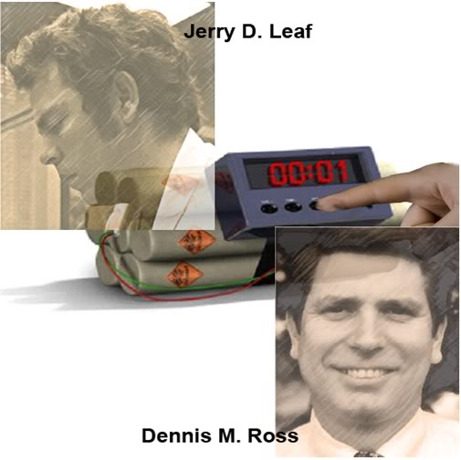
Saving Lives Now?
Coronary Artery Disease and Vasculopathy
I’ve been at pains here to emphasize that the primary purpose of the DSS is to alert cryonicists to the presence of a lethal or potentially lethal morbid process, so that we can make rational preparations for cryopreservation and avoid prolonged ischemia and autopsy. The question naturally arises, “Can this technology be used to extend or improve the quality of life now, during this life cycle?” In the case of atherosclerotic disease this seems likely, and several activist organizations within the conventional medical community are urging the adoption of cardiac CT calcium scans as screening to tool to allow for subsequent invasive, drug and dietary interventions, as necessary, to avoid heart attack. This is approach is not yet proven to reduce death from CAD, or to reduce the incidence of severity of heart attacks. However, it seems an eminently reasonable approach and, considering that each year 785,000 Americans experience their first heart attack, 470,000 more have a second, third…heart attack and 325,000 more experience sudden cardiac death. (Rogers, 2012)
In the past 48 hours I’ve learned that three acquaintances have died from SCA. Two of the three were a father and his daughter who suffered fatal heart attacks within a week of each other. Their brother had undergone coronary bypass surgery a few years previously, and the incidence of CAD and SCA in their family history was high. This case presents special irony, because the brother had undergone a CUS (which showed intimal thickening) and then a cardiac CT, which showed heavily calcified coronary vessels. It is hard not to believe that these diagnostic tests did not at least spare him a heart attack. He is now on aggressive drug and dietary treatment for his vasculopathy (he also has atherosclerosis in his peripheral vessels and in one renal artery). Whether a meaningful extension of life span will ensue can only be determined by large scale application of such screening, with accompanying long term outcome studies. However, from a cryonics perspective, it seems clear that, were this man a cryonicist, this technology would have granted him a clear opportunity to benefit in at least the following ways:
* Notify his CO, his physician and possibly his local coroner or medical examiner that he has a (superbly) documented history of severe CAD. Since he lives alone and the circumstances of his life are placid, if he does suffer SCA, this makes it much less likely he will be autopsied.
* Consider acquiring and using a wearable automatic defibrillator, at least until such time as (if) his CAD has shown demonstrated reversal by angiography as a consequence of drug/diet treatment.
* Relocate to near his cryonics service provider to minimize both cold and warm ischemic times following medico-legal death.
* Use an emergency alert system to signal either (or both) cryonics or medical personnel that he has experienced cardiac arrest. Possible options here are the Vitalsens system by Intelesens and the NUVANT Mobile Cardiac Telemetry System.
* Alert family and friends to “check & report” on him so that he is not ischemic for days, or longer, in the event of SCA.
*Acquire cryonics first aid supplies, such as ice, instant ice packs, a head ice positioner, and other items that might be appropriate to his circumstances.
The ability to engage in these preparations alone is a huge improvement from a cryonics standpoint.
The illustration that opens this article is of two of the finest men I’ve ever had the privilege to know: Jerry Leaf and Dennis Ross. Both were long time cryonicists. Jerry is, of course, well known for his enormous contributions to cryonics, both personally and professionally. Dennis was not so visible, but was an important and energizing presence in cryonics as well. Dennis was one of the founding members of the Cryonics Society of South Florida, and was a source of good advice and wise counsel for me, and I’m sure for others in cryonics as well. Both Jerry and Dennis deanimated as a consequence of vascular lesions that could arguably have been detected with the imaging techniques available today that have just been discussed here. In Jerry’s case, the technology was nascent in 1991 when he suffered his heart attack. In Dennis’ case, the technology was mature, readily available and easily affordable to anyone whose income is middle class, or better and who appreciates the need to access it.
This is ever the sad paradox of medical singularities, in that there is almost always a considerable lag time between their introduction, and their working acceptance. As we’ve seen in this article, there are many sound logistic and practical reasons for delays in the widespread application of novel medical technologies. The devil is in the details, as has certainly been the case with the PSA test. And bite back can be punishing, as can be the unforeseen adverse effects of the new modality; cancer in the case of x-rays, and cancer again in the case of hormone replacement therapy in menopausal women (a treatment that has caused many malignancies and deaths).
The uniquely attractive thing about quantum advances in areas of medicine like imaging, is that they offer such powerful advantages with such little potential for harm – if they are used intelligently. In this unusual case, we have a great deal of prior (bad) experience with screening technologies to guide us, and we also have the long history of experience with using these modalities in their less spectacular form. We know, for instance, about the adverse effects of ionizing radiation and we know about the relative safety of MRI. The “singularity” making aspects of medical imaging as discussed here are thus not the application of new imaging means, but rather are a result of the exponential growth in computing under the overdrive force of Moore’s Law.
Cancer & Others
Unlike atherosclerosis, neoplastic disease follows a course that is more nearly exponential than linear. The earliest phases of malignant transformation occur on the molecular and the microscopic level, with many tumors remaining very small for a consider period of the time course of the disease. Even where tumors are detected “early” via imaging techniques, the outcome is variable, depending upon the nature of the malignancy and the effectiveness of the treatments available.
With the notable exception of prostate cancer (Schroder, 2009), the majority of cancers are diagnosed when the disease is well advanced – usually late Stage II, or later. In the case of breast and colorectal cancer, earlier diagnosis has proved effective at improving long term survival. Early trials of lung cancer screening for smokers are also proving encouraging. While there is considerable debate about the utility of early screening in reducing deaths from other cancers, it seems reasonable in the current treatment milieu that the earlier the disease is diagnosed, the better the chances are for survival. (Hanley, 2010)
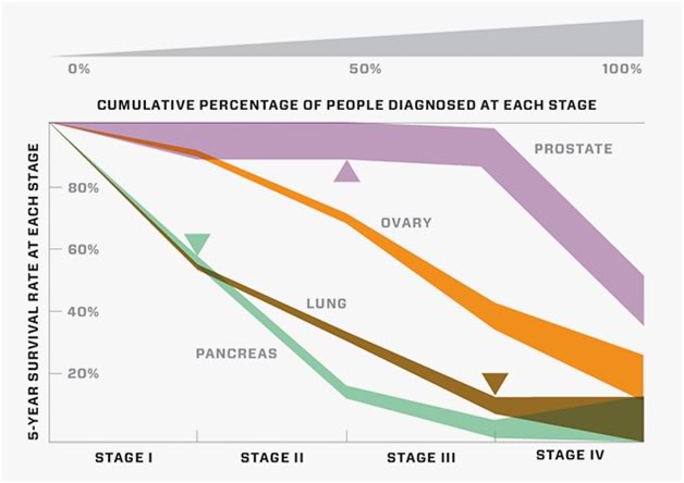
Figure 30 : Cumulative percentage of people diagnosed with prostate, ovarian, pancreatic and lung cancer at each stage of the disease. Source: Wired Magazine, 17:01;80-122, 2009.
To a great extent the value of early diagnosis may depend upon continuing advances in the treatment of cancer at the molecular level. The past decade has seen the emergence of “molecularly targeted” drugs, such as Gleevec, and more are in the pipe. If cancer treatment becomes more rationalized and targeted, it seems possible that earlier detection will be of greater value. Alternatively, definitive treatment for cancers that inhibit tumor cell proliferation or induce selective tumor cell death, may render the need for the “earliest possible diagnosis” a thing of the past.
In the case of cancers, it bears repeating that DSSing is not intended, nor is it likely to serve as more than a warning of impending deanimation. Any “saves” that occur as a consequence will thus be incidental, and the scans should not be relied upon to disclose treatable neoplastic disease.
Neuronal Attrition Disorder of Aging (NADA)
As was pointed out earlier, all interventions to extend life span by effectively treating or delaying non-brain degenerative diseases will ultimately result in “brain failure.” The question not asked by the legions of clinicians, activists, NGOs and others working to find a cure for AD (and the other dementias) is just exactly what will happen when they do? As they often point out, AD is a discrete pathology, and not a “normal” part of aging.
But curing it begs the question of what happens next, because brain cell death (both neuronal and glial) is a process that begins at ~ 2 years of age – at least for the neurons that comprise the gray matter of the cerebral cortex, and which proceeds relentlessly throughout the individual’s lifetime (Giorgio, 2010) Brain cell loss and degeneration become morphologically apparent in the brain’s white matter by the time we are in our early 20’s, although there is evidence that more subtle changes have been afoot for much longer. (Hedden, 2004) Losses in gray matter volume proceed approximately linearly with age in normal aging, and the average gray matter volume decreases from ~390 mL at age 22, to ~300 ml at age 82. (Courchesne, 2000) Total loss in brain mass between age 20 and age 80 is, on average, ~450 g, or roughly 1/3rd of our youthful brain volume.
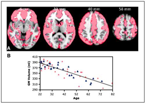
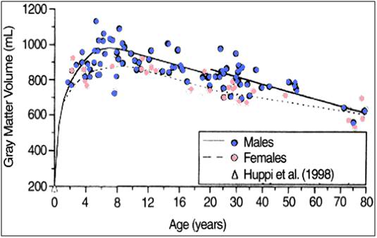
Figure 31: Gray matter loss with aging.
Top: Voxel Based Morphometry (VBM) analysis of gray matter changes in aging. (A) Colored voxels show regions demonstrating significant negative correlations between gray matter volume and age (p < 0.05, fully corrected for multiple comparisons across space). Clusters are overlaid on the MNI152 template brain. Images are shown in radiological convention. (B) Plot to illustrate relationship between age and mean gray matter volume across all significant voxels. The pink triangles represent female subjects. [From: Giorgio, A, Santelli, L, Tomassini, V, Bosnell, R, Smith, S, De Stefano, N, Johansen-Berg, H. Age-related changes in grey and white matter structure throughout adulthood. Neuroimage. 2010;51(3):943-51.Epub 2010 Mar 6.]
Bottom: Growth and aging changes in gray matter for 116 living healthy individuals. Gray matter volume reached maximum by 6 to 9 years of age and thereafter declined linearly. [From: Courchesne E, Chisum HJ, Townsend J, et al.: Normal brain development and aging: quantitative analysis at in vivo MR imaging in healthy volunteers. Radiology. 2000;216:672.]
Medicine currently has no name for the grotesque pathological state that will emerge when this failure mode is allowed to manifest itself as a result of the elimination of AD and the continued extension of the life span via various incremental advances in treating other, non-brain degenerative diseases. So that we can have a common shorthand for discussing this soon to be problematic malady, I have labeled it the Neuronal Attrition Disorder of Aging, or NADA, for short.
The near linear loss of gray matter volume and the accompanying heavy losses in gray matter neurons poses a severe problem for the aging cryonicist because they imply that ever more sophisticated advances in 1/2TM, and even HTM, exclusive of true brain rejuvenation, will lead to our becoming neurological struldbrugs,[1] and that is a condition from which not even cryonics can resurrect us.
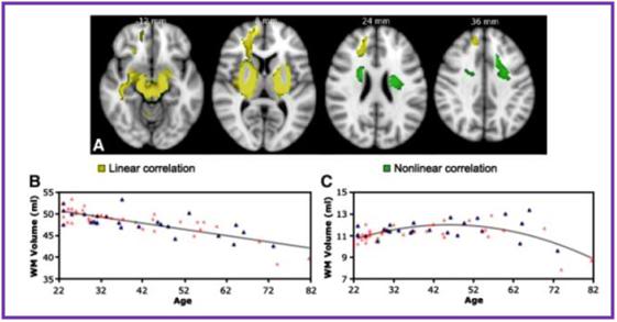
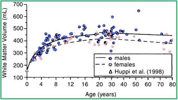
Figure 32: VBM-style analysis of WM changes with age. (A) Colored voxels show regions where WM volume shows a significant linear (blue) or non-linear (green) relationship with age (p < 0.05, fully corrected for multiple comparisons across space). Clusters are overlaid on the MNI152 template brain. Images are shown in radiological convention. (B, C) Plots to illustrate relationship between age and mean WM volume across all voxels showing a significant linear (B) or nonlinear (C) relationship with age. The pink triangles represent female subjects. Giorgio et al. The graph in the green bordered box below shows white matter volume as evaluated by conventional MRI using T1 weighted imaging. This data shows a steady increase in WM volume until age ~40, followed by a modest decline in advanced old age. However, using more sophisticated directional Voxel Based Morphometric imaging, as shown in the purple bordered box at the top of this page, WM changes are revealed to be complex, inhomogeneous between brain hemispheres, and begin in the early 20’s. As can be seen in the VBM white matter graph (purple box) there are, in fact, extensive loses in WM, however they are regional in nature as opposed to the global losses experienced by gray matter as a function of ‘normal’ aging. Growth and aging changes in white matter for 116 living healthy individuals. White matter volume rapidly increased until 12 to 15 years of age, and thereafter increased at a slower rate, plateauing at approximately the fourth decade of life. [From Courchesne E, Chisum HJ, Townsend J, et al.: Normal brain development and aging: quantitative analysis at in vivo MR imaging in healthy volunteers. Radiology. 2000;216:672.]
Beginning in middle age there is a very noticeable steady degradation in the integrity of the white matter tracts, particularly those in the hippocampus (the brain’s memory trafficking center). In particular, the perforant pathway (PP) is seriously affected, and there is typically a loss of upwards of 25% of PP axons with aging.(Hyman, 1986; Scheff, 2006)
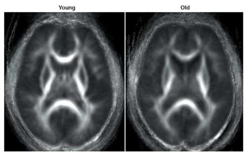
Figure 33: Group-averaged diffusion tensor images of anisotropy of white matter in young and normal elderly. Parallel movement of water molecules through white matter results in anisotropic diffusion, with greater anisotropy (and so greater white matter density) indicated by brighter areas. Older adults tend to show decreased white matter integrity compared with younger adults, with the greatest age-related declines occurring in anterior cortex. (Head, D. et al. Differential vulnerability of anterior white matter in non-demented aging with minimal acceleration in dementia of the Alzheimer type: evidence from diffusion tensor imaging. Cereb. Cortex (in press). This paper offers a comprehensive DTI study of white matter changes in normal and demented aging and demonstrates the loss of fiber tracts, gliosis and scarring that occur in the so called ‘healthy’ aging brain.
Until a scant few years ago, it was impossible to image the structural changes in long nerve processes in the brain. Now, with the advent of a technique called diffusion tensor imaging (DTI) (Dennis, 2007) it is not only possible to image these changes but also to quantify of alterations in white matter microstructure during aging. Thus, for the first time, literally within the past 2-3 years, we are getting a clearer picture of the neuropathology of ‘normal’ aging, and it isn’t a pretty one. (Augustinack, 2010; Yassa; 2010; Abe, 2002)
The development of DTI has been especially useful in documenting age-related changes in white matter, and there is now solid evidence that one of first areas of the brain to undergo age-related white matter decay is the medial temporal lobe (MTL),41 which is the area of the brain that is central to the formation of new memories, and in particular, to the acquisition of new factual information and to remembering events.(Wang, 2010; Sauvage, 2010, Bjornekbekk, 2010) Changes in the MTL are first observed (and remain most pronounced in) the perforant path (PP). The PP is so called because it perforates the subiculum[2] and carries input from the entorhinal cortex to the hippocampus, where memory consolidation and encoding are thought to be moderated.(Yassa, 2010; Burke, 2006)
The importance of NADA to cryonicists should be obvious, while perhaps the relationship between NADA and DISSing, is not as clear. Even if there is currently little or nothing we can do to halt NADA, we do need to know the speed and extent at which it is progressing. This will help us to plan more effectively about the conditions under which we would like to be cryopreserved, and it will also offer us an opportunity to determine if any interventions we try to slow, halt, or reverse NADA are working. We don’t get the luxury of a do-over in this situation. The way that DSSing will be of use in this respect is by providing both a baseline (if you are younger than ~ 35-40) scan of brain morphology and volume, as well scans progressively documenting brain structure and mass changes as we age.
In the coming decades it seems entirely possible, if not likely, that therapeutic and lifestyle approaches will be identified that slow NADA. There are currently a number of promising drugs in the laboratory (some already clinically available for other uses) which decrease or partially reverse the brain mass loss associated with aging. It is an irony of NADA that one of the first and most precious capabilities of which it robs many, is the ability to see that it is happening at all. The decrease in raw processing capability due to neuronal loss concurrently decreases our ability to perceive the deficits it is creating. While the positive offset of accumulated life experience provides a great deal of compensation for the functional losses, the result is that most aging people have very little conception of just how seriously their brains are being degraded over time. The very slow and subtle character of the changes also allows for “continuous adaptation” to a condition then interpreted as “normal.” In short, DSSing will provide a powerful source of objective, quantifiable feedback about the impact of aging on our brains.
FUD: “I have seen my death!”
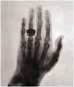 Figure 34: First x-ray of human hand; Anna Bertha Röntgen, 1895.
On 22 December, 1895 Wilhelm Conrad Röntgen made the first x-ray of human being. The subject was his wife, Anna Bertha, or more accurately, her hand. Anna Bertha’s reaction upon viewing the developed film was to exclaim, “I have seen my death!” (Hase, 1997) Prior to that time, there was virtually no way a living human being could see the skeleton of another, except after decomposition of the soft tissues was complete, following death. At that time to see one’s skeletal hand must have been a shocking reminder of mortality.
DSSing has the same potential psychological effect and it seems only fair to go further and speculate that the major obstacle to the effective use of this technology may not be the medical, ethical, financial or organizational ones, but rather, the fear uncertainty and dread (FUD) it may provoke. I have no answer to this. I would simply note that a major factor in even communicating about cryonics to the rest of the world is the FUD it provokes. Death scares the hell out people, as well it should. We cryonicists are extraordinary out of measure in our ability to either overcome that fear, or in some cases, to hardly perceive it at all.
As is the case with cryonics itself, DSSing provides us with an opportunity to extend our lives – but again, only at the cost of confronting our own mortality. The difference being that in the case of DSSing, it will be objectified, repetitive and incrementally worse with each passing interval of time. That’s not much of an advertisement for a technology, but again, as is the case with cryonics, it comes down to how acceptable you consider the alternative?
End
Footnotes
[1] In Jonathan Swift’s savagely satirical novel Gulliver’s Travels, the name struldbrug is given to those humans in the country of Luggnagg who are born normal, but are in fact immortal. Although the struldbrugs do not die, they do nonetheless continue aging. Swift describes the plight of the struldbrugs in terms almost any resident in an nursing home today (who is still compos mentis) would immediately understand: “when they have completed the term of eighty years, they are looked on as dead in law; their heirs immediately succeed to their estates; only a small pittance is reserved for their support; and the poor ones are maintained at the public charge. After that period, they are held incapable of any employment of trust or profit; they cannot purchase lands, or take leases; neither are they allowed to be witnesses in any cause, either civil or criminal, not even for the decision of meers and bounds.”
[2] The subiculum receives input from CA1 and entorhinal cortical layer III pyramidal neurons and is the main output of the hippocampus. The pyramidal neurons send projections to the nucleus accumbens, septal nuclei, prefrontal cortex, lateral hypothalamus, nucleus reuniens, mammillary nuclei, entorhinal cortex and amygdala and as such, is the principal routing network for information from the hippocampus. The subiculum is also critically involved in the formation of procedural memories.
