Liquid Assisted Pulmonary Cooling in Cardiopulmonary Cerebral Resuscitation, Part 2

Section 2:

Experimental Studies to Determine the Effectiveness of LAPC under Laboratory Conditions

Experimental Studies to Determine the Effectiveness of LAPC under Laboratory Conditions

 [This section is an edited version of an article authored by Steven B. Harris, Michael G. Darwin, Sandra R. Russell, Joan M. O’Farrell, Mike Fletcher and Brian Wowk entitled, Rapid (0.5°C/min) minimally invasive induction of hypothermia using ~4ºC perfluorochemical lung lavage in dogs, which first appeared in Resuscitation, 2001. 50: p. 189-204.)]

 1. Introduction

The potential utility of profound and ultra-profound hypothermia (0-5oC ) to arrest deleterious neurological changes has long been understood in both biology and medicine.[168],[169],[170],[171],[172] In 1959 Benson, et al., reported good outcome using profound hypothermia (10-22oC ) as a treatment following cardiac arrest. [173] This work was followed up by a number of clinicians [168],[174],[175],[176] who also reported favourable results. However, due to coagulopathy, arrhythmias, and the increased incidence of pneumonia and sepsis associated with such deep and prolonged cooling, post arrest hypothermia failed to gain acceptance and was abandoned. It was not until the work of Safar, et al., [160],[53],[82] that the utility of mild therapeutic hypothermia (MTH) (DT = −2 to −3°C) as an active treatment for the post-resuscitation syndrome was rigorously demonstrated, and subsequently validated by others. [177],[178],[55],[179-181]  As noted in Section 1, while CPB offers the most rapid core cooling possible, it is logistically unsupportable as currently practiced. Additionally, CPB carries the added risks of anticoagulation, further activation of the immune-inflammatory cascade, RBC aggregation, and the danger of gas embolism, as chilled, nitrogen-saturated blood is rapidly re-warmed as it perfuses warm tissues.[182]

As was also previously noted, less invasive modalities with the potential for in-field application, such as surface cooling and lavage of body viscuses with a balanced salt solution, are only effective in achieving cooling rates in the range of 0.10–0.15°C/min. The seemingly straightforward  experimental technique of ‘tidal liquid ventilation’ (TLV) with chilled, oxygenated PFC uses the ~20 m2 surface area of the lungs for heat exchange, but thus far has been no more effective in inducing hypothermia than surface cooling with ice bags or chilled water blankets.[155] After preliminary experiments demonstrated the technical adequacy of LAPC at achieving heat exchange in range of 0.25 to 0.35°C/min [183] a comprehensive study was undertaken by 21st Century Medicine Inc., (21CM) and Critical Care Research, Inc., (CCR), beginning in 1999, to define and validate this cooling modality in a canine model. The goals of this research were to, a) demonstrate the fundamental safety and efficacy of the technique, b) determine the optimum cycle and volume of liquid and gas fractional tidal liquid ventilations (FTLVs), and c ) attempt to determine safe airway pressures and define liquid and gas ventilation strategies that minimized or eliminated baro- and volutrauma.

This technique, developed at 21CM/CCR, was initially called ‘mixed-mode liquid ventilation cooling’ and was later renamed ‘gas-liquid ventilation’ (GLV). However, neither of these names adequately describes the technique, and this author (Darwin) has chosen to the use the term liquid assisted pulmonary cooling (LAPC) instead. In previous studies where large fractional tidal liquid volumes and shorter cycles of FTLV were used, the performance of LAPC deteriorated towards that seen when TLV-cooling (or warming) was used. In practice however, certain significant differences remained and understanding these differences proved essential to optimization of the technique.

In LAPC the critical elements of gas ventilation are retained allowing for flexibility in selecting ventilation parameters independently for heat and gas-exchange, allowing for liquid-mediated heat-exchange to be easily undertaken using existing ventilation systems[1]. The combination of gas and liquid FTLVs may also play a role in the surprisingly good thermal efficiency of LAPC as compared with TLV.

The following study explored the performance of LAPC using a prototype automated FTLV device, and discussed the basic mechanics and intrinsic limitations of heat-exchange using FTLV.

2. Materials and methods

These experiments were approved by 21st Century Medicine, Inc.’s Institutional Animal Care and Use Committee and were in compliance with the Animal Welfare Act and the National Research Council’s Guide for the Care and Use of Laboratory Animals. Fifteen mongrel dogs weighing 13.8–25.7 kg were used (Table 1). Dogs were pre-medicated with I.M. acepromazine (1.0 mg/kg) and atropine (0.02 mg/kg) prior to induction of general anesthesia using sodium pentobarbital (30 mg/kg I.V., with maintenance dosing). Anesthetized dogs were intubated with a reinforced 10.0 mm I.D. (Willy Rusch AG, Kernen, Germany) endotracheal tube (E.T.), and ventilated on room air using a Bennett MA1 or Siemens Servo 900 C ventilator. Ventilator parameters, unless otherwise noted, were 12 gas-breaths/min, gas tidal-volume of 15 ml/kg, I:E ratio of 1:3, and a maximal positive inspiratory pressure (PIP) limit of 26 cm H2O (2.5 kPa). Gas pressures were measured at the E.T. adapter. Gas minute-volume (Vg) was adjusted to maintain PaCO2 between 35 and 40 torr. Animals were maintained at ~37.5°C prior to LAPC, using a temperature-controlled water blanket. Rectal and bilateral tympanic temperatures (Ttym) were monitored continuously using a type-T thermocouple system (Cole-Parmer, Vernon Hills, IL) with a response time constant (to) of 5 s.

Combination pressure, blood sampling, and temperature-probe catheters were constructed from rigid polyethylene pressure-monitoring catheters, threaded centrally with 0.05 in. O.D. Teflon™-sheathed type-T thermocouples (to=0.3 s, Physitemp Instruments, Clifton, NJ). In order to reduce the risk of catheter-associated clot formation, I.V. sodium heparin was given to adjust activated clotting times to 300–500 s, prior to central line placement. Femoral vessels were isolated surgically, and arterial and venous catheters placed and advanced to a level above the renal vessels, as confirmed by X-ray. During surgery, bupivacaine (0.5%) was infiltrated into wounds to mitigate post-operative pain.

In one dog (Trial I-2), a femorally-placed pulmonary artery thermodilution catheter replaced the venous combination catheter. Blood and ventilator pressures were acquired through a Hewlett Packard 78532-B monitor/transducer system.

Immediately prior to LAPC, dogs were assessed for adequacy of general anesthesia, and then given pancuronium bromide (2 mg) to inhibit shivering and spontaneous breathing. FIO2 was increased to 100% and external temperature control discontinued. To serve as a cannula for both delivery and removal of PFC liquid, a 19-Fr. flat-wire reinforced Bio-Medicus® venous catheter (Medtronic, Eden Prairie, MN) was introduced through the suction port of the E.T. adapter, and advanced ~45 cm to approximately the level of the carina (Figures 2-1 and 2-2) as confirmed by X-ray. This cannula was connected to the LAPC apparatus described below. LAPC was performed using the PFC liquid ‘FC-75’ (3M Corporation, St. Paul, MN), a perfluorinated butyl-tetrahydrofuran isomer mixture.[184]
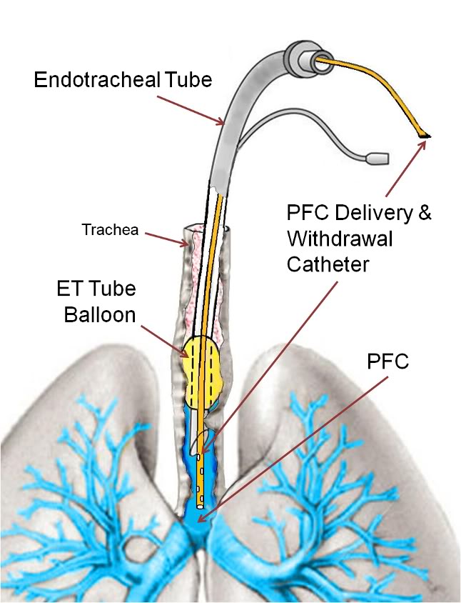
Figure 2-1 (right): PFC delivery and withdrawal catheter threaded through the endotracheal tube with the tip positioned at the level of the carina.

A two reservoir circuit (Figure 2-3) was used to deliver and remove PFC from the lungs (FTLV) via the cannula, in cycle periods of 37 s (Trial I) or 16 s (Trial II). During timed PFC infusions (tin=20 s for Trial I, or 10 s for Trial II), PFC was pumped through the cannula by a continuously-operating Travenol CPB roller-pump (Sarns, Ann Arbor, MI). A bypass loop, open during suction, allowed the roller-pump to divert (recirculate) PFC flow back into the storage reservoir whenever flow was not directed by line clamps V1–V3 into the animal. PFC was pumped continuously through an in-line 0.2 μ ‘pre-bypass’ filter (Pall PP 3802, Pall, East Hills, NY), a primary heat-exchanger (Torpedo-T, Sarns, Ann Arbor, MI), and a combination silicone membrane oxygenator/heat-exchanger (SciMed II-SM35, SciMed Life Systems, Minneapolis, MN). The oxygenator was supplied with 5–6.5 l/min O2 (maximal device design rate), and the reservoir PFC was allowed to circulate and equilibrate with heat-exchangers and O2, before LAPC was initiated. The circuit tubing was constructed of S-50 HL TYGON® 3/8 and 1/2 in. I.D. class VI tubing (Norton/Performance Plastics, Akron, OH) with the exception of a length of silicone tubing (Masterflex® 96410-73, Barrant Co., Barrington, IL) used in the roller-pump head in order to allow flexibility at low temperatures. PFC suction was driven by a vacuum pump (model 107CAB18B, Thomas Compressors, Sheboygan, WI), and suction reservoir negative pressure was limited to −35 torr by a vacuum relief valve. Figure 2-2: Detail of LAPC PFC introduction and removal catheter and ET Tube and gas ventilator configuration. A Biomedicus CPB venous return catheter was threaded through the suction port of a standard 16 mm respiratory ET tube swivel connector.
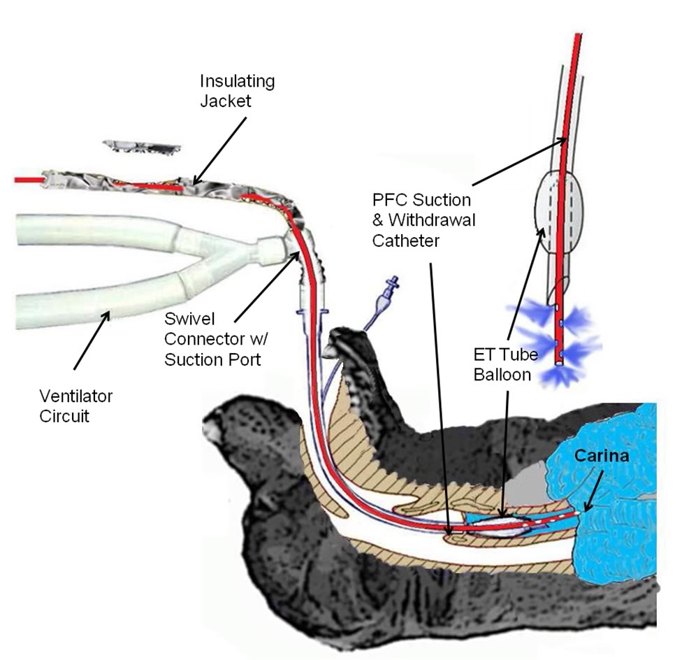
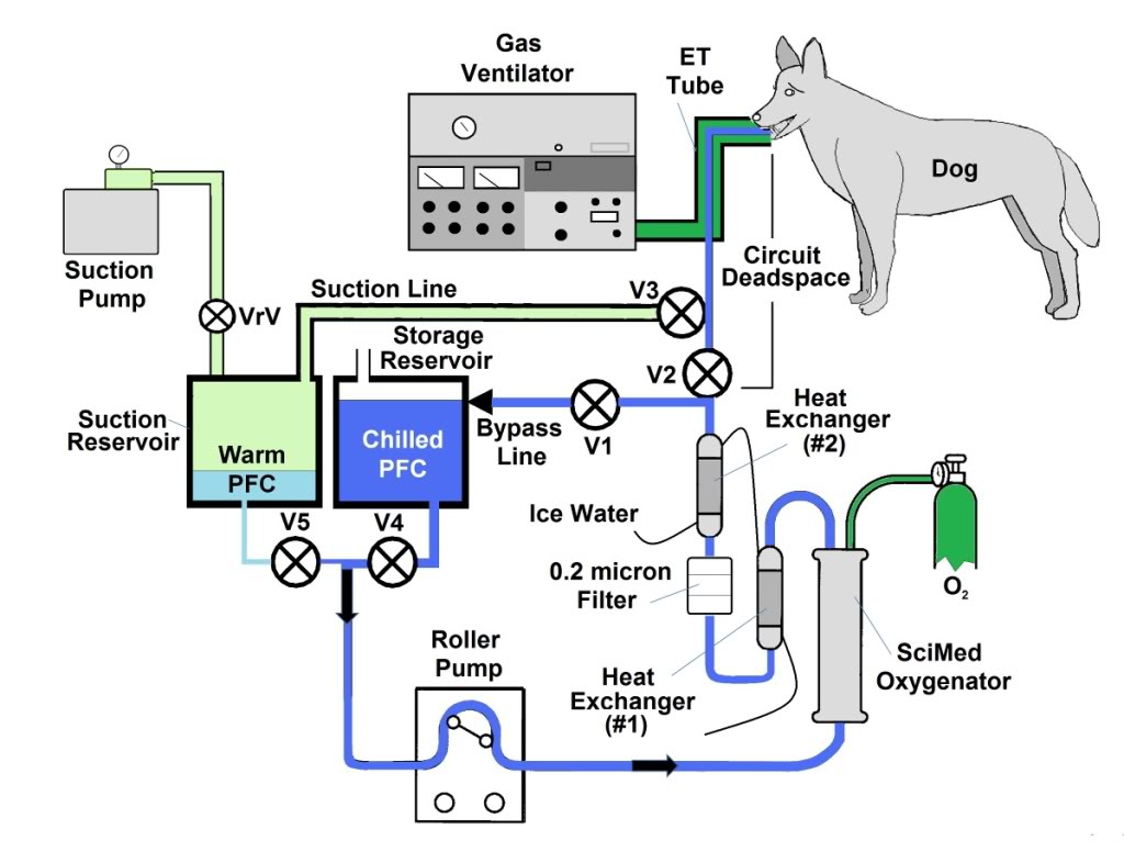
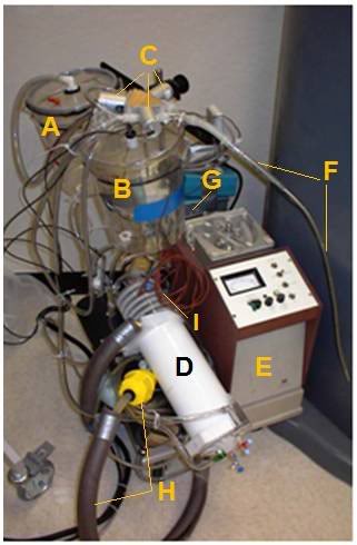
Figure 2-3: The LAPC system. The LAPC system was connected to a catheter inserted into the suction port of the E.T. adapter. PFC flows were directed by manual or mechanical clamps at V1–3. During the suction phase, FC from the lungs was removed into a sealed ‘suction’ reservoir, for later addition to the primary circuit (via adjustment of V4 and V5), while ‘infusion ready’ PFC was re-circulated through a bypass loop. Negative pressure was limited by a vacuum relief valve (VrV). Photo (right) A) Suction Reservoir, B) Storage Reservoir, C) Solenoid Valves (V1-V3), D) SciMed Oxygenator & Heat Exchanger, E)Sarns Roller Pump, F) PFC Suction/Delivery Catheter, G) Pump Controller, H) Heat Exchanger Return Line with weighted water diffuser (yellow), I) Thermocouple Probes.
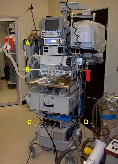
Figure 2-4: Gas ventilator and respiratory monitoring equipment used in the LAPC experiments; a) Novametrix CO2SMO respiratory function monitor and capnograph, b) Siemens Servo 900 C ventilator, c) Korr Medical, Inc., automated device used to perform rapid-cycle LAPC, d) LAPC apparatus.

  Concurrent FC-75 FTLV and gas ventilation (LAPC) was performed for 18 min in Trials I and II (n=12). This time was chosen, on the basis of preliminary work (data not shown), to achieve rapid systemic-cooling of greater than 5°C. For Trials I and II, the PFC recirculation rate within the LAPC device (=PFC infusion rate, ˙V inf), was set at 50 ml/kg per min rate, VFTLV) was set at 50 ml/kg per min.

Immediately after a timed FTLV, PFC was removed as rapidly as possible. Infusion of PFC for the next FTLV began immediately after suction was discontinued. In LAPC experiments, PFC was chilled to ~4°C prior to FTLV (Table 1), whereas in normothermic (control) dogs, isothermic PFC was delivered to the dog within ~2°C of tympanic temperature (Ttym). The PFC inflow and outflow temperature was measured continuously by a thermocouple inserted into the PFC path at the base of the delivery/removal cannula. Temperature data was collected throughout LAPC, and for 22 min after LAPC was completed. Arterial blood gas (ABG) samples were taken from the femoral arterial line before the start of LAPC, and every 2 min during LAPC. Following the post-LAPC equilibration period, monitoring devices were removed and incisions closed.

Table 1:
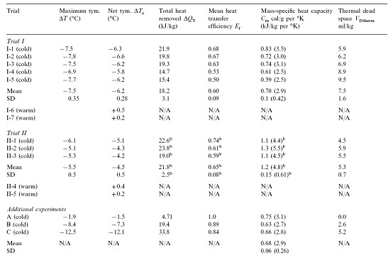
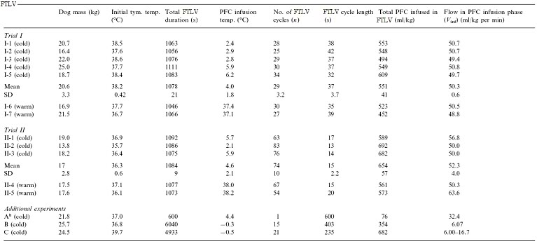
Table 2:
2.3. Trial I (manually-controlled LAPC)

Trial I was designed to investigate the variability in the response of individual animals to LAPC and to investigate the physiological effects of the LAPC technique with and without cooling (i.e., ~4ºC PFC vs. isothermic PFC FTLV). Either isothermic (near-body temperature) or ~4ºC PFC FTLV was administered using a manually-controlled system (V1–V5 in Fig. 1 represent CPB tubing-occluders in this Trial). One FTLV (period tc ~37 s) was composed of a timed FTLV (tinf = 20 s), followed by PFC suction (ts ~17 s). Suction was stopped when PFC liquid return became sparse, or gas pressure in the ventilator circuit fell below −5 cm H2O (−0.5 kPa). Five dogs received ~4ºC FTLV (Trial I-1–5), while two controls received the same protocol using isothermic FTLV (Trial I-6 and 7).

 2.4. Trial II (machine-controlled LAPC)

Trial II assessed the utility of using an automated device (custom manufactured by Korr Medical, Inc., Salt Lake City, UT) to perform rapid-cycle LAPC. Computer-controlled solenoid clamp-valve occlusion of circuit lines at V1–V3 allowed smaller FTLV volumes (VFTLV) and smaller tc. While tinf was decreased to 10 s in Trial II, VFTLV remained constant, and the effective PFC FTLV rate (VFTLV) remained in the range of VFTLVfor Trial I. Table 1 gives relevant trial parameters. In Trial II, suction of PFC from the lungs began immediately after infusion, and was automatically stopped whenever a ventilator circuit pressure of −5 cm H2O was reached (ts ~6 s, giving tc ~16 s). Three dogs received ~4ºC PFC (Trial II-1–3), while two controls (Trial II-4 and 5) received isothermic PFC.

 2.5. Animals A, B and C

 Selected data from three dogs in an earlier method development series was used. These dogs had been prepared as above, then manually given 1, 15 and 21 FTLVs, respectively with ~4ºC PFC, at much slower rates than in Trials I and II (Table 1). Data from these animals allowed independent measurements of FTLV volume heat-contents and temperatures, and thus the heat capacities and heat transfer efficiencies, by a more thorough thermal accounting method (Table 2, Appendix A).

2.6. Data collection and correction, statistical methods, graphical display and presentation

 Temperature and pressure data were collected using a PCI E series data acquisition board and LabVIEW™ software (National Instruments, Austin, TX). Graphical analysis and display of temperature data, and curve fitting, was done using the software package Origin™ (Microcal Software, Northhampton, MA). Statistical comparison of Trial group values was done using GraphPad Prism (GraphPad Software, San Diego, CA). Group means are reported ± standard
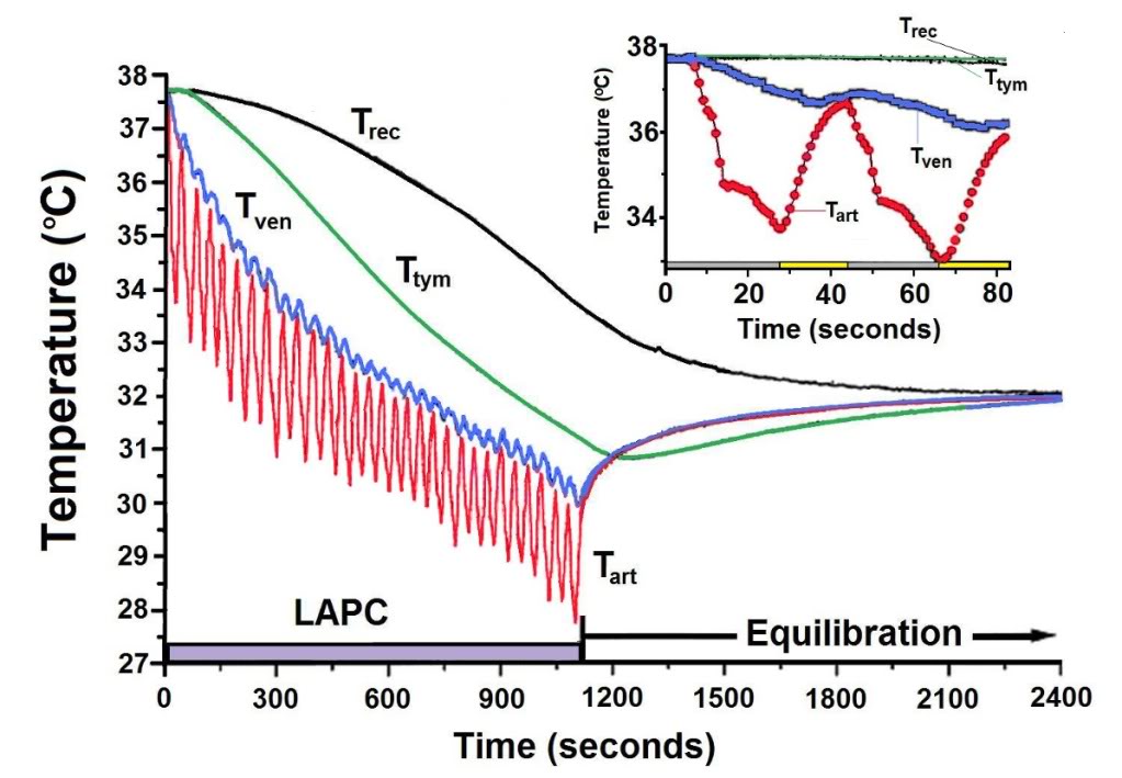
Figure 2-5 (above): Body temperature changes observed during LAPC (Method of Trial I). In this illustrative experiment from Trial I (I-4), FTLVs of ~4ºC FC-75 were infused (~20 s) and removed (~17 s) from the lungs. LAPC was performed for 18 min (hatched bar), then stopped to allow thermal equilibration (22 min). Arterial temperature (˜Tart), central venous temperature (˜Tven), tympanic temperature (˜Ttym), and rectal temperature (˜Trec) are shown. Inset: Enlarged view of temperature changes recorded during the first two cycles of PFC infusion (gray bar) and removal (yellow bar).

 deviation (SD) except as otherwise noted. For each animal, the Ttym from whichever probe cooled most rapidly, was used (right probe in 12/15 dogs). In order to facilitate comparison of cooling rates between sites in the same animal, temperatures at all probe sites were corrected to the baseline aortic temperature (Tart), as measured immediately prior to the start of LAPC. For ease of description, LAPC-cooling is presented in terms of thermal-deficit (‘cold’) moving from the lungs into successive body compartments. A compartmental analysis of thermal transfer in this model, and a glossary of notation and equations used, is given in Appendix A.

 3. Results

 LAPC allowed FTLV of dogs during concurrent gas ventilation. Suction from the submerged catheter tip at the carina allowed collection of PFC even during forced gas inspiration. It was discovered that a long suction catheter was necessary to insure that adequate suction pressure could be used to withdraw PFC throughout the liquid removal sequence, without prolonged exposure of the gas filled portion of the airways to the negative pressure of the suction system/reservoir. Additional protection of the airways against excessive negative pressure during the relatively brief time after liquid no longer filled the suction line was provided by incorporating a negative pressure relief valve on the suction reservoir. Suction in this manner was efficient, although FTLV volume measurements showed that the lungs retained ~12 ml/kg PFC (approximately FRC) between FTLVs.

The PFC pump circulation/infusion rate (˙V inf), measured volumetrically preceding and following LAPC, was stable to within 1% over the duration of LAPC, and was not significantly different between trials (P = 0.28). The VFTLV, calculated as tinf  inf/tc, was 30.7±2.3 ml/kg per min (Trial I) and 36.4 ± 3.2 ml/kg per min (Trial II). The ˙VFTLV was significantly (P = 0.023) larger in Trial II because machine-controlled suction made more efficient use of available non-infusion time, resulting in faster net PFC removal.
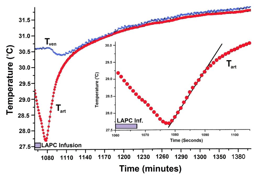
Figure 2-6: Thermal equilibration after LAPC. Mean Tart and Tven values (Fig. 2) are shown for Trial I, dogs 1–5. To highlight equilibration changes, Ttart curve nadirs (n=5) were superimposed before calculation of means, and Tven data (n = 4) for each dog was adjusted with its corresponding Tart curve. Incompatible Tven data from a pulmonary artery thermodilution catheter in I-2 has been omitted. Inset: The sigmoidal mean (N = 5) Tart recovery during the first ~12 s after final LAPC. FTLV is approximated by linear fitting.
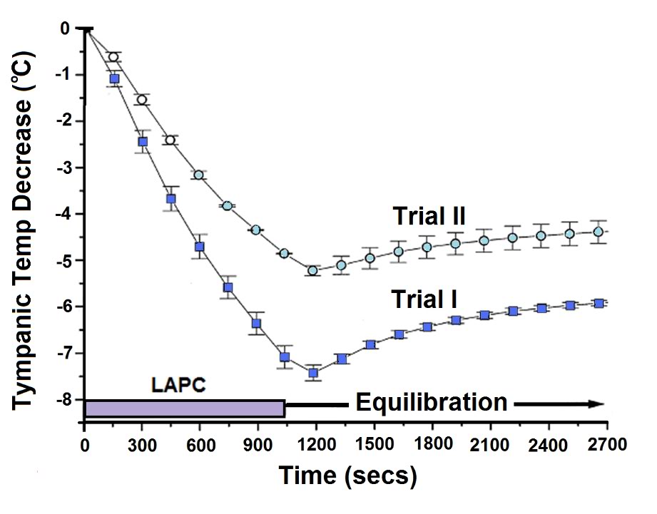
Figure 2-7: Body temperature changes during manual and mechanical LAPC (Trial I vs. Trial II). The relative rates of core body cooling in dogs undergoing 18 min (hatched region) of manual (Trial I, solid squares) or machine-driven (Trial II, open circles) ~4ºC LAPC, were assessed by comparing changes in group mean Ttym. Symbols represent the mean and SEM (n = 5 for manual, and 3 for machine groups).

 3.1. Thermal results of LAPC

3.1.1. Cooling time delay

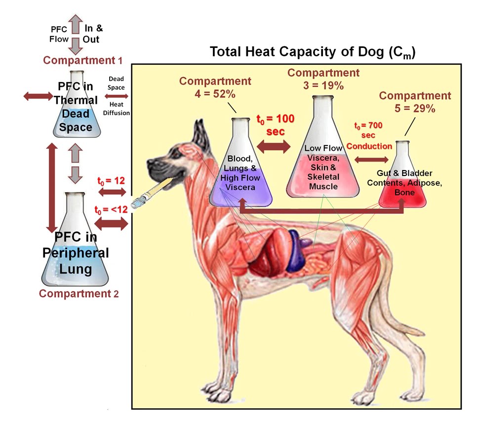
 Figure 2-5 illustrates LAPC cooling in a representative dog (I-4) from Trial I. The Tart began to decrease 3–6 s after the start of each PFC FTLV. Since this delay included circulation delay from lungs to aorta, the transfer of thermal-deficit from newly-introduced PFC to pulmonary blood was very rapid. The venous temperature (Tven) began to decrease 10.4 ± 6.9 s after Tart decline, representing the minimum systemic circulation time. Though exhibiting delay, damping, and broadening behavior (presumably due to peripheral heat-exchange and varying systemic blood-return path lengths), Tven transients from FTLVs mirrored Tart transients. Ttym temperatures, presumably reflecting brain and viscera temperatures, were non-oscillatory. The Ttym did not begin to decrease until ~24 s after the start of LAPC. This decrease occurred in three phases: an initial phase lasting ~100 s, an exponential phase lasting for ~900 s, and a final linearly-decreasing phase lasting until the end of LAPC. Core cooling as measured by Ttym continued for about 120 s after the end of LAPC (Figure 2-5), then exhibited a marked rebound effect [185] with exponential dampening (t ~20 min, Figures 2-5–2-7). These phases of cooling and equilibration were consistent with a five-compartment thermal model, in which the three compartments representing animal tissues corresponded roughly with (1) the blood and vasculature; (2) the classical thermal core; and (3) the classical thermal periphery (Figure 2-8). Modelling equations and estimation of compartment sizes are given in the Appendix A.

3.1.2. Cooling rate

 Crude cooling rates were determined numerically from appropriate T vs. t graph segments. The mean cooling rate from LAPC initiation, or DTtym/Dt, reached a maximum value in Trial I at −0.49±0.09°C/min (t=6.6 min). The differential cooling rate d (DTtym)/ dt = dTtym/dt reached a maximum (max) value of -0.59 ± 13°C/min at t ~100 s, near the end of the initial heat exchange development region. (This value is comparable to analytic d (DTtym)/dt  (max) from (Eq. (1)) = DTk/to  = −0.63°C/min). Corresponding cooling rates in Trial II were DTtym / Dt (max) = −0.33 ± 0.02°C/min (at t = 7.3 min) and dTtym / dt (max) = −0.37 ± 0.06°C/min (at t = 100 s).

3.1.3. Mean cooling power

 The mean heat removal rate (cooling-power) P over the entire duration of LAPC, for each animal, was estimated from DTe according to P = m Cm DTe/t (total).

Here t (total) is the entire LAPC application time = ~1080 s. (Note: for this calculation, the more accurate Trial I mean Cm is used for all Trial II animals.) The mean cooling power of Trial I was 336 ± 60 watts, while that of Trial II (using the Trial I value of Cm) was 207 ± 49 watts (P = 0.02). Variation in animal size was the major source of intra-group variability.

Figure 2-8: Heat transfer among body compartments during LAPC. Heat transfer during LAPC in the dog may be modeled using 5 thermal compartments. Heat transfer between compartments (which is by blood circulation, except as noted) is shown in the box diagram as double-headed arrows. The pair of arrows connecting Compartments 2 and 3 represents the different processes of lung equilibration with (1) pulmonary artery flow; and (2) with the complete blood volume and selected viscera.

 3.2. Gas exchange

 ABG measurements demonstrated that infusion of ~4ºC PFC stabilized PaO2 and PaCO2 during LAPC. In contrast, LAPC using isothermic PFC failed to maintain baseline PaO2 or PaCO2 levels (Figure 2-9). In Trial II-4, hypercarbia during the first 13 min of isothermic LAPC was abolished by increasing the tidal volume from 15 to 25 ml/kg (final Vg = 375 ml/kg per min). In Trial II-5, Vg was pre-set to 375 ml/kg per min in an attempt to avoid hypercarbia, and no significant ABG changes were observed.
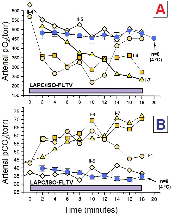
Figure 2-9: LAPC does not maintain normocarbia at isothermic temperatures without alteration of the gas ventilation parameters. Animals in Trials I and II underwent LAPC using either ~4ºC (˜) or isothermic ( ™¯) FC-75. Both arterial PaO2 (Panel A) and PaCO2 (Panel B) levels were affected by FTLV temperature. ~4ºC FTLV data from Trials I and II were very similar in magnitude, and therefore, have been combined (n=8). Isothermic LAPC is shown as four separate experiments (Trial I-6 and 7, and Trial II-4 and 5). Gas tidal volume was increased from 15 to 25 ml/kg in Trial II-4 at t=13 min, and at t=0 in Trial II-5, normalizing PaO2 and PaCO2 in both animals. Declining PaO2 in Trial I-7 was due to inadvertent failure to pre-oxygenate PFC.
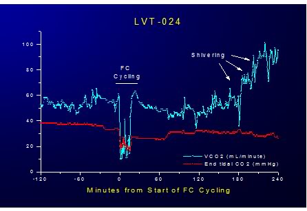
Figure 2-10: Effect of LAPC on VCO2 and EtCO2 during 20 minutes of ~4ºC FTLV. LAPC allows superior CO2 removal due to gas ventilation because ·V PFC = no more than 30 mL/kg/min of liquid, allowing gas ventilation of at least 200 mL/kg/min, resulting in a maximum ·VCO2 removal rate of  >8 mL/kg/min or a minimum of 400% of basal metabolic rate.

3.3. Clinical observations and gross pathology

 With the exception of one dog, animals subjected to LAPC displayed mild tachypnea and increased expiratory sounds, but otherwise exhibited unremarkable recovery from anesthesia, including the ability to walk and drink. The exception was an eosinophilic animal (Trial II-1) which had normal oxygenation during LAPC, but developed severe hypoxemia shortly after LAPC.

Chest X-ray pre- and post-procedure showed no (comparatively) remarkable features. This dog was sacrificed at 9 h.

Necropsy revealed a mass of D. immitis (heart- worm) embolized into the pulmonary arterial circulation, possibly as a result of local chilling of the parasite mass due to LAPC (this animal had been heartworm seronegative). Necropsies performed on nine remaining Trial I and II animals sacrificed 24 h post-procedure revealed diffuse spongy, resilient hyperinflated non-collapsible lungs (HNCL) seen in animals exposed to a high-vapor pressure PFC at high PIP pressures.[186]  HNCL was most prominent in the anterior, least dependent areas of the lung lobes. This trapped intra-alveolar PFC was thought to be the cause of broncho-constriction and wheezing found in post-LAPC animals. There was also evidence gross, dependent-lung damage evidenced by pulmonary edema with consolidation in both isothermic and ~4oC PFC-FTLV animals.

Other organ systems in this series were grossly normal. Two animals in Trial II (II-3 and II-4) were not sacrificed, and were held for long term evaluation. They were neurologically normal at 1 year post-LAPC.

3.4 Impact on Hemodynamics

 FTLV with both ~4ºC and isothermic PFC resulted in an almost immediate modest decrease central venous pressure (CVP) which persisted for the duration of the FTLV and recovered to pre-FTLV values at the conclusion of each FTLV cycle (Figure 2-11 – 2-13).
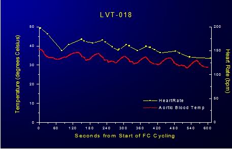
Figure 2-11: Impact of LAPC on HR.

In animals subjected to FTLV with ~4ºC PFC there was an immediate, transient reduction in heart rate (HR) and mean arterial blood pressure (MAP) and a corresponding increase CVP during the FTLV cycle. In animals undergoing ~4ºC FTLV these effects could be attributed to the acute, cyclical chilling of the coronary blood supply during each FTLV with chilled PFC. The temperature of the blood entering the coronary os was 10o to 15oC colder than systemic blood (as measured by pulmonary artery catheter), and this would be expected to have an immediate depressive effect on myocardial contractility due to the transient hypothermia the myocardium would experience as a consequence of perfusion with chilled blood. In fact, consonant with this interpretation, HR decreased steadily during ~4ºC FTLV, recovering progressively less after each FTLV cycle, as systemic hypothermia was induced (Figure 21).
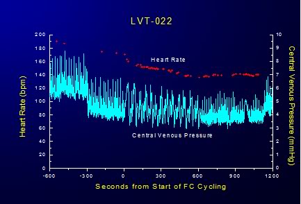
Figure 2-12: Cyclical variation in CVP is response to FTLVs with isothermal PFC.
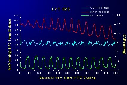
Figure 2-13: Effect of FTLV with ~4ºC PFC on MAP and CVP over the course of 6 minutes of LAPC.  MAP is transiently markedly depressed and CVP is concurrently increased in response to loading with ~4ºC PFC; this effect is reversed when the PFC load is suctioned from the lungs; although there is increasing depression of MAP in response to the induction of systemic hypothermia. Cardiac output (CO) was not measured in these studies and mathematical analysis of the MAP waveforms generated during isothermic FTLV were not done. Thus, the precise extent to which FTLV with isothermic PFC (i.e., without the thermal-metabolic effects of chilled blood on the myocardium as occurs in LAPC) impairs cardiac output or coronary perfusion pressure is unknown.

Interestingly, the cyclical increase and recovery of the CVP remained constant during both ~4ºC and isothermic FTLV. Initially, the effect of FTLV on CVP was thought to be due to compression of the thoracic vena cavae by the relatively dense PFC load in the lungs. It was hypothesized that this effect might be more pronounced in the dog due to the V-shape of the canine thorax with the cavae resting at the bottom of the thoracic ‘trough’ in a dependent position under the lungs. To test this hypothesis, isothermic FTLV was carried out with a dog in the prone position. Pronation had no effect on the transient, cyclical depression of HR and increase in the CVP associated with FTLV. It thus seems possible that loading of the lungs with dense PFC liquid results in increased pressure on the thoracic vasculature, in particular on the thoracic venous vasculature, in much the same way gas PEEP reduces thoracic venous capacitance and raises CVP; reducing right ventricular preload and right ventricular output (cardiac output). These effects would seem the most likely explanation for the reduction in MAP observed during maximal PFC loading during both ~4ºC and isothermic FTLV.

CO was not measured during these LAPC studies nor was mathematical analysis of the aortic pressure waveforms undertaken to determine with precision the degree to which FTLV depressed MAP. Crude analysis of MAP during isothermic FTLV suggests that cyclical PFC loading is responsible for ~15-20% reduction in MAP over baseline.  Mean CVP is increased ~35% over basal levels during FTLV. To what extent CO will be impacted as a result of FTLV with PFC during CPR will have to be determined experimentally (see discussion in 4.5.3. Overcoming Increased Intrathoracic Pressure and Preserving CO, below).

4. Discussion

 4.1. Apparent effect of temperature on gas exchange

 Isothermic LAPC in this model was surprisingly poor at removing CO2, considering that the CO2 carrying capacity in FC-75 decreases by only ~23% from 0 to 40°C (extrapolated from [184]). A useful observation was that pO2 values decreased even in isothermic animals, indicating an influence on total ventilation and possibly also reflecting decreased CO as evidenced by the (average) decline in MAP and increase in systemic vascular resistance during LAPC.

Capnographic analysis of LAPC in Trials I and II (data not shown) indicated that isothermic LAPC had a much larger negative effect on pressure-limited total gas ventilation Vg, as compared to ~4ºC LAPC using the same technique and the same gas ventilator settings. Since LAPC at a ˙VFTLVr of 30–36 ml/kg per min relies on gas ventilation Vg for ~50% of total alveolar ventilation, a differential loss of pressure-limited Vg with temperature appeared to be the basis of CO2 retention in isothermic LAPC. The mechanism of the implied differential change in lung compliance may be related to the depressive effects of FTLV on CO, and presumably, on perfusion. Thus, gas ventilation adjustments similar to those in Trial II-4 and 5 may be required if LAPC is used as a re-warming technique, and it may additionally be necessary to abolish gas PEEP, or even apply continuous negative airway pressure [187],[188] to counteract the PEEP-like effects of PCF loading and to generally improve CO and coronary perfusion pressure (CPP) during CPR.[189]

4.2. Thermal transfer efficiency and kinetics

 The optimal LAPC cooling (or warming) protocol remains unknown. However, the finding that the thermal equilibration of non-dead space PFC and local pulmonary blood flow proceeds very rapidly (to <12 s) suggests that FTLV infusion times need to be no longer than this time scale. When PFC lung dwell times exceed this duration, the FTLV load is in place longer than is required to transfer the most labile part of its thermal potential to the pulmonary blood and parenchyma.
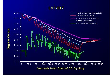
Figure 2-14: Relative insensitivity of PFC dwell time to heat exchange was demonstrated by progressively shortening the duration of FTLVs and increasing their frequency. Even at the maximum achievable rate of FTLV at ˙VFTLV  rates of 30 ml/kg per min and ˙VFTLV  rates of 50 ml/kg per min no deterioration in the efficiency of heat exchange was observed. This suggests that thermal equilibration at the level of the alveolus is practically instantaneous and that the shortest FTLV dwell time should be used for optimum heat exchange.

Since FTLV ventilation rates (˙VFTLVr) in the present study are already at least a third of the maximal rates possible in TLV, it seems probable that PFC infusion rates and pressures, rather than heat transfer rates from PFC to lung, will be the fundamentally limiting factor to power transfer in LAPC. These observations suggest that, as least to ˙VFTLV r rates of 30 ml/kg per min and ˙VFTLV  rates of 50 ml/kg per min, the total cooling power (cooling rate) in LAPC will be greatest if no PFC dwell time is allowed, and all available time during the FTLV cycle is used to either introduce, or remove, PFC.

4.3. Question of diffusion dead space in LAPC

 Mammalian lungs depend on simple gas diffusion for CO2 transport through the acinar airways during normal tidal ventilation. An intractable problem in experimental TLV has been that simple diffusion is not sufficient to similarly move CO2 through liquid PFC at physiologic CO2 partial pressure gradients. This limitation appears in TLV as a ‘CO2 diffusion dead space’ which effectively lowers alveolar ventilation. In part due to such extra physiologic dead space, TLV of adult humans has been estimated to require liquid minute-volumes near 70 ml/kg per min.[190] This value is at the upper bound of realistically attainable liquid flow rates [191],[192] and leaves little leeway for treating hypercarbia, hypermetabolic states, or lung disease. Such difficulties are not a theoretical limitation in LAPC, however, since LAPC does not require high liquid flow rates for ventilation. In the most rapid-cooling LAPC protocol used in this trial, ˙VFTLV was 31 ml/kg per min—a low baseline value which permitted the addition of 10 times this minute-volume of gas ventilation (see Fig. 4, Trial II-4, 5). Moreover, since normal gas minute volumes were required to maintain normocapnia in Trial I, there is as yet no evidence for any CO2 diffusion limitations caused by intrapulmonary PFC in LAPC. Possible reasons for this are discussed below.

Thermal-diffusion limits in TLV have not been studied per se, but their presence is suggested by the results of Shaffer and co-workers.[155] In their cat TLV model using a ˙V FTLV of 75 ml/kg per min, a decrease in PFC inspiration temperature from 20 to 10°C (increasing the thermal gradient by a factor of 1.6) increased the cooling rate from −0.13 to −0.15°C/min. This small rate change represented a significant loss of efficiency. By contrast, in the present LAPC study using PFC at 4°C, there was no evidence of a thermal-diffusion limit at rates up to 4 FTLVs/min. Notably, in Trial I, where 100% of the VFTLVr, and 40% of the ˙VFTLVr of the cat TLV model was used, cooling rates for LAPC were more than three times those reported for cats subjected to TLV at 4.5 liquid breaths/min at 10°C.[155]

The possible quantitative presence of a thermal diffusion limit for LAPC at 4 FTLVs/min may be evaluated using a modified version of the concept of gas-exchange dead space (VD). The respiratory system of a dog undergoing LAPC heat-exchange may be considered, by analogy with gas exchange dead space (VD), to also contain a ‘thermal exchange dead space’ (VDtherm). Each thermal FTLV volume ˙VFTLV of PFC then also contains a VDtherm , which by definition does not participate in heat-exchange. Thus, cycle thermal transfer efficiency Ef may be expressed as (VFTLVr VDtherm)  / ˙VFTLV, and any measured value of mean Ef may be expressed as an equivalent mean VDtherm = ˙VFTLVr  (1− Ef). For Trial I (Ef = 0.6, Appendix A for calculation), mean VDtherm was then seen to be 7.5±1.6 ml/kg, and in Trial II (using Eq. (6) Ef  Value = 0.40), VDtherm was 5.3±0.8 ml/kg (P = 0.072). The absence of an increase in VDtherm in Trial II vs. Trial I indicated that the size of VDtherm in these LAPC protocols was non-dynamic at time-scales of one FTLV cycle, providing evidence against the presence of a ‘thermal diffusion dead space’ (analogous to a CO2 diffusion dead space) at these FTLV rates. In absolute terms, it may be useful to compare calculated VDtherm in the LAPC dog model to the expected physiologic gas-exchange dead space, VDCA, which in healthy animals is close to the dog anatomic VD = ~6.5 ml/kg.[193]

In thermal diffusion, as in gas diffusion, diffusion physiologic dead space would be expected to significantly add to anatomical dead space. However, the sum of mechanical-VD in the LAPC circuit (~1.5 ml/kg) plus the anatomic VD for dogs is found to be more than the calculated VDtherm in either trial in this study, leaving little room for a large heat-diffusion contribution to VDtherm. For these reasons it is suggested that the loss of cooling power observed in Trial II was not due to heat diffusion limitations, but instead due to a loss of efficiency effect similar to that seen with low tidal volumes in ordinary gas ventilation. In these terms, low FTLV volumes in LAPC result in an increase in ‘thermal dead space ventilation’ at the expense of PFC flow involved in active heat-exchange, resulting in a larger ‘wasted’ FTLV VDtherm / ˙VFTLV. VD in heat transfer (VDtherm) that is analogous to VD in gas-transfer; in as much as all dead space is ‘diffusion dead space’ at long-enough time-scales. However, some of the mechanisms for diffusion modification of VDtherm are unique. By contrast with gas molecules, heat diffuses rapidly through device tubing into the PFC in the LAPC circuit dead space, and heat also diffuses directly through the tracheal wall into the anatomic-VD. Thus, heat diffusion from dead space liquid at sufficiently slow FTLV rates might be expected to have a pronounced effect on Ef in LAPC, due to slow heat-diffusion reduction in VDtherm.

Some evidence for such a process was found, though at FTLV dwell times too long to be of interest for rapid cooling. At the relatively small tc of Trials I and II, the calculated VDtherm was found to be ~VDCA; but in animal B, with a much longer tc of 7 min, the VDtherm was only 2.6 ml/kg. The limit of this process was reached in animal A, in which the VDtherm of a single retained ‘breath’ of highly-oxygenated PFC fell to nearly zero after 10 min. Disappearance of VDtherm by thermal equilibration, estimated from individual cycle Ef variations in animals B and C, was estimated to occur with a half-time of ~5 min (data not shown).

This process was slow enough to be neglected when the duration of FTLV intervals (tc) was less than several minutes.

Thus, at the FTLV rates of Trials I and II, a full-sized VDtherm of ~6 ml/kg appeared, and accounted for significant loss of cooling power at low ˙VFTLV (e.g. Trial II where VFTLV was only 8.8 ml/kg). The characteristic size of VDtherm at all but the slowest FTLV rates (~1 FTLV per 5 min) implies that the only thermally-efficient solution for performing LAPC at faster rates is maintenance of [˙VFTLVr  / VDCA] or [˙VFTLVr / VDtherm] ratios >3, in order to avoid excessive ‘wasted’

VDtherm  ventilation. This requires a ˙VFTLV of ~18 ml/kg in dogs. In humans, where the anatomic-VD is <3 ml/kg, less than half the value for dogs, both the VDtherm, and therefore, most-efficient ˙VFTLV values, might also be expected to be correspondingly less. In any case, it is clear that rapid-cooling LAPC techniques cannot wait for the relatively slow thermal equilibration of PFC within the anatomical VD, since equilibration in the remaining non-VD parts of the lung is so rapid (i.e. less than Trial II tc of 16 s).

4.3.2. Possible synergy of combined gas and liquid ventilation in assisting mass (CO2) and heat transfer

 The absence of expected heat-diffusion and gas-diffusion limitations in LAPC suggests that some assistive process for both gas and heat transfer through PFC in the peripheral lung may occur in LAPC. The authors’ fluoroscopic observations (made with the non-brominated and relatively radiolucent FC-75) have been that each gas breath in PLV produces a flash of fine bubbles which spread uniformly throughout the lung. As compared to the more familiar behavior of water, the low surface tension of PFCs (15 dyne-cm for FC-75, about 1/5th that of water) lowers the energy barrier to producing small bubbles in forced gas/liquid flows. Such

bubbles moving within small airways may induce eddies and turbulence in laminar liquid flows at small scales, contributing significantly to heat and mass (CO2) transport though PFC liquid by means other than diffusion.

It is hypothesized that the lack of bubble-induced turbulence in TLV may account for the large diffusion-dead-space for heat and CO2 which seems to be present in TLV at even low breathing rates – an effect which is apparently absent in both PLV and LAPC.

4.4. Potential development of clinical LAPC

 Rapid cooling of the CNS has now become a primary goal in the clinical management of the post resuscitation syndrome [49] and potentially in the acute management of spinal cord injury.[194],[195],[196] Based on their work, Safar, et al., have noted that clinical implementation of mild resuscitative hypothermia, which was highly effective in the dog model of SCA, will depend upon the development of truly rapid MTH.[197]  A recent editorial in Stroke [83] commented on the striking ability of the combination of  (33-35oC ) [198] and pharmacological pre-treatment to ameliorate ischemic brain damage in the rat middle cerebral artery occlusion model, and  then addressed similar concerns: “A problem for use of this technique for acute stroke therapy is that the time required to induce hypothermia in patients is likely to be considerably longer than for rats. […]. To substantially increase the rate of hypothermia induction in humans, it will almost certainly be necessary to use some sort of invasive procedure, such as a heat-exchanger, to cool the circulation.”

The technique of LAPC may eliminate the need for such invasive measures. For example, in the cited trial [199], rats were cooled from 37 to 33°C (−4°C) over 40 min, using surface cooling with ice packs. By contrast, the present study demonstrates cooling of the canine body core and brain by −4°C in less than 10 min.

4.5. Challenges Ahead

 4.5.1. PFC Selection

Development of clinical LAPC awaits identification of suitable PFCs for various LAPC applications. For example, the pharmaceutical PFC perfluorooctylbromide (Perflubron™ Alliance Pharmaceuticals) would presumably not be suitable for rapid LAPC cooling due to its freezing point of +6°C, but might be useful for slower cooling or for LAPHE facilitated re-warming. Some industrial PFCs have pour-points low enough to make them potentially useful as LAPC rapid-cooling media; however, most of these agents also have unacceptably high vapor pressures at 37°C or are not chemically defined in terms of chain length or even precise chemical composition (see Section 3: Perfluorchemicals). PFCs with such high vapor pressures exacerbate barotrauma by causing HNCL.

High vapor pressure PFCs may also increase the danger of long lasting lung collapse as a result of PFC, secondary to pneumothoraces, entering the pleural space and vaporizing (‘perfluorothorax’). FC-75, (formerly FX-80) was the first PFC used in liquid ventilation [10], but its relatively high vapor pressure (31.5 torr) makes it an undesirable LAPC agent. Assuming that a PFC with the desired biophysical properties is identified and produced to medical standards, LAPC should be easily scalable to adult humans. For example, the viscosity of FC-75 is similar to water [11], and under standard suction a 19 Fr. An adult pulmonary toilet catheter will remove FC-75 at ~2 l/min. As in the system described, a LAPC system may interface with a conventional gas ventilator system via a simple liquid-carrying catheter which extends through the endotracheal tube adapter suction port.

 4.5.2. Coronary Perfusion during LAPC

The effect of LAPC on coronary perfusion in the setting of ROSC following cardiac arrest due to myocardial infarction (MI) or in the presence of coronary artery disease (CAD) is unknown but is a possible cause of concern. One possible adverse effect is the potential compromise of the coronary circulation in CAD due to perfusion of the heart with profoundly chilled blood (i.e., blood temperature  @10°C below systemic temperature) leaving the pulmonary circulation and entering the coronary os. One of the authors (Darwin) has observed the onset of severe, acute angina in two hemodialysis patients with stable angina who were inadvertently dialyzed using very cold dialysate (QB of 250 ml/min at a blood temp of ~10° to 15°C) which resolved only when the dialysate temperature was increased to 30°C or above.

It is well established that exercise in cold environments, with associated inhalation of cold air, can trigger angina and lead to cardiac arrest in patients with coronary artery disease.[200],[201]  There has been considerable debate as to whether the cause of angina in this setting is due to cold air inhalation per se, or to the effects of a cold environment. It has been suggested that exposure to a cold environment is the primary factor in inducing cold weather angina, presumably by increasing peripheral vascular resistance resulting in an increase in cardiac workload at any given level of exercise [202],[203] in the same way that the cold pressor test produces acutely increased afterload and thus increased left ventricular wall stress.[204],[205],[206],[207] Hattenhauer and Neill [208] studied 33 male patients with coronary artery disease,11 of whom gave a positive history of angina when exposed to cold winter air. Seventeen of these patients were subjected to inhalation of cold air at -20°C for 4 min, and this resulted in angina at rest in four of these patients.

Cold air inhalation also produced angina in four of the seven patients who were paced at a heart rate which was well below the threshold for angina at room temperature. Cold air inhalation did not significantly increase myocardial oxygen consumption, or alter coronary blood flow as determined by the xenon clearance method. In a separate arm of the Hattenhauer and Neill study, cold air inhalation for 90 s in 18 patients produced no detectable-constriction of coronary arteries visualized angiographically. The investigators concluded from these findings that cold air inhalation induced angina could not be explained by an increase in cardiac work and myocardial oxygen consumption.

This study also demonstrated that there was no evidence of large or intermediate coronary artery or coronary arteriole constriction in response to the inhalation of very cold air. As a consequence, the authors proposed that cold air inhalation induced angina might be the result of constriction of minute coronary collaterals, or to other vessels compromising blood flow to potentially ischemic regions of the diseased myocardium. The work of Lassvik and Aveskog [209], confirmed the effects of cold air inhalation in inducing angina and failed to demonstrate any decrease in workload at either the onset of angina or at maximal workload during inhalation of moderately cold air (-10°C) in a room at 20°C. Dodds, et al., [210] evaluated 12 male patients with stable angina inhaling cold air (-8.8°C) during exercise to investigate if the vasoconstrictor peptides endothelin-l (ET-1) and angiotensin-II (AT-lI) played a material role in the etiology of this phenomenon. They concluded that neither ET-1 nor ATII had any significant role in the pathophysiology of cold air inhalation induced angina.

In contrast to Hattenhauer and Neill, and Lassvik and Aveskog, these investigators documented decreased myocardial oxygen consumption during peak exercise in cold air inhalation and concluded that the cause of this was a centrally operating mechanism such as a reduction in coronary flow. These investigators noted that the response to cold air inhalation was biphasic, and posit that the initial response; earlier onset of angina during exercise while breathing cold air, was due to activation of cold receptors in the upper airways stimulating a systemic increase in peripheral vascular resistance; and thus the observed accompanying rise in blood pressure and increased cardiac workload (i.e., the cold pressor test response). The secondary response; reduction in total exercise time, which occurred at a significantly lower rate-pressure product compared with the same patients breathing ambient temperature air, was thought to be due to peripheral reflex responses. Thus, these investigators hypothesize that the early onset of angina during exercise is due to sympathetic stimulation from inhaled cold air, but as exercise continues, central mechanisms play an increasing role in the pathophysiology of cold air inhalation induced angina.

The implications of cold air induced angina for LAPC in the setting of coronary artery disease are troubling and certainly bear careful investigation in animal models of compromised myocardial circulation. Patients in all of these studies developed significant ST depression (³ 1 mm) concurrent with the onset of cold air inhalation induced angina, suggesting clinically significant myocardial ischemia. What is unclear is whether the concomitant profound reduction in myocardial temperature seen in LAPC (both transient and long-term) will be protective against any perturbation in myocardial perfusion induced as a result of the sympathetic or central effects of cold FTLV.

Such a protective effect is suggested by the well established finding that ‘cold’ (~34°C) hemodialysis (HD) is protective against both intra-dialytic hypotension and angina [211], [212],[213],[214], as a result of sympathetic stimulation from the return of ‘cool’ blood to the systemic circulation. [215],[216],[217] In addition to its protective effect on hemodynamics during aggressive ultrafiltration, cold HD also attenuates the hypoxemia leucopoenia, [218] and the production of Complement 5a induced by blood exposure to the dialyzer membrane.[218],[219] It should also be noted that catecholamine administration (mimicking profound sympathetic stimulation) in the form of epinephrine is still a mainstay treatment of ventricular fibrillation and aystole in SCA.[59, 220],[221]

Finally, it is essential to point out that mild and even moderate induced hypothermia not only do not interfere with defibrillation from cardiac arrest, but actually greatly facilitate conversion of VF to perfusing NSR.[222],[223],[224],[225],[226]

 4.5.3. Potential for Regional (Myocardial) Overcooling

Under the low flow conditions of CPR, the possibility exists that due to thermal compartmentalization myocardial temperature could be reduced to below the fibrillation threshold or to adversely affect contractility. In the laboratory and the clinical setting moderate (28-32 oC ) and deep (10-22 oC) hypothermia are known to induce both benign and malignant cardiac arrhythmias; [227],[228] and  ventricular fibrillation is the most common cause of death in accidental hypothermia. [229],[230] In addition to the arrythymogenic effect of deep hypothermia (J waves, prolonged PR, QRS, QT intervals, and atrial arrhythmias) there is evidence that defibrillation becomes increasingly problematic as myocardial temperature decreases below 30oC.[231],[232],[233]

The temperature of blood leaving the pulmonary circulation under normal flow conditions (during spontaneous circulation) within 5 minutes of the start of LAPC can reach temperatures of 17-20oC (see Figure 2-14, FC Suction Reservoir Temperature; this temperature closely approximates the PA blood temperature during the FTLV cycle). This is benign under high flow conditions because heat is being rapidly and continuously transferred between body compartments (Figures 2-5 – 2-7). However, under low flow conditions, and especially in the presence of pharmacologically induced severe peripheral vasoconstriction, rapid, selective core over-cooling could occur.

Disproportionate cooling of the body core happens under normal conditions in humans given large volumes of intravenous fluid chilled to 4oC, and this is the basis for cold IV fluid induced hypothermia for cardiac arrest.[234],[235],[236],[237] This surprisingly durable core cooling, well beyond that predicted on the basis of calculations for the whole body, occurs because the thermal distribution volume in humans given rapid  cold IV infusions turns out to be much lower than total body volume. The result is that chilled IV fluids are ~3 times more effective in inducing hypothermia than suggested by all-compartment equilibrium calculations.[234]  Several explanations have been offered for this apparent thermodynamic inconsistency; most plausibly that peripheral vasoconstriction and inherently slower kinetics of heat exchange between central and peripheral compartments act to keep core temperature below the all-compartment equilibrium for at least 60 min after the conclusion of the cold IV infusion [238],[239] which is long enough for external cooling to begin contributing to cooling and for endovascular cooling to be initiated under ideal conditions.

These findings provide additional reason for vigilance in avoiding excessive myocardial or core cooling during CPR and suggest that a surrogate for myocardial temperature be sought and that the use of warmer PFC FTLVs be explored; trading off rapidity of temperature systemic reduction against the danger of over-cooling the heart.

 4.5.4. Overcoming Increased Intrathoracic Pressure and Preserving CO

Following the development of active compression decompression CPR (ACD-CPR) by Cohen, et al., in 1992 [240] the critical importance of maintaining negative intrathoracic pressure during the decompression phase of the CPR duty cycle has become increasingly understood.[241],[242]  There is a rapidly growing body of both animal and clinical CPR research documenting improved survival and decreased neurological morbidity when the intrathoracic pressure is kept negative during the  decompression (release of chest compression) phase of CPR by the use of inspiratory impedance threshold devices and active ACD-CPR.[188],[243],[244],[241] Similarly, there is accumulating evidence that the increased intrathoracic pressure that results from excessive positive pressure ventilation (PPV) during CPR dramatically reduces CO and causes increased morbidity and mortality.[245],[246],[247],[248],[249],[250]

In 2004, Yannopoulos, et al., reported the development of a device which allows for the continuous application of negative intrathoracic pressure (Figure 2-15) by applying controlled suction to the airway.[187]  This device, called the intrathoracic pressure regulator (ITPR) allows PPV to be delivered as needed during ACD-CPR, while maintaining negative intrathoracic pressure at all times when PPV is not being administered. The device effectively transforms the thoracic cavity into a low negative pressure (vacuum) chamber; increasing venous return from the body and consequently increasing preload and cardiac output. The ITPR also markedly increases CPP while at the same time decreasing intracranial pressure (ICP). In CPR ICP is typically elevated from the basal value of 12-16 mm Hg to 22-30 mm Hg (as the result of pressure transmission by blood in non-valved veins and by transmission of intrathoracic pressure via the cerebrospinal fluid) further compromising already inadequate cerebral perfusion.[251] Reduction of ICP during CPR has been shown to improve both survival and neurological outcome in an animal model of CPR.[252]
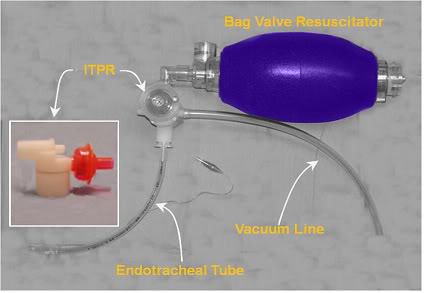
Figure 2-15: Prototype ITPR (Advanced Circulatory Systems, Inc.) in position in a typical bag-vale resuscitator – ET tube set-up.

 The ITPR has been shown to dramatically improve gas exchange, hemodynamics, blood flow, vital organ perfusion, and short-term survival rates during VF cardiac arrest in a porcine model of SCA and CPR [187] The ITPR is able to not only overcome the high intrathoracic pressures. associated with CPR (45 to 55 mmHg or ~61 to 75 cmH20 [253]) but to both create and sustain negative intrathoracic pressure (determined indirectly by measuring the ET tube pressure) continuously during prolonged periods of ITPR-CPR, even in the presence of induced hypovolemia.[189]   In hemorrhaged (hypovolemic) pigs, Yannopoulos, et al., were able to sustain CPP at >15 mm Hg (the accepted threshold for successful defibrillation in human SCA) and the isovolemic VF animals in the study maintained CPP at >25 mm Hg throughout the full 15 minutes of ITPR-CPR. In both groups, ETCO2 was consistently maintained >25 mm Hg, and the 1-hour survival was 100%, as contrasted with 10% in control animals receiving AHA standard CPR (P = 0.0001).

By comparison, after 3 minutes of conventional CPR the control animals had a mean coronary perfusion pressure of <15 mm Hg and all had developed pseudo-respiratory alkalosis indicative of the V/Q mismatch of standard CPR.[254]  Blood gases in VF animals were strikingly preserved during ITPR-CPR; paO2, which was 96±2 mm Hg at baseline, was   214±12.37 mm Hg after 10 min and 198±6.75 mm Hg after 15 min of ITPR-CPR. These findings would seem to suggest that ITPR-CPR may be reducing or eliminating the pulmonary edema that accompanies CPR and the high intrathoracic (and thus pulmonary arterial and venous pressures) generated during CPR.  ITPR is similarly effective at improving both hemodynamics and survival in a swine model of severe hypovolemic hypotension.[255]

The use of an ITPR during LAPC offers the prospect of reducing or abolishing the undesirable hemodynamic effects of FTLV with PFC. These effects, while not clinically significant in the healthy animal with spontaneous circulation undergoing LAPC (and the ability to dynamically respond to alterations in preload, CVP and SVR), may be unacceptable in the setting of cardiac arrest and CPR.

While it is clear that FTLV with PFC reduces MAP and elevates the mean CVP,[2] the extent to which these effects will reduce CO during CPR is unknown and will necessarily require further experimentation in animal models of SCA and CPR that closely approximates those experienced in humans under clinical conditions. The mechanics of CO and perfusion in CPR are radically different than those that pertain under conditions of spontaneous circulation.[256],[257],[258] In the healthy beating heart, modest increases in CVP (~5-15 mm Hg) result in increased cardiac output via the Frank-Starling mechanism (Starling’s Law of the Heart). Increased CVP increases left ventricular diastolic end pressure (LVEDP) increasing ventricular volume and stretching the ventricular myofibrils. Myofibrillary stretch results in sarcomere extension thereby increasing the affinity of troponin C for calcium, causing a greater number of cross-bridges to form within the myofibrils; this increases the contractile force of the heart increasing CO. This biochemical mechanism is not operational during cardiac arrest and CPR. Indeed, as outlined in the discussion below, little of the mechanics of perfusion under normal physiological conditions pertain in the setting of CPR.

4.5.5. Consideration of the Mechanics of Blood Flow in CPR and Implications for LAPC

During CPR in humans ventricular volume is not a primary determinant of CO, at least not in an appreciable fraction of patients undergoing CPR.[258] Despite the fact that it has been forty-eight years since the invention of closed chest CPR – the mechanics of blood flow during CPR in humans have not been definitively established – and a great deal of controversy surrounds the subject. Broadly, three mechanisms of antegrade blood flow have been proposed in (human) CPR:

  • The Direct Cardiac Compression (Cardiac Pump) Theory posits that mechanical compression of the ventricles between the sternum and vertebral column creates a pressure gradient between the ventricle and the aorta (or pulmonary artery in the case of the right ventricle); as a consequence the mitral and tricuspid valves are closed, and blood is moved antegrade out of the ventricle. The ventricle then refills during the decompression phase of CPR and the process is repeated with each compression cycle.[259]  The cardiac pump theory was most definitively challenged with the publication of case data documenting the ineffectiveness of CPR in patients with flail chest. This could be reversed only be restoring elastic recoil to the chest by binding it; indicating that it was the generation of a net negative intrathoracic pressure upon recoil of the chest during the decompression phase of CPR that was essential for blood flow.[260]
  • The Thoracic Pump Theory asserts that chest compression increases intrathoracic pressure forcing blood to flow from the thoracic to the extrathoracic circulation[3]. Retrograde flow from the right heart to the systemic veins is prevented by the venous valves; with the heart serving only as a passive conduit, and having no function as a pump.[261], [262] In the thoracic pump paradigm blood circulates because increased intrathoracic pressure is transmitted more or less equally to all of the intrathoracic vascular structures [256] and the atrioventricular valves remain open during the compression phase of CPR.[263]  However, these intravascular pressures are not equally transmitted to the extrathoracic arterial and venous beds, thus creating an extrathoracic arterial-venous pressure gradient resulting in antegrade blood flow during the period of high thoracic pressure. During the decompression phase of CPR, blood flows into the lungs as a result of the extrathoracic venous-to-intrapulmonary pressure gradient.  Angiographic [256] and echocardiographic studies in dogs documented static ventricular volumes during CPR [262], and case reports of human patients with cardiac tamponade recovering successfully after closed chest CPR both supported the thoracic pump mechanism as the explanation for antegrade flow in CPR.[264]
  • In the Lung Pump Theory, as in the thoracic pump theory, the increased intrathoracic pressure during the compression phase of CPR is similarly transmitted equally to all intrathoracic vascular structures and there is insignificant regurgitation of blood from the pulmonary artery into the right ventricle and vena cavae until the pulmonary valve closes. [265] Thus, blood under pressure within the pulmonary vasculature will flow out of the lungs via the left side of the heart.[263],[266] During the decompression phase of the cycle the intrathoracic pressure drops below that present in the extrathoracic vasculature and blood flows into the thorax via the vena cavae and aorta. The pulmonary vascular volume is replenished by blood flowing from the right side of the heart through the open tricuspid and pulmonary valves. [266], [267] Retrograde aortic flow is prevented by competent closure of the aortic valve.[268],[265]

Transthoracic echocardiographic studies reported in the early 1980s during CPR in humans supported the thoracic pump theory.[268],[265] However,more recent studies employing transesophageal echocardiography (TEE) have concluded that because the mitral valve was observed to close during the compression phase, and open during the decompression phase, and the left and right ventricular volumes decreased during the compression phase, the  mechanism of antegrade flow during CPR was consistent with the direct cardiac compression theory.[269],[263]

Perhaps the most likely explanation of these seemingly incompatible findings is that all three mechanisms are responsible for antegrade flow, but not in every patient. In other words, the mechanics of forward flow may differ from patient to patient.  In fact, in the 1981 paper in which they proposed the thoracic pump theory of antegrade blood flow in CPR, Weisfeldt and Chandra stated, “It is not essential in the human to think about these mechanisms in an exclusive fashion. Direct cardiac compression is useful when possible; when it is not potent enough to maintain cerebral perfusion, manipulation of intrathoracic pressures would likely have a favorable additive effect on carotid blood flow.”[261]

In support of this view is the study by Ma, et al., which evaluated 17 patients undergoing CPR using TEE to measure both pulmonary and trans-mitral flow.[270]  Five of the 17 patients demonstrated closure of the mitral valve during the compression phase of CPR, with associated mitral regurgitation and forward aortic flow occurring which is consistent with the cardiac pump theory.

In the remaining 12 patients, the mitral valve remained open during both the compression and decompression phases of CPR with maximal antegrade mitral flow occurring during the compression phase of CPR. Eight of these 12 patients demonstrated antegrade mitral flow during the compression phase that was accompanied by antegrade pulmonary vein flow; consistent with the classic ‘thoracic pump’ mechanism. The last 4 patients in this study evidenced retrograde pulmonary vein flow concurrent with antegrade mitral flow during the compression phase; which is most consistent with lung pump theory as the primary cause of antegrade flow, at least in these patients.

5.0 Review of the Literature on LAPC

From the foregoing, it should be easy to understand the difficulty of establishing by experiment, let alone extrapolating on any theoretical basis, what the hemodynamic impact of FTLV or LAPC will be under clinical conditions in humans undergoing CPR. Similarly, the effect of LAPC on regional myocardial blood flow, myocardial irritability, coronary electrophysiology, and susceptibility to defibrillation will require further laboratory and, likely, experimental clinical investigation as well. Since publication of the 21CM/CCR LAPC research in 2001 there have been 4 subsequent studies to evaluate various aspects of the utility and safety of LAPC for application in CPR, or as a therapy in MI.

The first of these studies was published by Hong, et al., in 2002 and announced, Our study is the first to demonstrate an induction of hypothermia by adopting PLV (no need of an extracorporeal circuit) and 0°C PFC.” [271] failing to cite the previously published work of either Darwin, et al., or Harris, et al. [272] These investigators attempted to validate the utility of LAPC using FTLV to achieve rapid reduction in core temperature and also studied the physiological impact of the procedure. The cooling rate achieved appears to have been ~0.11°C/min and this slow rate was almost certainly an artifact of the small volume of PFC used for each intrapulmonary exchange (~20 ml) and the small number of exchange (10 – 12 exchanges over 38 minutes). In fact, the cooling rate was faster in the animals in this study that were cooled externally by repeated application of ice water slush (0.17°C/min).

The most interesting result of this investigation were that MAP, mean PA pressure, HR, and CO, CBC and lactate of the LAPC treated animals were not significantly different than in the surface cooled animals. One variable that was altered in the LAPC group was an increase in pulmonary vascular resistance which appeared immediately after liquid loading and did not begin to return to baseline until halfway through the LAPC period. It is also remarkable that 1 animal in both the LAPC and the surface cooled groups (n = 7 for both groups) developed pulmonary hypertension and lethal pulmonary edema. The authors speculate that the observed pulmonary hypertension could have been a result of pulmonary venous constriction and/or increased pulmonary microvascular blood sludging due to the profound local cooling of lung vasculature resulting from instillation of 0°C PFC. They also raise the possibility that the observed elevation in PAP may have resulted from the mechanical effects of the PFC load; but offer no explanation for this.

Otherwise, hemodynamics were unaffected by LAPC. The paCO2 became progressively elevated during the hypothermic interval in the LAPC group, perhaps due to inadequate PEEP or inappropriate parameters of mechanical ventilation in the face of evaporating PFC (which was not replenished during the course of the experiments).

The second study to be published was by Ko, et al., in 2002.[273] The purpose of this study was to evaluate the effect of PLV on pulmonary blood flow under low blood flow conditions designed to simulate those encountered during CPR in order to further validate the use of LAPC as an adjunct to cardiopulmonary cerebral resuscitation. Isolated perfused rat lungs were subjected to 20 min of PLV with room temperature Perflubron™ and segmental (i.e. pre-capillary, capillary, and post-capillary) hemodynamics were studied at a perfusate flow rate of 6 ml/min (~5% normal cardiac output [274]). Lungs received either gas ventilation or 5 or 10 ml/kg PLV. Segmental pressures and vascular resistances were determined, as was transcapillary fluid flux. PLV at both the 5 and 10 ml/kg Perflubron™ dose produced no detectable changes in pulmonary blood flow or in transcapillary fluid flux and the investigators concluded that, “These data support further investigation of this technique as an adjunct to cardiopulmonary resuscitation.”
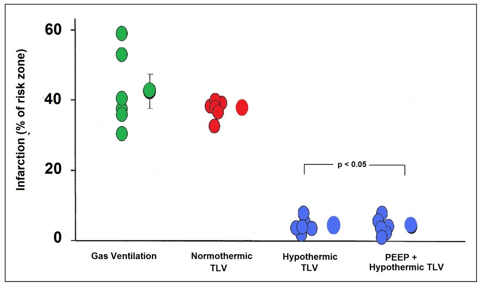
 

Figure 2-16: Dramatic reduction in myocardial infarction size in rabbits rapidly cooled to ~34 by the use of TLV LAPC: infarct volume was 4.0 ± 5% in the LAPC groups vs. 37.7 ± 1.3% in the normothermic gas ventilated group (p = 0.001). Redrawn from Tissier, et al.[275]

Unfortunately, this study did not explore the effect of instilling cold PFC into the lungs, nor did it employ FTLV. Because it was an ex vivo study it was not possible to determine the effects of PFC loading, cold or isothermic, on the sympathetic response, coronary blood flows or other physiological parameters of concern in LAPC.

In 2007 a study by Tissier, et al., [275] used a rabbit model of evolving MI to evaluate the efficacy of TLV LAPC administered during the ischemic interval in achieving rapid core cooling to reduce infarct size and provide myocardial protection [276],[277],[278] even after substantial delay  [279] and when induced during reperfusion. [280],[281] It is well established that intra- and post-ischemic myocardial hypothermia dramatically reduces infarct size in animal models of MI and this observation has recently been extended to humans in the clinic.[282]

These investigators note that in the US the time to coronary revascularization following infarction is 120 min in 41.5% of patients admitted during off-hours and that 27.7% of patients presenting during normal business hours still averaged delays to revascularization in excess of 120 min.[283]  Rapid induction of hypothermia in these patients could be effective in providing substantial myocardial salvage during the delay between presentation and revascularization. This study demonstrated a remarkable reduction in infarct size in the LAPC treated group; 4.0 ± 5% vs. 37.7 ± 1.3% in the normothermic, gas ventilated animals (p = 0.001). The cooling rate achieved with TLV was 1.32°C/min; 6.6°C in 5 minutes. The effectiveness of TLV LAPC is not consistent with results obtained in larger animals in this author’s experience. It may be that the relatively short distances between the large and small airways and the very small diameter of even the largest airways in the rabbit as compared with the dog or human, may have improved the efficiency of heat exchange.

The most recent study by Staffey, et al., [284] is more on-point and provides considerable reassurance that cold PFC loading of the lungs during CPR and subsequent resuscitation is not only not deleterious, but in fact is markedly beneficial. In this study swine were subjected to 11 min of VF and treated either with a static intrapulmonary infusion of PFC chilled to -12 C to ~75% of total lung volume (40 ml/kg), static infusion of isothermic PFC (33oC) under the same conditions, TLV with -15oC PFC at 6-cycles per minute during 10.5 minutes of the arrest interval, and a control group that was arrested with no intervention during the ischemic interval. The animal were then given AHA standard CPR with defibrillation and advanced cardiac life support (ACLS). The specific objectives of this study were to determine what the effects LAPC; tidal and static, administered during the period of cardiac arrest would be on hemodynamics, CPP, gas exchange, defibrillation, and hemodynamic stability during a 1 hour period of evaluation post ROSC. The endpoint was ROSC for 1 hour without ionotropic support.

The global objective of the study was to determine if LAPC could be used to rapidly induce cardiopulmonary hypothermia during the arrest period as opposed to cerebral or systemic hypothermia. Work by Rhee, et al., and Boddicker et al., also using swine models, had previously demonstrated that systemic hypothermia induced before resuscitation from cardiac arrest resulted in more rapid and consistent defibrillation from VF as well as earlier and more stable ROSC. Since the purpose of this study was to cool only the thoracic viscera to determine the effect of local cardiopulmonary hypothermia on resuscitation; ‘targeted cardiopulmonary intra-arrest moderate hypothermia (28-32oC),‘ LAPC was not continued beyond the period of cardiac arrest.

Both static and TLV LAPC succeeded in reducing the pulmonary artery temperature to the desired temperature of 34oC by 6 and 10 min post arrest, respectively. Eighty two percent of the animals in both LAPC groups were resuscitated successfully and survived the 1 hour evaluation period without pharmacological support, as contrasted with only 27% of the controls animals (p = 0.03).  Interestingly, 73% of the isothermic  TLV swine achieved and maintained ROSC as opposed to 27% of the swine in the control group (p = 0.09), suggesting that the presence of PFC in the lungs during either the arrest or resuscitation interval may improve the odds of successful defibrillation.
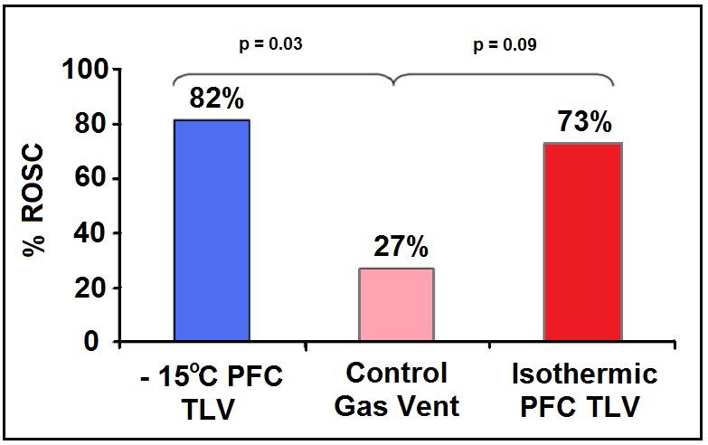
Figure 2-17: Results of the study by Staffey, et al., to evaluate the effect of cold and isothermic PFC infusion and TLV on resuscitability of swine after 11 min of electrically induced VF cardiac arrest. PFC loading, and in particular cold PFC loading or TLV improved 1 hour stable hemodynamic survival by 73% versus 27% in controls. Infusion of isothermic PFC to near vital capacity also showed a trend towards increased survival. Redrawn from Staffey, et al.,[284].

Chamberlain et al., have recently proposed that dilation of the right ventricle (RV) that occurs after the first ~5 min of cardiac arrest results in compression of the left ventricle (LV) within the confines of the pericardium. The effect of this would be to reduce myocyte stretch and reducing the contractile strength of the left ventricle in response to defibrillation (see discussion of the Frank-Starling curve above). They posit that an arrested heart that has a reduced contractile state, and in which left ventricular volume is reduced by the right ventricle over-distended with venous blood will not be able to initiate effective contraction even if coordinated electrical activity is restored. Distension of the RV occurs post-arrest due to centrally mediated sympathetic contraction of the vasculature, as well as movement of blood into the venous circulation as arterial and venous pressure equilibrate; this is a commonplace finding at both necropsy and autopsy in normovolemic subjects.

Unloading the RV prior to defibrillation results in an immediate improvement in successful ROSC independent of any known metabolic that might accrue from ~10 to ~15 sec of coronary perfusion that might also result. Chamberlain, et al., believes that it is the decompression of the left ventricle and restoration of a more physiologic morphology that facilitates or even enables defibrillation under these conditions. Instillation of a large volume of dense PFC may antagonize distension of the RV and prevent LV volume loss and compression of the LV to below the threshold required to established effective contractile activity in response to defibrillation. PFC to vital capacity (VC) should effectively preclude this post-arrest pooling of blood in the thoracic caval and pulmonary vessels and may act to preserve left ventricular morphology during prolonged cardiac arrest.

Especially encouraging findings from the Staffey, et al., study were the absence of any noticeable adverse hemodynamic impact from either isothermic PFC loading or from cold PFC loading or cold TLV. MAP, CVP, CPP, pH and blood gases were not statistically different between the four groups of animals in the study.

These four studies of LAPC aimed at answering questions bearing on the feasibility of LAPC in MI, SCA and CPR are very encouraging. They indicate that LAPC is being seriously considered as a potential therapeutic application in humans. That this research is being undertaken in independent academic and medical research both in the US and elsewhere is especially heartening and would seem to indicate that the enormous therapeutic potential of LAPC-induced hypothermia has been successfully communicated.

6. Conclusions

LAPC is capable of inducing hypothermia in a fraction of the time that it takes to prepare a patient for cooling via CPB. In addition, automated LAPC need not have the spatial and technical restrictions of the hospital setting. Although relatively simple methods of continuous arterio-venous shunt heat-exchange that do not require a blood pump or carry the most of the risks attendant to CPB have been described which might be potentially applicable in the field [285], these techniques also have the drawback of requiring skilled personnel for cannulation of a major artery and vein. Intracaval heat exchange catheters can reduce core temperature,  but only at rates of ~1.46 ± 0.42°C/h [286]; far too slowly to achieve the maximum benefit from post-reperfusion hypothermia
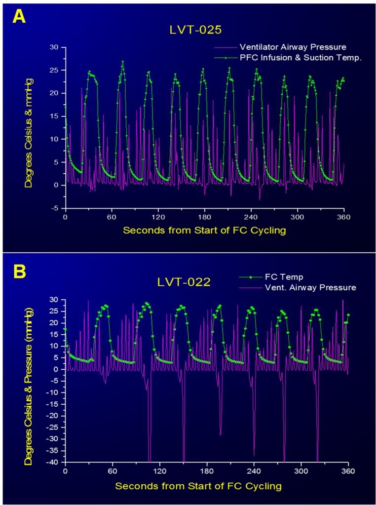
Figure 2-18: Large negative excursions in airway pressure occurred during suctioning of PFC at the end of most FTLVs when this operation was carried out manually (A). This occurred because when the PFC suction catheter was no longer filled with PFC, evacuation of gas from the airways occurred very rapidly owing to the much lower viscosity of gas compared to PFC. It was impossible for the operator to anticipate when the last of the bulk liquid would be removed, or to react rapidly enough when this occurred. Computerized sensing and control eliminated this potential source of baro-injury (B).

By contrast, LAPC may be a candidate for a much wider range of emergency field-uses in civilian and military settings since the primary technical skill required to initiate LAPC in the field is endotracheal intubation; a skill possessed by paramedical personnel throughout Canada, the U.S. and Europe. LAPC has also potential as a very rapid treatment for heatstroke and malignant hyperthermia. While  not the subject of this report, LAPHE clearly also holds promise for core rewarming in severe hypothermia, although an absolute maximal PFC temperature of 42°C would in theory limit the re-warming rate to about one-third of that possible in cooling.

Computer control of both gas and FTLVs is effective at eliminating excessive positive and negative airway pressures during LAPC (Figure 2-16) and computer control can easily be extended to encompass cooling rate, depth, and duration and can also be extended to control gas ventilation, as necessary.

Whether used inside or outside hospital, successfully implemented LAPC might more generally serve as a neuroprotective bridge [287] in order to gain time for more technically sophisticated supportive or definitive treatment (e.g. neurovascular thrombolysis or interventional thrombectomy, emergency CPB, spinal cord decompression or definitive management of hemorrhagic shock in trauma or surgery).

Although some modalities of liquid ventilation have been clinically evaluated [288], the safety parameters of rapid and cold liquid delivery to the lungs remain to be determined. As noted in this study, LAPC can cause pulmonary injury. The mechanism of such damage suggest by the location of lesions indicates that both barotrauma (dependent lung) and volu-trauma (nondependent lung) are the primary, if not the sole factors. It has been observed that LAPC causes little permanent lung injury in long term survival animals. Similar pathology seen in lungs exposed to either isothermic or ~4ºC LAPC in the present study (data not shown) suggest that thermal/chilling-injury per se is not the major insult. Although more subtle biochemical and immune problems secondary to hypothermia itself are suggested by reports from some longer duration studies of MTH (pneumonia and sepsis), it is not clear that the short duration of treatment necessary to achieve the benefit of post-resuscitative MTH will pose such problems. It is hypothesized that the pulmonary injury observed in LAPC may be reduced with better control of LAPC pressure and volume limits, and by use of PFC liquids having more physiologically suitable properties.

A significant, unresolved concern is the potential negative impact of LAPC on myocardial perfusion and the danger of overcooling the heart during the low flow conditions of CPR with possible adverse effects on achieving ROSC and maintaining a stable rhythm following defibrillation. As already noted, these questions can only be resolved with further study.

Acknowledgements

 The authors thank Saul Kent and William Faloon for support, and Casey Brechtel for helpful discussions. Several of the authors have applied for LAPC device patents. This trial was funded by a grant from the Life Extension Foundation (Hollywood, FL).

Appendix A

 (The abbreviations contained in this appendix are also reproduced in the table of abbreviations and acronyms at the beginning of this document.)

 A.1. Abbreviations and notation

 In the text, volumes (V) are given in ml/kg, and flows (dV/dt =V’= ˙V ) in ml/kg per min. Since all V and ˙V are expressed in mass-specific (per kg animal) terms, derived quantities DQ and Cm are automatically mass-specific. Cm and Cvf are given in calories / (g or ml) per °K for easy comparison with water.

PFC = perfluorochemical; hydrogen-free organic molecule in which most of the peripheral atoms are fluorine.

TLV = tidal liquid ventilation is a modality in which liquid completely fills the lungs and ventilator.

PLV = partial liquid ventilation is a modality in which all gas exchange is via gas ventilation, with ~1/2 FRC of PFC liquid present in the lungs to recruit dependent lung in ALI or ARDS.

LAPC = liquid assisted pulmonary cooling is a heat-exchange modality in which ventilation occurs via both gas and liquid ventilation proceeding concurrently.

Ttym = tympanic temperature

Tart = arterial temperature

Tven = central venous temperature

Trec = rectal temperature

DTenet DTtymp = resulting from LAPC, after equilibration at t=40 min

TFTLV  = FTLV cycle infusion time

ts = FTLV cycle suction time

tc = FTLV cycle period (=tinftc +ts)

VFTLV = single-cycle PFC FTLV infusion volume=tinf V_ inf

 VS = single-cycle PFC FTLV suction-return volume

VD  = ventilatory dead space (any type)

VDCA = expected gas ventilation VD=sum of circuit (mechanical) VD plus anatomic VD

VDTherm = thermal or heat-exchange VD (ml/kg, in reference to liquid PFC infusion)

˙V inf = PFC infusion rate (set to _50 ml/kg per min in Trials I and II)

˙VFTLV = effective PFC FTLV rate=LAPC liquid FTLV minute-ventilation (ml/kg per min) = VFTLV/ tc

˙Vg = gas minute-ventilation (ml/kg per min) m animal mass Ch heat capacity

 CT = total heat capacity of the animal (= mCm)

m = mean mass-specific heat capacity of the animal ( = DQT / DTe)

Cvf = volume-specific heat capacity of FC-75 (mean of 0 and 25°C values) = 0.45 cal/ml per °K

DQT = total heat removed during LAPC (kJ/kg animal) = S DQc

DQc = heat removed during one FTLV cycle?????

Ef = mean cycle heat transfer efficiency=mean of [DQc / (theoretic DQc (max)] for all cycles in a single experiment

n = number of FTLV cycles in LAPC experiment

S = sum entire quantity following, for all cycles i = 1 through n

 Tinf   = PFC infusion temperature

TS = PFC suction removal temperature (time-averaged PFC suction flow temperature)

TSM= PFC mixed suction return-volume temperature (temperature of mixed VS)

 A.2. Thermal kinetics

 During LAPC cooling and equilibration, the blood and tympanic temperature changes in the animals were modelled by a simple five compartment model (Figure 2-8). During the initial ~100 s of LAPC (value used as empiric time mark), full development of heat-exchange behavior is established between the lungs, blood volume, and the thermal core of the animal, as suggested by the characteristic half-times for equilibration of these systems (see below).

Modelling of cooling during LAPC:

After the initial ~100 s of cooling, the data for tympanic DT(t) = DTtym during LAPC in Trial I and II were modelled by a single time-constant exponential decline.

Mean DTtym data for each trial from times t=100 to 1080 s were fit using (Eq. (1)).

DT (t) = T100 + DTk [1− exp (−t / to) ],

DT(t), total Ttym change from baseline Ttym at start of LAPC; t, =time in seconds after empiric time mark, 100 s after start of LAPC; T100, observed DT at empiric time mark, 100 s after start of LAPC; DTk, observed temperature-interval constant, specific to each LAPC method; to, observed natural-base time-constant, in sec (to = halftime / ln 2).

Best-fit values for Trial I data were: T100 = −0.52 ± 0.02°C; DTk = −11.2 ± 0.02°C; and to = 1064 ~3 s. Trial II values were T100 = −0.24 ± 0.02°C; DTk = −8.14 ± 0.02°C; and to = 1107 ± 5 s. The relatively long time-constant associated with this thermal phase, which was similar in the two trials, presumably reflects the long time-constant (~700 s, see below) associated with heat transfer from the thermal core of the animal to the thermal periphery; thus the exponential phase represents full development of heat exchange between the LAPC cooling device and the entire animal. The final linear segments of cooling occurring after this phase, measured at −0.29°C/min (Trial I) and −0.21°C/min(Trial II), represent the final relatively simple state which exists after heat exchange equilibrium between cooling device and animal has been fully established.

Cooling in blood and tympanic sites during LAPC, and thermal evolution in these sites during equilibration phase after LAPC was discontinued, was in accordance with a five-compartment thermal model (Figure 2-8). In this model, the tissues of the animal are divided into three thermal compartments, corresponding loosely with the vascular system, the thermal core, and the thermal periphery.

Modelling of equilibration after LAPC:

Perfusion-driven convection is the major heat transfer mechanism in very rapid systemic cooling processes. This fact allowed Tart and Tven changes to be used to quantify some features of heat transfer between body thermal compartments during the equilibration period after LAPC. The mean Tart curve in Trial I increased nearly linearly (R2 = 0.9976) for 12 s after the end of LAPC, rising at a rate of 7.9°C/min. After this initial 12 s, Tart departed from linearity (Figure 2-6 inset), and was modelled by the sum of three exponential terms with respective time constants (t0) of 12 ± 0.4, 102 ± 2 and 701 ± 8 s. These t0 times differed to a large enough extent that their respective influences could be considered to be controlling over discrete time periods of about twice their value. Thus, the four equilibration phases seen after the end of LAPC lasted approximately 12, 24, 200, and 1400 s (23 min), respectively and represented 34, 14, 25, and 27% of the 5.1°C rise in Tart during equilibration after LAPC.

These data may be interpreted as follows: during each phase of the equilibration process, one or more thermal compartments in the animal equilibrated with the next-most closely-connected compartment (Figure 2-8). Afterwards, the newly captured compartment(s), as part of a larger unit bound together by blood mediated convection, equilibrated with the next-most closely connected compartment, and so on. The 12 s linear first equilibration phase (Figure 2-7, inset) most likely represents development of heat transfer from the lungs to local pulmonary blood flow. This phase was not associated with blood recirculation since it was seen as a rise in Tart but not Tven. The second equilibration phase (duration ~24 s) was characterized by an increase in dTven / dt to the value of dTart  /dt, indicating that the lungs, blood-volume, and certain other well-perfused viscera, such as the kidneys, were now evolving into a single thermal system. Since the observed to for this phase was 12 s, less than the animal’s mean circulation time (= cardiac output / blood volume ~30 s), this process appeared to be driven by blood circulation via the most rapid paths (e.g. renal circulation). Such short paths for circulatory heat transfer were evident in the relatively small lag times (10.4 ± 6.9 s) noted between Tart and Tven changes in these animals.

During the first two equilibration processes, the pulmonary circulation added thermal potential to the blood-volume more rapidly than it could be removed by the systemic circulation. By the end of the second equilibration phase, however, lung-to-blood heat transfer no longer dominated, and the gap between Tart and Tven was set by the magnitude of heat transfer from the circulating blood volume to the tissues that comprise the ‘thermal core’ of the animals. In this third equilibration phase (duration ~200 s), the viscera and blood-volume, as a unit, equilibrated with the remainder of the ‘well-perfused’ tissues of the body (thermal core, comprising about 70% of the animal’s heat capacity). Heat capacities for thermal compartments are calculated below. The to for this process is seen most directly in the ~2 min. delay between maximal DTven and maximal DTtym (Figure 2-7).

Finally, heat transfer within well-perfused tissues fell to a new minimum, and the Tart to Tven gap decreased to a value set by the fourth equilibration phase (duration ~23 min) during which the well-perfused tissues equilibrated, as a unit, with a succession of the more poorly-perfused compartments, e.g. gut contents, fat, and other tissues comprising the thermal ‘periphery’ [12]. These processes could be consolidated into a single exponential term. Due to the long time-scale, heat transfer during phase four was probably partly conductive. Estimates of basal metabolism in the anesthetized, non-shivering dog (@90 J/kg per minute) indicate that as much as 0.6°C of warming per 20 min in this model may be due to metabolism.

A.3. Thermal accounting

 Heat transfer efficiency:

Although machine-LAPC allowed 2.3 times the FTLV frequency of the manual method, and resulted in a larger ˙VFTLV by a factor of 1.2, the cooling magnitudes and rates for machine-LAPC significantly (P < 0.001) fell short of those obtained with manual-LAPC (Fig. 3). The strategy of increasing FTLV frequency (1/tc) and decreasing ˙VFTLV, in order to arrive at approximately the same FTLV rate (˙VFTLV), therefore, significantly decreased the fraction of thermal potential which was transferred from each FTLV (= heat transfer efficiency, Ef). Unexpectedly, when the Ef for each of the 8 LAPC cooled animals of Trials I and II (Table 2) was calculated using (Eq. (2)), the value did not differ (P = 0.46) between trials; nor did total heat removed per kg (DQT), as calculated using (Eq. (3)), differ (P=0.14) between Trials. Both of these quantitative methods were therefore inaccurate for some dogs. Calculation of whole-animal mass-specific heat capacities Cm (= DQT / DTe) suggested that the Trial II values of ~DQT and Ef principally were inaccurate, since the mean Cm value of 0.70 ± 0.1 cal/g per °K for Trial I was consistent with the Cm reported in the literature for mice and humans [289], whereas Cm values calculated for Trial II using (Eq. (2)) were unrealistic, being greater than the Cm of water.

E(method 1) = 1/nS (TSTinf) / (TvenTinf),     (2)

DQT (method 1) = ˙VFTLV Cvf STSTinf.                     (3)

To independently check the accuracy of Trial I values, we computed DQT and Cm for three dogs from an earlier study (Tables 1 and 2: dogs A, B, and C) that had been given ~4ºC PFC with FTLV times sufficiently long to allow the volumes and mixed-temperatures of suction-return liquid to be measured for each FTLV.

This allowed computation of DQT and Ef by a more detailed method (Method 2, Eqs. (4) and (5)), which used the extra thermal data (not available for Trials I and II) to more directly estimate FTLV heat transfer.

DQT (method 2)

= Cvf S VFTLV (TvenTinf) − VS (TvenTSM),                              (4)

When this was done, the mean Cm for dogs A, B, and C was found to be 0.68 ± 0.06 cal/g per °K, consistent with the Cm in Trial I (P=0.80). Method 1 (Eqs. (2) and (3)) required the assumptions that PFC suction volume equalled infusion volume, that suction flows remained constant, and that thermal hysteresis was negligible. These assumptions apparently held true at the larger ˙VFTLV and tc values of Trial I, but not for the smaller values of Trial II.

With this information, a new Ef for Trial II was estimated using method 3 (Eq. (6)), which employed an estimate for the heat required for the observed DTe, vs. the total PFC thermal-deficit theoretically available.

This estimate required a presumed value of Cm. However, if the mean Trial II Cm was assumed to be the same as that of Trial I, then the true Trial II Ef could be calculated (Eq. (6)) to be 0.40 ± 0.06.

This value agreed with the rough estimation that since Trial II achieved only 73% of the DTe of Trial I, despite using 1.19 times more total PFC (Table 1), the

Ef in Trial II was expected to be about 73%/1.19 = 61% that of Trial I.


Thermal compartment size:

Heat removal for each cycle (DQc) in Trial I was calculated from the individual terms of Eq. (3), and individual-cycle mean cooling power calculated as DQc/tc. The latter parameter was useful since thermal compartments in the dog are relatively isolated at short time scales (Figure 2-8), and thus the ratio of cooling-power to cooling rate (Eq. (7)) at a given probe site was expected to give the heat capacity (Ch) of the system of thermal compartments that were in equilibrium with each other, and with the site, at the time of the measurement:

Ch (Comp N), total heat capacity of thermal compartments N = 2 + 3, or N = 2 + 3 + 4.

Both tc and dT/dt values were chosen at a time t, of interest when compartment system N has not yet equilibrated with slower half-time compartment(s). Thus, in

Trial I, near the end of FTLV cycle c1 (t = 30 s), the FTLV thermal-deficit had equilibrated within thermal Compartments 2 + 3 (PFC/viscera/blood-volume), but had not yet significantly reached Compartments 4 or 5.

If the cooling rate of Tven at t = 30 s (−1.9 ± 0.8°C/min) was then taken as the cooling rate of the system of PFC/viscera/blood-volume, the Ch for this compartment system could be estimated from (Eq. (7)) as 20 ± 9% of CT, the total heat capacity of the animal (CT = m Cm). Subtracting the Ch contribution of lung PFC (=m ˙VFTLV Cvf) allowed estimation of the remaining tissue Ch for Compartment 3 as ~19 ± 9% of CT.

Similarly, the Ch of Compartments 2, 3, and 4 together, was estimated at t = ~140 s (cycle 4) as 71 ± 17% of CT, corresponding to the classical whole-body ‘thermal core.’ The Compartment 5 Ch was then calculated to be the remainder (100−71%) = 29 ± 17% of CT, corresponding to the classical ‘thermal periphery.’


[1] In fact, a bag-valve ventilator can be effectively used to carry out LAPC.

[2] FTLV causes the CVP to oscillate and as a consequence to take on a pulsatile character so discussion of the CVP under these conditions must be in terms of the mean CVP..

[3] Perhaps the most lucid explanation of the hemodynamics of the lung pump theory was that given by its originator’s, Wiesfeldt and Chandra in their paper proposing the idea. That explanation is reproduced as Appendix B.

Posted in Cryonics Technology (General), Ischemia-Reperfusion Injury, Medicine, Uncategorized | Tagged , , , , , , , , , , , , , , , , , | Leave a comment

Liquid Assisted Pulmonary Cooling in Cardiopulmonary Cerebral Resuscitation – Part 1

Table of Contents

Introduction………………………………………………………………………………………………………. 6

Table of Abbreviations, Symbols and Acronyms………………………………………….. 9

Section I: Introducing Liquid Assisted Pulmonary Cooling………………………… 14

Liquid Assisted Pulmonary Cooling in Cardiopulmonary Cerebral Resuscitation, Introduction

………………………………………………………………………………………………………………….. 15

Hypothermia as an Active Therapeutic Agent

………………………………………………………………………………………………………………….. 17

The Benefits and Limits of ‘Delayed’ MTH: Real World Experience

………………………………………………………………………………………………………………….. 26

The Problem of Heat Exchange

………………………………………………………………………………………………………………….. 29

The Pathophysiology and Biophysical Limitations of External Cooling

………………………………………………………………………………………………………………….. 33

Consideration of Invasive Core Cooling Methods

………………………………………………………………………………………………………………….. 35

Exsanguinating Trauma Resulting Cardiac Arrest

………………………………………………………………………………………………………………….. 37

The Lungs as Heat Exchangers

………………………………………………………………………………………………………………….. 38

A Brief Précis of the History and Development of Liquid assisted Pulmonary Cooling (LAPC) for Induction of Hypothermia during CPR

………………………………………………………………………………………………………………….. 41

What LAPC Can Potentially Deliver

………………………………………………………………………………………………………………….. 46


Section 2: Experimental Studies to Determine the Effectiveness of LAPC under Laboratory Conditions ……………………………………………………………………………………………………… 51


1. Introduction

………………………………………………………………………………………………………………….. 52

2. Materials and methods

………………………………………………………………………………………………………………….. 53

2.3. Trial I (manually-controlled LAPC)

………………………………………………………………………………………………………………….. 59

2.4. Trial II (machine-controlled LAPC)

………………………………………………………………………………………………………………….. 59

2.5. Animals A, B and C

………………………………………………………………………………………………………………….. 59

2.6. Data collection and correction, statistical methods, graphical display and presentation

………………………………………………………………………………………………………………….. 59

3. Results

………………………………………………………………………………………………………………….. 60

3.1. Thermal results of LAPC

………………………………………………………………………………………………………………….. 62

3.1.1. Cooling time delay

………………………………………………………………………………………………………………….. 63

3.1.2. Cooling rate

………………………………………………………………………………………………………………….. 63

3.1.3. Mean cooling power

………………………………………………………………………………………………………………….. 63

3.2. Gas exchange

………………………………………………………………………………………………………………….. 64

3.3. Clinical observations and gross pathology

………………………………………………………………………………………………………………….. 66

3.4 Impact on Hemodynamics

………………………………………………………………………………………………………………….. 67

4. Discussion

………………………………………………………………………………………………………………….. 69

4.1. Apparent effect of temperature on gas exchange

………………………………………………………………………………………………………………….. 69

4.2. Thermal transfer efficiency and kinetics

………………………………………………………………………………………………………………….. 70

4.3. Question of diffusion dead space in LAPC

………………………………………………………………………………………………………………….. 71

4.3.2. Possible synergy of combined gas and liquid ventilation in assisting mass (CO2) and heat transfer

………………………………………………………………………………………………………………….. 73

4.4. Potential development of clinical LAPC

………………………………………………………………………………………………………………….. 73

4.5. Challenges Ahead

………………………………………………………………………………………………………………….. 74

Acknowledgements…………………………………………………………………………………… 81


Appendix A……………………………………………………………………………………………….. 81


Section 3: Perflurochemicals………………………………………………………………………… 96


The Perflurochemicals

………………………………………………………………………………………………………………….. 97

Physical Chemistry and Synthesis

………………………………………………………………………………………………………………….. 97

Physical Properties

………………………………………………………………………………………………………………….. 98

Commercially Available PFCs

………………………………………………………………………………………………………………….. 99

Toxicology

………………………………………………………………………………………………………………… 102

Environmental Impact and Future Availability

………………………………………………………………………………………………………………… 104


Section 4: History of Liquid Assisted Ventilation and Implications for LAPC

………………………………………………………………………………………………………………………109


History of liquid Ventilation

………………………………………………………………………………………………………………… 110

Partial Liquid Ventilation (PLV)

………………………………………………………………………………………………………………… 114

Unanticipated Effect of Lung Protective Ventilation Strategies

………………………………………………………………………………………………………………… 119

Defective Translational Research Models

………………………………………………………………………………………………………………… 119

Failure to establish a Dose-Response Curve

………………………………………………………………………………………………………………… 120

Gas Trapping and Selection of Appropriate PFCs

………………………………………………………………………………………………………………… 120

The PFC-Air Interface and Shear Effects in Small Airways

………………………………………………………………………………………………………………… 121

PLV and the Law of Laplace

………………………………………………………………………………………………………………… 122

The Best as the Enemy of the Good

………………………………………………………………………………………………………………… 127

Possible Implications for SCA and LAPC

………………………………………………………………………………………………………………… 127

__________________________________________________________

This work is dedicated to David W. Crippen, MD, FCCM, who, literally, made it possible. The computers this work was written on, the myriad books and journals consulted, and the liaison with many of the professionals required for such work were all directly facilitated by Dave Crippen. But, these are the least important elements he contributed. By far the most critical ingredients were and are his faith and friendship. For these he has my unending thanks.

___________________________________________________________

 
Intellectual Property Rights: The intellectual property rights to the technology of liquid assisted pulmonary cooling (heat exchange) (LAPC or LAPHE) using combined gas and fractional tidal liquid ventilation are controlled by Critical Care Research, Inc., under US Patent 6,694,977, “Mixed-mode liquid ventilation gas and heat exchange.” Inquiries regarding access to this technology should be directed to Critical Care Research, Inc., 10743 Civic Center Drive, Rancho Cucamonga, CA 91730-3806. Telephone: (909)987-3883.


Liquid Assisted Pulmonary Cooling in Cardiopulmonary Cerebral Resuscitation

By Michael G. Darwin

Introduction

Liquid assisted pulmonary cooling (LAPC), or liquid assisted pulmonary heat exchange (LAPHE) will not likely be applied in the West any time in the foreseeable future for many reasons; not a few of them having to do with regulation and consumer perception of what constitutes acceptable risk versus benefit.

LAPC relies completely upon the unique properties of the perflurochemicals (PFCs). PFCs are other-worldly molecules; neither soluble in water or lipids, twice as dense as water, but much less viscous; and chemically so inert as to make gold seem a highly reactive metal by comparison. It is impossible to work with the PFCs suitable for introduction into the lungs of mammals without immediately appreciating how truly amazing they are. PFC chemistry exists in a world of its own outside of conventional chemistry, and in fact, conventional chemists refer to PFC chemists as ‘unnatural’ chemists; with the all the double entendre that moniker implies. PFCs do not occur in nature, and if you want to find a marker for intelligent, industrial life elsewhere in the universe, one approach might be to look for the spectra of PFCs in the atmosphere of the candidate planet; they occur only as a result of deliberate, intelligent industrial activity. Thus, they are what I call, ‘the thinking species’ molecule.’

There is an ancient Chinese curse: “May you live in interesting times and come to the attention of important people.” The medical-molecular equivalent is: “May you be synthesized in interesting times and come to the attention of important medical (and environmental) bureaucrats.” This perfectly describes the fate of the medically useful liquid PFCs. They are dramatic molecules and they are at once both therapeutically paradigm changing and physiologically outrageous. There is a strong emotional reaction – one part wonder and one part shock – at seeing a patient’s lungs filled up with liquid, any liquid. Bulk liquid does not belong in mammalian airways, and in every clinical instance where it occurs, or is introduced into the lungs – in pulmonary edema, drowning and inhalation of organic liquids – it is an unmitigated disaster. The ‘breathable’ PFCs thus attracted a lot of attention.

So, PFCs had this strike against them, and they came about as putative therapeutic agents in ‘interesting’ times in the most Chinese sense of the word. The explosion of technological innovation that has occurred since the Industrial Revolution has brought not only powerful advantages, but also often damaging and not infrequently frightening downsides. We live in an age when people, at least in the West, have become acutely sensitized to the dark side of technology and increasingly unwilling to accept risk unless it is precisely quantified, small, and will almost certainly not affect them.

The PFCs present some unfortunate challenges in this regard. First, while they are among the most chemically inert substances known to man, they are biologically active in fentogram quantities. Most of the medically useful PFCs abolish white blood cell chemotaxis in picogram to fentogram concentrations.  While their very inertness and lack of chemical reactivity make them indispensable as a liquid ventilating medium, these same properties mean that, should they escape the lungs due to pneumothorax (and enter the mediastinum or other closed body viscuses or capsules) they will likely remain there for the rest of the patient’s life. This may seem ample reason to eschew these molecules as therapeutic agents. However, I believe these facts must be taken in the context of how we use and abuse similar (and in some cases nearly identical) molecules both in medicine and in our daily lives.

Introducing liquid into patients’ lungs will never be a trivial matter, or something undertaken lightly. Such therapies and their enabling molecules will not be broadly used (or abused) in medicine. Indeed, their use is necessarily confined to the emergency medical and critical care setting in intubated and mechanically ventilated patients. Foreseeable off-label medical use of neat PFCs is virtually nil. And yet, these molecules will ‘seem unlikely to be approved for medical use in the West in foreseeable future.

All of the existing molecules are either unsuitable for use in the induction of hypothermia (such as Perflubron™ which freezes at +6 deg C) or are chemically heterogeneous, as I explain in this manuscript. It would thus be necessary to synthesize and purify new classes of molecules and then vet them individually for safety and efficacy. The cost of doing this is more than any Western drug company could bear when weighed against even the most optimistic projections of financial return on the investment. There is very little place for a drug that a patient will use once during a lifetime and that, if it works, will leave him more than able to sue all involved in its application to him should he develop some adverse effects, even if these occur only after decades of additional, healthy and productive life which would not have been possible otherwise.

I hasten to add that I do not think the PFCs will be free of long term side effects. Most of the chronic survival dogs in our studies that underwent PFC ventilation and/or cooling have since grown old and died. We saw nothing unusual; they died of heart failure, cancer and the usual things old dogs die of, and seemingly at the usual rates, and at the usual ages. But, our sample size was ‘microscopic;’ and I think it likely that large populations of people treated with PFCs may have a statistically significant increase in conditions related to the immunomodulating and immunosuppressive effects of these molecules; possibly more infections, more serious infections, and more neoplasms. My answer to that is, ‘so what?’ We accept adverse effects in oncology, chronic hemodialysis, chronic circulatory support (left ventricular assist devices) and solid organ transplantation that are, by comparison, the stuff of nightmares. We know and accept that children who are given radiation and chemotherapy have a good chance of surviving many childhood cancers, but very often at the expense of lowered IQs and a lifetime complicated by greatly increased risk of infection and of cancers unrelated to their original diagnosis. The liquid ventilating PFCs are benign compared to OTC aspirin which claims ~50,000 lives each year; mostly in treating nothing more life threatening than the pain of osteoarthritis, headaches and joint and muscle pain. And yet, the PFCs will not be approved for liquid ventilation or heat exchange applications in the foreseeable future; not here in the US, and not anywhere else in the West.

In the Russian Federation these very same molecules are approved and are being used parenterally in the form of any oxygen carrying ‘blood substitute’ called Perftoran.[1] It isn’t the best such oxygen therapeutic possible (Alliance’s Oxygent™ was far superior[2]), but it works and it is an acceptable risk; at least Russians physicians and regulators think so (and for what it is worth, I agree). The Chinese, vastly more Westernized than the Russians, have still to approve the first PFC-based blood substitute (or liquid ventilating media, for that matter). Nevertheless, it will be in one of these places, or someplace like them, not in the US, Australia or Europe, where LAPC/LAPHE is first clinically applied. So, the information present here will necessarily be of mostly theoretical interest. Hopefully it will find distribution in parts of the world where it will be of some practical value as well.

 Mike Darwin,

22 October, 2008


TABLE OF ABBREVIATIONS, SYMBOLS & ACRONYMS


S = sum entire quantity following, for all cycles i = 1 through n
DQc = heat removed during one FTLV cycle
DQT = total heat removed during LAPC (kJ/kg animal) = S DQc
DTe net DTtymp = resulting from LAPC, after equilibration at t=40 min
˙VFTLVr  = effective PFC FTLV rate = LAPC liquid FTLV minute-ventilation (ml/kg per min) = VFTLV / tc
˙Vg = gas minute-ventilation (ml/kg per min) m animal mass Ch heat capacity
˙VS = single-cycle PFC FTLV suction-return volume
µ = micron
21CM = 21st Century Medicine, Inc.
ABB = airway-blood barrier
ACD-CPR = active compression-decompression cardiopulmonary resuscitation
AECC = American-European Consensus Conference on acute respiratory distress syndrome
AHA = American Heart Association
AIF = apoptosis inducing factor
ALI = acute lung injury
ALS = advanced life support
ARDS = acute respiratory distress syndrome
ARDSnet = acute respiratory distress network
atm = atmosphere or 760 torr (mmHg)
ATP = adenosine triphosphate
BBB = blood brain barrier
BLS = basic life support
CAD = coronary artery disease
CCR = Critical Care Research, Inc.
cmH20 = centimeters of water
CNS = central nervous system
CO = cardiac output
CO2  = carbon dioxide
ConA = Concanavalin A
 cp  = heat capacity
CPB = cardiopulmonary bypass
CPP = coronary perfusion pressure
CPR = cardiopulmonary resuscitation
CPR = conventional closed chest cardiopulmonary resuscitation
CRRT = continuous renal replacement therapy
cS = centistoke
CT = computerized tomography
CT = total heat capacity of the animal (= mCm)
Cvf = volume-specific heat capacity of FC-75 (mean of 0 and 25°C values) = 0.45 cal/ml per °K
CXR = chest X-ray
DAD = diffuse alveolar damage
DARPA = Defense Advanced Research Projects Agency
Deep Hypothermia = 10-27oC
ECF = extended care facility
ECMO = extracorporeal membrane oxygenation
Ef = mean cycle heat transfer efficiency=mean of [DQc / (theoretic DQc(max)] forall cycles in a single experiment
EM = electron microscope or electron microscopy
EMS = emergency medical system
EPR = emergency preservation and resuscitation
ERV = expiratory reserve
FC = flurochemical, an abbreviation for the Fluorinert™ liquids produced by 3M
FiO2 = fraction inspired oxygen
FRC = functional residual capacity
FTV = fractional tidal ventilation
HD = hemodialysis
HFOV = high frequency oscillating ventilation
HMD = hyaline membrane disease
HNCL = hyperinflated non-collapsible lungs; a condition thatoccurs when PFC becomes trapped in the alveolar surfactant layer.
HOCl- = hypochlorite
HSP = heat shock protein
HU = Hounsfield Units
ICAM = intercellular adhesion molecule
ICP = intracranial pressure
IIC = immune inflammatory cascade
IL = interleukin as in IL-1, IL2, etc.
IL = interlukin
ILCOR = International Liaison Committee on Resuscitation
iNOS = inducible nitric oxide synthasenNOS = neuronal nitric oxide synthase
IPC = intrapulmonary cooling
IRDS = infant respiratory distress syndrome
IRV = inspiratory reserve
ITPR = intrathoracic pressure regulator (regulation)
J g–1 K–1 = joule per gram-Kelvin
LAPC = liquid assisted pulmonary cooling
LAPC = liquid assisted pulmonary cooling is a heat-exchange modality in whichventilation occurs via both gas and liquid ventilation proceeding concurrently.
LAPHE = liquid assisted pulmonary heat exchange
LiquiVent™ Perflurooctylbromide, Perflubron™
LVAD = left ventricular assist device
m = mean mass-specific heat capacity of the animal (=DQT / DTe)
m2 = square meter
MAP = mean arterial pressure
MH = mild hypothermia; 33-35oC
MI = myocardial infarction
MinH = minimal hypothermia 36oC
mmHg = millimeters of mercury
MMP = matrix metalloproteinase
MOD = multiple organ dysfunction
ModH = moderate hypothermia; 28-32oC
MRI = magnetic resonance imaging
MSOF = multiple system organ failure
MW = molecular weight
n = number of FTLV cycles in LAPC experiment
NF-kB = nuclear transcription factor kB
NMDA = n-methyl d-aspartate
NMR = nuclear magnetic resonance
NOS = nitric oxide synthase
NSE = neuron specific enolase
NSR = normal sinus rhythm
O2 = oxygen
P = pressure
paCO2 = arterial partial pressure of carbon dioxide
PAF = platelet activating factor
PAM = pulmonary alveolar macrophage
paO2 – arterial partial pressure of oxygen
PAP = pulmonary artery pressure
PEEP = positive end exhalation pressure
Perflubron™ perflurooctylbromide
PFC = perfluorocarbon
PFC = perfluorochemical, hydrogen-free organic molecule in which most of the peripheralatoms are fluorine
PFOB = Perflubron™ or perflurooctylbromide
PH = profound hypothermia; 5-9oC
PHA = phytohemagglutinin
PIP = peak inspiratory pressure
PKC = protein kinase
PLV = partial liquid ventilation is a modality in which all gas exchange is via gas ventilation,with ~1/2 FRC of PFC liquid present in the lungs to recruit dependent lung in ALI or ARDS
PMNL = polymorphonuclear lymphocyte
Pplat = plateau pressure
PPS = palliative performance scale
PPV = positive pressure ventilation
PRCT = prospective randomized clinical trial
PVS = persistent vegetative state
PWM = pokeweed mitogen
Q = perfusion
QB = blood flow
r = radius
RBC = red blood cell(s)
RCT = randomized clinical trial
RDS = respiratory distress syndrome
RV = residual volume
SAH = subarachnoid hemorrhage
SCA = sudden cardiac arrest
SCI = spinal cord injury
SpO2 = saturation of peripheral oxygen
STP = standard temperature and pressure
SvO2 = central venous oxygen saturation
TAH = total artificial heart
Tart = arterial temperature
TBI = traumatic brain injury
tc = FTLV cycle period (= tinftc +ts)
TEE = transesophageal echocardiography
TFTLV   = FTLV cycle infusion time
TFTLV  = PFC infusion temperature
TLC = total lung capacity
TLV = tidal liquid ventilation or total liquid ventilation. Modality in which only liquidfills the lungs and ventilator
TNF = tumor necrosis factor
torr = 1 mm Hg
Trec = rectal temperature
ts = FTLV cycle suction time
TS = PFC suction removal temperature (time-averaged PFC suction flow temperature)
TSM= PFC mixed suction return-volume temperature (temperature of mixed VS)
Ttym  = tympanic temperature
TV = tidal volume
Tven = central venous temperature
UPH = ultraprofound hypothermia; 0-5oC
V = ventilation
VC = vital capacity
VD  = ventilatory dead space (any type)
VDCA = expected gas ventilation VD = sum of circuit (mechanical) VD plus anatomic VD
VDTherm = thermal or heat-exchange VD (ml/kg, in reference to liquid PFC infusion)
VF = ventricular fibrillation
VFTLV = single-cycle PFC infusion volume = tinf / Vinf
Vinf = PFC infusion rate (set to ~50 ml/kg per min in Trials I and II)
Vt = tidal volume
VT = ventricular tachycardia
WHO = World Health Organization
WOB = work of breathing
WWI = World War l
WWII = World War II
γ = gas-liquid interface
ρSATP = standard ambient temperature and pressure (25°C and 100 kPa)


Section 1:

Introducing Liquid Assisted Pulmonary Cooling

Liquid Assisted Pulmonary Cooling in Cardiopulmonary Cerebral Resuscitation

By Michael G. Darwin

Introduction

Each year in the United States there are ~450,00 deaths from myocardial infarction (MI) [1] (with 310,000of these deaths occurring before the patient reaches the hospital) as a result of a non-perfusing arrhythmia, principally ventricular fibrillation.[2] This mode of sudden cardiac arrest[3] (SCA) is also responsible for the majority of the 190,000 in-hospital deaths from MI, which typically occur within the first 24 hours following admission.[3]   Especially tragic is that 50% of these deaths occur in persons ~60 years of age or less.[4]  An estimated additional 20,000 incidents of SCA occur as a result of asphyxiation, drowning, electrocution, and genetic or developmental predisposition to lethal arrhythmias (Wolf-Parkinson’s White Syndrome, congenital thickening of the interventricular septum, and idiopathic arrhythmic disease) and other non-atherosclerosis causes. This latter category of SCA typically occurs in individuals whose mean age is less than 35.[5],[6]

At this time the principal treatments for SCA consist of initiation of manual, ‘bystander’ cardiopulmonary resuscitation, so-called Basic Cardiac Life Support (BCLS or BLS) followed by ‘definitive’ treatment of the arrhythmia beginning with defibrillation and the application of Advanced Cardiac Life Support (ACLS or ALS).[7]

 Figure 1-1 (right): Mortality from sudden cardiac arrest (SCA)in 2004  as a result of myocardial infarction compared to death from other ‘high profile’ causes of mortality in the US.
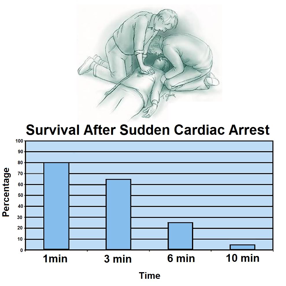
ACLS consists of the application of an algorithm of manual CPR, electrical defibrillation and pharmacologic therapy aimed at restoring a perfusing cardiac rhythm and adequate blood pressure and cardiac output to sustain life until definitive treatment of the underlying cause of the cardiac arrest can be achieved (e.g., coronary revascularization, implantation of an automatic defibrillator, or life-long anti-arrhythmic therapy).

 Figure 1-2 (right): Probability of survival as a function of time following cardiac arrest.[8]
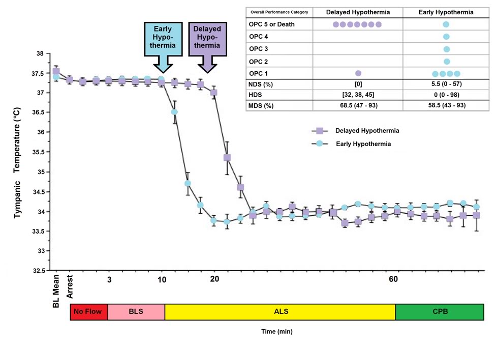
As is shown in Figure 1-6 below, the time to survival without neurological deficit following cardiac arrest in the absence of BCLS declines rapidly following a sigmoid curve with survival without neurological deficit being ~80-90% following 1 minute of arrest time, and less than 10% following 9 minutes of arrest.[8] Put another way, 50% of patients will experience significant morbidity or death following 4 minutes of circulatory arrest (Figure 1-2).

What is not shown in this graph is that the effect of immediate bystander CPR on survival is negligible in most studies [9],[10] with the primary benefit being observed in patients who’s time from the initiation of BCLS to successful cardiac resuscitation was greater than 8 minutes.[11] There is evidence in the literature that morbidity is improved with prompt by-stander CPR [12] providing that EMS response is also rapid, although this remains controversial.[11],[13]  A corollary of this is that the overall survival rate following SCA, with or without serious neurological morbidity, ranges between 1% (New York City, NY) [14] to 17% (Seattle, WA).[15] The mean survival (defined as survival to discharge from the hospital) in the United States as a whole is generally agreed to be at best 15%  [16] with ~70% of these patients experiencing lasting neurological morbidity (ranging from ‘mild’ cognitive impairment to total incapacitation in the Persistent Vegetative State (PVS).[17],[18],[19]

The primary cause of non-survival in patients experiencing SCA is failed cardiac or cerebral resuscitation. Arguably, it is failed cerebral resuscitation, since most underlying causes of refractory cardiac arrest could be treated by ‘bridging’ supportive technologies such as emergency femoral-femoral cardiopulmonary bypass (CPB) until myocardial revascularization and hemodynamic stabilization were achieved.[20] When emergency CPB is applied to patients who are candidates for good neurological outcome, the survival rate is increased.[21],[22],[23],[24] However, these technologies are not typically used on patients who are unsuccessfully resuscitated (restoration of adequate cardiac rhythm and perfusion) because of the justified perception that irreversible brain damage would have occurred during the prolonged period of cardiac arrest or CPR/ACLS.[21]  Similarly, it is for this reason that most attempts to achieve cardiopulmonary resuscitation in hospitalized patients who are not hypothermic or intoxicated with sedative drug are terminated after 15 minutes.[25],[26]

Within medicine it is widely understood that ‘CPR doesn’t really work’ and that if the return of spontaneous circulation (ROSC) is not achieved within ~ 5 minutes of cardiac arrest, the chances for survival are slim, and the chances for survival absent neurological impairment are slimmer still.[8] The principal reasons that conventional CPR is not effective are that it fails to supply an adequate amount of flow at an adequate pressure. Cardiac output (CO) is typically ~1/3rd of the at-rest requirement (~1.5 versus ~4.5 liters per minute), and mean arterial pressure (MAP) is typically 25 mm Hg to 45 mm Hg; well short of the 60 mmHg required to sustain cerebral viability.[27],[28]

The condition of the typical sudden cardiac arrest (SCA) patient and the circumstances under which he experiences cardiac arrest are far from the ideal of a patient who is a candidate for emergency cardiopulmonary bypass (CPB) in hospital. The typical SCA patient is middle aged or elderly, often suffering from one or more co-morbidities (diabetes, obesity, COPD, hypertension), and if subjected to prolonged CPR will invariably have impaired gas exchange due accumulation of fluid in both the parenchyma and the air-spaces of the lungs (pulmonary edema with alveolar flooding). This occurs because closed chest CPR quickly causes pulmonary edema.[29],[30]  As previously noted, even when the SCA patient is a ‘good’ candidate for salvage; someone who is relatively young and free of co-morbidities, CPR will likely prove futile due to cerebral ischemia-reperfusion injury and the post-resuscitation syndrome.

Over the past 25 years a vast number of therapeutic interventions have shown great promise in animal models of regional and global cerebral ischemia in the laboratory.[31],[32],[33],[34] In the last 6 years alone, over 1000 experimental papers and over 400 clinical articles on pharmacological neuroprotection have been published.[35],[36] However, with one exception, none of these interventions has been successfully applied clinically despite many attempts. [37],[38],[39],[40],[41],[42],[43],[44] The sole exception to this frustrating debacle has been the introduction of mild therapeutic hypothermia (MTH) as the standard of care for a select (and very small) minority of SCA patients.[45],[46],[47],[48],[49],[50],[51]

Hypothermia as an Active Therapeutic Agent

Since the demonstration by Safar, et al., of the neuro-salvaging effects of mild systemic hypothermia after prolonged cardiac arrest in dogs [52],[53] there has been an explosion of translational research which has lead to a transformation in our understanding and application of mild hypothermia.[54], [55] Once seen solely as a protective tool which conferred benefit by reducing metabolism, it has become clear that mild hypothermia (33°C–35°C) [56] has therapeutic effects which appear to be primarily anti-inflammatory and anti-apoptotic in nature, and which operate independently of hypothermia’s effect on metabolic rate.[57],[58] Table 1-1 reviews some of the known pro-inflammatory factors inhibited or moderated by mild therapeutic hypothermia (MTH) and documents the supporting literature.

 Table 1-1: Inhibition of Injury Cascades by Mild Therapeutic Hypothermia (MTH)


Reference

Model

Species

T (oC)

Factors

Takeda et al (2003)

Global

Gerbil

31 and 34

Anoxic depolarization
Busto et al (1989b)

Global

Rat

30 and 33

Glutamate
Dietrich et al (1990)

Global

Rat

30 and 33

BBB
Kawanishi (2003)

Hemorrhage

Rat

35

Edema; BBB; PMNL
Kawai et al (2000)

Focal

Rat

33

ICAM-1 mRNA; PMNL
Wang et al (2002)

Focal

Rat

30

ICAM-1; neutrophil and monocyte; microglia
Hamann et al (2004)

Focal

Rat

32 and 34

MMP-2; MMP-9; m-PA; t-PA
Karibe et al (1994a)

Focal

Rat

33

Ascorbate; glutathione
Kader et al (1994)

Focal

Rat

33

NOS; nitrite
Toyoda et al (1996)

Focal

Rat

30

Neutrophil
Chopp et al (1992)

Global

Rat

30

HSP-70
Mancuso et al (2000)

Focal

Rat

33

HSP-70; C-fos
Tohyama et al (1998)

Focal

Rat

30

PKC
Shimohata et al (2007a)

Focal

Rat

30

ePKC
Harada et al (2002)

Global

Rat

32

CaM kinase II; PKC-a,b,g synaptosome
Tsuchiya et al (2002)

Global

Mouse

33

Zn2+
Phanithi et al (2000)

Focal

Rat

33

Fas; caspase-3
Zhao et al (2007)

Focal

Rat

33

Cytochrome c and AIF
Karabiyikoglu et al (2003)

Focal

Rat

33 intra or

post

iNOS; nNOS
Wagner et al (2003)

Focal

Rat

33 post

BBB; MMP-9
Inamasu et al (2000)

Focal

Rat

34.5 post

Neutrophil infiltration; microglia
Horstmann et al (2003)

Stroke

Human

33 post

MMP-9
Horiguchi et al (2003)

Global

Rat

32 post

Hydroxyl radical
Han et al (2003)

Focal

Rat

33 post

NF-kB; iNOS; TNF-a
Van Hemelrijck et al (2005)

Focal

Rat

34 post

Caspase-3; nNOS
Inamasu et al (2000)

Focal

Rat

34.5 post

Bax
Friedman et al (2001)

Global

Rat

30 intra/post

GluR1A; GluR2B; GluR3C; NMDAR1
Ohta et al (2007)

Focal

Rat

35 post

Inflammatory genes: osteopontin, earlygrowth response-1, and macrophage inflammatory protein-3a
Luo et al (2007)

Focal

Rat

33 post

Base-excision repair pathway
Preston & Webster (2004)

Global

Rat

32 post

BBB
Liebetrau et al (2004)

Focal

Rat

32 post

Calpain
Hu et al (2008)

Global

Rat

32 pre/post

of GluR6-PSD95-MLK3 signaling module
Deng et al (2003)

Focal

Rat

33 post

ICAM-1
Karabiyikoglu et al (2003)

Focal

Rat

33 post

nNOS; iNOS and peroxynitrite
AIF, apoptosis-inducing factor; BBB, blood–brain barrier;; HSP-70, heat-shock protein-70; iNOS, inducible nitric oxide synthase; intra, intraischemic hypothermia; MMP-9, matrix metalloprotease-9; M, mouse; NF-kB, nuclear transcription factor kB; NOS, nitric oxide synthesis; nNOS, neuronal nitric oxide synthase; PKC, protein kinase C; PMNL, polymorphonuclear leukocytes; post, postischemic hypothermia; R, rat; S, species; T(1C), intraischemic temperature, unless specified; TNF-a, tumor necrosis factor-a.

Reproduced with modifications from Zhao, H., Steinberg, GK, Sapolsky, RM., General versus specific actions of mild-moderate hypothermia in attenuating cerebral ischemic damage. J Cerebr Blood Flow Metab, 2007. 27: p. 1879-1894.

Table 1-1:  Intraischemic hypothermia delays or attenuates both ATP depletion (Ibayashi et al, 2000; Sutton et al,1991; Welsh et al, 1990) and anoxic depolarization (Bart et al, 1998; Nakashima and Todd, 1996; Takeda, et al, 2003), it also blocks glutamate release (Busto et al, 1989b; Patel et al, 1994; Winfree et al, 1996), suppresses inflammation (Kawai et al, 2000; Wang et al, 2002), maintains the integrity of the BBB (Dietrich et al, 1990; Huang et al, 1999; Kawanishi, 2003), reduces free radical production (Maier et al, 2002), inhibits protein kinase C translocation (Cardell et al, 1991; Shimohata et al, 2007a, b; Tohyama et al, 1998), inhibits matrix metalloproteinase expression (Hamann et al, 2004), and blocks both necrosis and apoptosis. Intraischemic hypothermia also preserves the base-excision repair pathway, which repairs oxidative damage (Luo et al, 2007). In addition to those cascades directly associated with neuronal injury, hypothermia further blocks astrocyte activity and inhibits white matter injury (Colbourne et al, 1997; Dempsey et al, 1987; Kimura et al, 2002). Similarly, postischemic hypothermia blocks free radical generation (Horiguchi et al, 2003), attenuates inflammation (Horstmann et al, 2003; Ohta et al, 2007), prevents BBB permeability (Preston and Webster, 2004), and suppresses caspase activities (Van Hemelrijck et al, 2005). Indeed, a browse through the literature gives an overwhelming impression that hypothermia seems to block every damaging event associated with necrosis or apoptosis. One reason for this impression of pan-inhibition may lie in the causality of ischemic damage.  For example, is the inflammatory response the cause of tissue damage or is it induced by brain injury? If it is the latter, then since hypothermia prevents tissue damage, it certainly also prevents the inflammatory response.

 – Zhao, H., Steinberg, GK, Sapolsky, RM., General versus specific actions of mild-moderate hypothermia in attenuating cerebral ischemic damage. J Cerebr Blood Flow Metab, 2007. 27: p. 1879-1894.

 The journey from the laboratory to the clinic for MTH has been long and difficult. Seven years after the publication of the prospective randomized trials clearly showing that MTH improves survival and neurological outcome in out-of-hospital cardiac arrest patients, and 6 years after the ILCOR and AHA Guidelines  [59] recommended that: “Unconscious adult patients with spontaneous circulation after out-of-hospital cardiac arrest should be cooled to 32°C to 34°C for 12 to 24 hours when the initial rhythm was ventricular fibrillation (VF),” [49] only a minority of SCA patients are being treated with MTH. In surveys of emergency and critical care physicians conducted in 2005 and 2006, 74% of those responding in the US [60] and 64% of the international respondents indicated they had never used MTH.[61],[62] The use of pre-hospital, in-field MTH, is virtually nonexistent.[63]

No doubt, the commonly cited ‘obstacles’ of lack of institutional protocols, lack of physician education about the benefits and guideline changes, as well as the inevitable inertia that accompanies any paradigm shift in treatment are playing a significant role in the failure of MTH to become the practiced standard of care for the post resuscitation syndrome.[64],[60] However, what is not being said, or considered, is that while MTH as currently practiced represents a large relative improvement in outcome, the benefits are still modest in absolute terms. Only a miniscule subgroup of SCA patients currently can benefit from MTH; and even in its best clinical implementation MTH still fails to rescue ~60% of that sub-group of SCA patients to whom it is applied.[65],[66],[67],[68],[69] This is in stark contrast to what can be achieved with MTH in ameliorating post-ischemic encephalopathy in the laboratory, where post-resuscitation MTH consistently provides rescue with stunning efficacy.[70],[71]

Figure 1-3: The impact of a delay of 10 min in inducing MHT is a dog model of cardiac arrest followed by 3 min of systemic ischemia, 7 minutes of mechanical CPR and 50 minutes of advanced life support. Hypothermia to 34oC was induced beginning at 10 min post arrest in the early hypothermia group˜ and at 20 min post arrest in the delayed hypothermia group ¢.  In the early hypotherrmia group group, 5 of 7 surviving dogs were functionally normal (OPC 1 or 2), 1 had OPC 3, and 1 had OPC 4 (coma) at 96 hours of recovery. Histologically, 4 of 8 dogs in this group were normal (HDS 0), 1 had HDS 16, 1 had 22, and 1 had 98. The only surviving dog in the DH group was functionally normal at 96 hours (OPC 1, NDS 0) with an HDS of score of32 (mild injury) Due to early mortality only two other dogs in the delayed hypothermia group were evaluated histologically and their  HDS scores 38 and 45, respectively. Dogs in this study were scored by ‘overall performance categories’ (OPC; 1=normal, 2=moderate disability, 3=severe disability but conscious, 4=coma, and 5=death) Neurological function and  neurological deficit scores (NDS; 0% to 10%=normal, 100%=brain death). [72],[73] Histological damage scores were obtained by neuropathological examination of 19 distcrete brain regions for severity and extent of ischemic neuronal changes, infarcts, and edema.  A total brain histological damage score (HDS)  >40 represented moderate damage, and HDS >100 represented severe damag.[74] Redrawn from Nozari, A., et al., Critical time window for intra-arrest cooling with cold saline flush in a dog model of cardiopulmonary resuscitation. Circulation, 2006. 113(23): p. 2690-6.
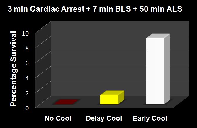
The primary obstacle to realizing this bonanza in translation research has been the practical impossibility of achieving systemic cooling within the narrow therapeutic window demonstrated in animal models of SCA and resuscitation.[75],[67],[65],[66],[67],[68],[69] If the clinical outcome of MTH was even half that achievable in the laboratory, widespread application would likely have been rapid and uniform; there is rarely resistance to the ‘miraculous’ if it is simple, easy to understand, biophysically well characterized and highly cost-effective. MTH applied immediately post ROSC would be all of these things.[4]

Figure 1-4: The results of the Nozari, et al., [71] study on the effects of delayed MTH are presented graphically at right with the addition of historical controls from the literature treated similarly, but with no hypothermia (no survivors). This graphic illustrates the potency of truly rapid post arrest hypothermia in ncreasing survival.
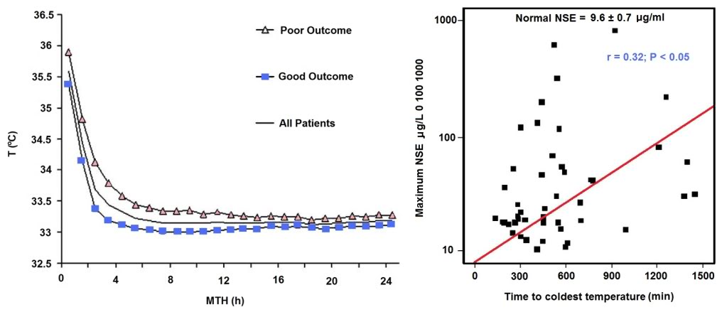
The data in Figures 1-3 and 1-4 exemplify what is possible when MHT is induced within its optimum therapeutic window of 0-15 min post ROSC versus a delay of even 10 minutes. In this study by Nozari, et al., of PeterSafar’s group, [71] VF was electrically induced in 17 dogs all of whom were subjected to a period of 3 minutes of no flow beginning when the MAP dropped below 30 mm Hg, followed by 7 minutes of mechanical CPR and 50 minutes of advanced life support during which time VF was maintained and mechanical CPR was continued. Nine animals were treated with rapid (early) induction of MTH to 34oC starting at 10  min post arrest (EH group) (concurrent with the start of ALS to simulate the time course of arrival of EMS paramedics) using a combination of cold IV saline and veno-venous heat exchange. Induction of hypothermia was not

Figure 1-5: Results of a study of 48 cardiac arrest patients treated with MTH via endovascular cooling. A strong correlation was found between rapidity of cooling and both neurological outcome and serum neuron specific enolase levels. Left: Time course of MTH among patients with good and poor neurological outcome. The curves indicate the course of mean body core temperature during MTH among patients with good (¢) and those with poor (p) outcome as well as in the entire (▬) patient group. Right: Correlation between time to coldest temperature (minutes) and the maximum NSE values (μg/L). Normal serum NSE is 9.6±0.7 μg/L. Redrawn from Wolff B, et al., Early achievement of mild therapeutic hypothermia and the neurologic outcome after cardiac arrest, Int J Cardiol (2008).
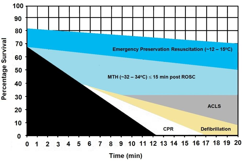
begun until 20 min post arrest in the delayed hypothermia group (DH group) which consisted of 8 dogs. Target core temperature was achieved at 6.0±2.7 minutes after the initiation of cooling (3.5 minutes after the start of veno-venous cooling) in both groups. The delay from arrest to reaching ~34oC  was 16.6 min in the EH group and 25.4 minutes in the DH group.

After 60 minutes of VF, ROSC was achieved with cardiopulmonary bypass for 4 hours, and intensive care was given for 96 hours. In the early hypothermia group, 7 of 9 dogs survived to 96 hours, 5 with good neurological outcome. By contrast, in the delayed hypotherrmia group 7 of 8 dogs died of multiple organ failure within 37 hours (P=0.012);  3 animals in secondary VF that was resistant to CPR with antiarrhythmic treatment and repeated defibrillations. Only one dog in the EH group died, and that animal succumbed to single organ failure; pulmonary edema with hemoptysis. This study extends the previous work by this group documnting an optimum therapeutic window for MHT (in dogs) of ~10-15 min.[76],[76],[77] The therapeutic window of MTH after cardiac arrest has been demonstrated to be similarly short in other species.[78],[79],[80],[81]

The dramatic efficacy of MTH in the laboratory made quick converts of the pioneering researcher-clinicians who forged ahead with the application of MTH to SCA in the clinic precisely because it was dramatic; indeed it was as close to the miraculous as interventions in medicine come. The real barrier to translating that ‘miracle’ to everyday practice has been the seemingly intractable problem of achieving cooling over the same time course that has proven so effective in the research setting.

The problem is that the optimum therapeutic window for the treatment of cerebral ischemia-reperfusion injury appears to be in the range of 0 to 15 minutes post ROSC. One of the first follow-up studies on MTH carried out by Safar, et al., demonstrated that in a standardized model of cardiac arrest in dogs a delay in the application MTH of as little as15 min after ROSC abolished most of the benefit.[77],[82] While the work of Bernard, et al., [46] and that of the Hypothermia after Cardiac Arrest Study Group [48] demonstrated that delays in cooling of up to 2-3 hours post ROSC in humans still have sufficient clinical utility to justify the routine application of MTH in a selected group of SCA patients, this benefit is marginal when contrasted to that achievable in the laboratory when MTH is rapidly induced during the first 15 minutes after ROSC.[77] Thus, the optimum clinical benefit of MTH in ischemic and very likely traumatic, CNS injury requires the ability to achieve very rapid core cooling.[83]

Figure 1-6: Survival after cardiac arrest declines rapidly as a function of time to ROSC, exhibiting the sigmoidal curve shown at left, above (¢>¢), with essentially all patients failing to survive with normal mentation after arrest intervals of @ 10 min. Application of MTH (¢) within the window of @ 15 min offers the promise of squaring the survival curve in SCA of @ 10 min duration yielding a survival rate of ~65-70% with little or no neurological deficit. Application of deep (10-22oC) or profound hypothermia (5-9oC) (¢) may allow survival after intervals of as long as 1-2 hours of CPR. Graphic by M.G. Darwin

In the clinical arena the time to reach the target core temperature under ‘good’ circumstances is in the range of 3-4 hrs; not 10 to 30 min, as is the case in the laboratory. Even with such long delays in cooling the adverse effect of delay is still present. Wolf, et al., recently published a study of 49 out of hospital cardiac arrest patients who were treated with MTH (32.0-34.0°C; with a target temperature of 33.0°C) of 24 h duration using endovascular cooling.[69]  The study endpoints were neurological outcome on discharge from hospital and serum neuron specific enolase (NSE) levels (a sensitive and specific marker of neuroinjury) at 24 h intervals to 3 days (Figure 1-5).

Figure 1-7: The graph above shows the hypothesized relative effect on survival of effectively administered CPR started at 5 min post-arrest followed by defibrillation at 6 min and ACLS at 8 min post arrest (~30% survival). The light blue shaded area of this graph shows the expected improvement in survival if MTH is induced at the start of ACLS (8 min post-arrest) and target temperature is reached by 15 min post ROSC. The dark blue shaded area shows the potential of Emergency Preservation resuscitation (EPR) using moderate (10-22oC ) or profound (5-9oC) hypothermia to not only square the curve of survival with CPR, but to facilitate survival in patients who would otherwise not benefit from either BCLS or ACLS (i.e., refractory to defibrillation, hypovolemic, etc.). Graphic by M.G. Darwin
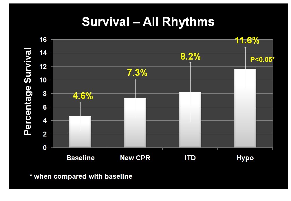
 As is the case in laboratory studies of MTH in cardiac arrest in dogs, Wolff, et al., found that neurological outcomes were binary, with no patients who survived experiencing moderate degrees of disability; patients either recovered well with no or /mild neurological impairment, or experienced severe disability (n = 1) coma or PVS (n = 6). Twenty-eight patients were discharged with a good outcome and a strong correlation was found between good outcome and the time interval from the start of cooling to the lowest temperature (p =.035) and a less robust correlation with the time to reach target temperature (p=.071). Similarly,

NSE levels were found to correlate well with the time required to reach the lowest temperature achieved in each patient (Figure 1-5).

Even with delays in the start of cooling that averaged 2.5 hrs; and a mean time to reach target core temperature of 6.8 hrs, additional injury accruing from slowness in cooling was still clinically and biologically apparent. Despite the homogeneity of the patients, their arrest times, and their course of treatment, ~60% of the patients in this study either did not survive, or were comatose or PVS.

The Benefits and Limits of ‘Delayed’ MTH: Real World Experience

To understand the benefits and limits of MTH when it is aggressively and competently implemented with currently available technology, it would be hard to find a better example than that of Wake County, SC. Wake County, is located in the northeast central region of North Carolina and is part of the Research Triangle metropolitan area, which consists of Raleigh, Durham, Chapel Hill, and surrounding urban and suburban areas. The area serviced by the Wake County Emergency Medical System (Wake EMS) has a population of 832,970 (as of 2007). The Wake County EMS operates 35 ambulances from 23 locations with 825 ALS personnel; ambulances are staffed with two paramedics 95% of the time and there is always one paramedic responding.[84] In 2002 the Wake EMS answered more than 50,000 medical requests for service. Based on the latest national data Wake County ranks third in the US for recovery from “survivable” cardiac arrests (primarily ventricular fibrillation-ventricular tachycardia). Nationally, the average survival rate is 17% for patients presenting with these arrhythmias.

 Figure 1-8: Improvement in overall survival of patients in cardiac arrest in response to the phased introduction of CPR per AHA 2005 Guidelines, use of an impedance threshold device (ITD) and in-field induction of MTH.[85]
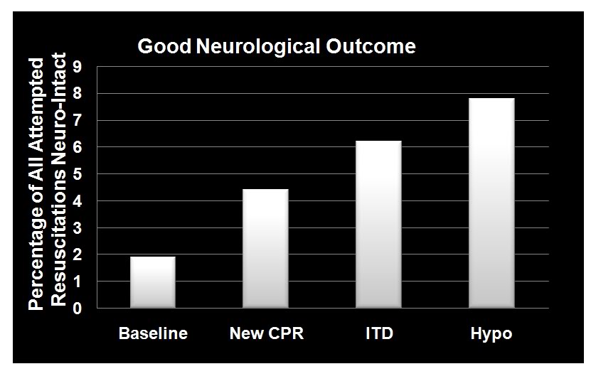
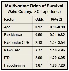
 Beginning in January 2004 Wake EMS initiated a study to evaluate the efficacy of CPR as they then practiced it, and to evaluate the effectiveness of the impending change in the American Heart association (AHA) guidelines for CPR, the introduction of an impedance threshold valve (the ResQPod™) and Wake EMS’ planned implementation of the ILCOR Guidelines for post-arrest MTH.[85],[86] From January 2004 until April of 2005, Wake EMS personnel employed the then extant AHA guidelines, which mandated an emphasis on intubation and a 15:2 compression-to-ventilation ratio; with interruption of chest compressions for ventilation. This period constituted the baseline of the study, and data were collected per protocol; not gathered retrospectively.

During the baseline period survival to discharge from hospital was 2.4% for all patients given CPR and 12.1% for patients with ventricular fibrillation-ventricular tachycardia (VF-VT) arrhythmias. In April 2005 Wake EMS implemented continuous cardiac compression CPR with a 30:2 compression-to-ventilation ratio with emphasis on no, or very minimal, interruption of chest compressions.  After 12 months, the overall survival rate had risen to 4%; and had more than doubled to 21.8% for patients who presented in VF-VT.

In April 2006, Wake EMS added the use of an impedance threshold device (ITD) to improve cerebral and coronary perfusion during CPR. Introduction of the ITD resulted in an increase in overall survival to 4.5% and an increase in the survival of patients with VF-VT to 28.5%.

  Figure 1-9: Dramatic improvement in neurologically intact survival as a result of the phased introduction of CPR per AHA 2005 Guidelines, use of an impedance threshold device (ITD) and in-field induction of MTH.[85]

 The final phase of the investigative protocol began in October of 2006 when Wake EMS added in-field induction of MTH to the two previous interventions. MTH was induced using a combination of external cooling employing ammonium nitrate-water eutectic ‘instant cold packs’ applied to the axilla and groin, and  cold IV saline (1-2oC, 30mL/kg to a maximum of 2 liters) given rapidly via two large bore catheters and/or intraosseous infusion. Criteria for induction of hypothermia were that the patient have ROSC and show no return of consciousness (Glasgow Coma Score (GCS) <8). Induction of hypothermia was initiated two to three minutes after ROSC. There was heavy emphasis on avoiding over-ventilation and on attempting to maintain end-tidal CO2 (EtCO2) at a minimum of 40 mm Hg.  Patients undergoing MHT were sedated with etomidate, paralyzed with vercuronium and given a titrated dopamine drip to maintain mean arterial pressure (MAP) between 90-100 mm Hg. The mean time to target temperature (34oC) in this study was extraordinarily short: 68 minutes (95% CI 47 to 88); compared to the 2-3 hours typically required to induce MTH.

With the combination of continuous compression CPR, use of the ITD and prompt application of MTH, survival rates for the 12 months from October of 2006 to October 2007 had increase to 6.7% overall and 37.4% for patients with VF-VT. The odds of overall survival increased three-fold (95% CI 1.7 to 5.0) and the odds of survival for patients in VF-VT increased 4.3-fold (95% CI 2.2 to 8.6) from the beginning of the study (Figures 1-8 and 1-10).The probability of a good neurological outcome increased from 20% at baseline to 80% at the conclusion of the study (Figure 1-9). In a multivariate analysis, the odds ratios for survival for each phase of implementation were as follows:

Figure 1-10 (right): Multivariate odds for all factors in outcome evaluated during the Wake EMS study. MTH was by far the most powerful intervention. As in most previous studies of survival factors associated with CPR age and residence (home versus extended care or assisted living facility) had only modest impact on survival.[85]
 

  • New CPR protocol: 2.13 (95% CI 1.12 to 4.04)
  • Addition of impedance threshold device: 2.33 (95% CI 1.09 to 5.00)
  • Addition of early hypothermia: 3.99 (95% CI 2.19 to 7.27)
  • Patients who received bystander CPR 1.79-fold (95% CI 1.18 to 2.72) more likely to survive.

Figure 1-11 (right): The Engel-15 portable, (compressor-type) refrigerator/freezer has a 14 L capacity, weighs 11.5 kg and can maintain 12-13 liters of saline at 1-2oC at ambient temperatures as high as 40oC . It retails for ~$380 US. [Photo courtesy of Engel, Ltd., Australia]
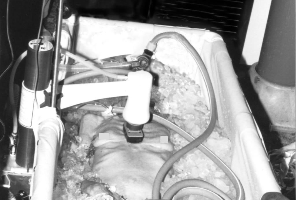
 

Interestingly, all three elements of the Wake EMS protocol were implemented at a cost of less than $200 per patient. Due to budget constraints, Wake EMS chose simple, inexpensive commercial products for refrigeration of IV fluid and implementation of external cooling, as opposed to more costly products developed specifically for medical application, such as the EMCOOLS surface cooling system (Emergency Medical Cooling Systems, AG, Austria). Saline was kept at the requisite temperature of 1-2oC with a compact, 12V operated, consumer travel refrigerator/freezer (Figure: 1-11, Engel-15: http://www.i-m-d.com/) and surface cooling was with generic ammonium nitrate cooling packs.

The Wake County EMS program is extraordinary in every way. It represents the best application of the best available technology by arguably some of the best medical and paramedical personnel in the US. The mean time to target temperature of 68 minutes is unprecedented in any clinical study employing MTH. Of the 359 patients who participated in the study (all comers) after MTH was in place; 25 survived. In the subgroup of 93 patients who presented with VF-VT; 34 survived, with 78% or 27 patients being discharged with a good neurological outcome. Put another way 92% of patients who presented under the most favourable circumstances (VF-VT), treated with the best currently available interventions, at the fastest rate of cooling so far reported, failed to survive or did so with profound neurological debility.

The primary difference between the survivors and the profoundly disabled or dead was the development of the post-resuscitation syndrome and the primary reason for this complication was not comorbidity, or delay in paramedical assistance, but rather delay in the rapidity of cooling which, if achieved within the first 15 min post ROSC, would have offered the prospect of neurologically intact survival in the range of 70-80% in patients presenting with VF-VT, and 30-40% in all comers.

These interventions, remarkable achievements that they are, do not escape from the harsh reality that the 400% increase in survival from cardiac arrest in Wake County, when expressed in absolute terms, means that the number of lives saved increased from ~5 to 25 – out of 395 SCA patients; a huge relative gain, but a comparatively small increase in the absolute number and percentage of lives saved, and minds salvaged. The true life saving potential of MTH remains elusive by virtue of its exceedingly small therapeutic window.

 The Problem of Heat Exchange

Because of this minute therapeutic window, there is a pressing need to achieve rapid and durable core cooling of patients during CPR by simple, easily accessible means.  External cooling is only effective at reducing core temperatures by 0.15 to 0.25ºC/min in the average patient undergoing CPR (Figure 1-12) and this is achieved only by complete immersion of patients in a stirred ice water bath.


The Efficacy of External Cooling in Four Cryopatients[5]

 Figure 1-12:  Comparison of the cooling rates of four cryopatients. Immediately following pronouncement of medico-legal death patients were given closed chest mechanical cardiopulmonary support and placed in a stirred ice water bath for induction of hypothermia. Epinephrine was administered as per ACLS guidelines; thus peripheral vasoconstriction would be expected to be comparable to that seen in the typical SCA patient undergoing cardiac resuscitation.
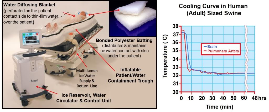
The most effective external cooling achieved by a commercial system using direct, whole body surface cooling employing circulation of ice water (ThermoSuit,™ Life Recovery Systems, Kinnelon, NJ)  is probably the work of Janata, et al., using human human-sized swine.[87]

They were able to achieve core cooling at a rate of 0.3oC/min; however it is important to note that the animals in this study were not in cardiac arrest while undergoing CPR in the presence of profoundly peripherally vasoconstricting agents, such as epinephrine or vasopressin; as would usually be the case during ACLS in humans [59] and which is known to further slow surface cooling.[88]

 Figure 1-13: The Life Recovery Systems ThermoSuit™ employs direct ice water contact with the patient’s skin to achieve the maximum possible rate of cooling by external means. The system consists of an inflatable insulating and water containment patient enclosure inside of which the patient rests on a mat of Dacron bonded polyester ‘wool’ which acts to diffuse and film water pumped over the dorsal surface of the patient’s body. Water at 2-4oC is thin-filmed over the ventral surface of the body by a thin, transparent blanket with many hundreds of small perforations through which water under pressure pours out and over the patient. Cold water is recirculated over crushed or cubed ice in an insulated reservoir containing a disposable liner and pumps. Cooling is computer controlled via a thermistor which can be placed in any desired anatomical location. All patient contact items are single-use and disposable (including, as previously mentioned, the pumps) http://www.life-recovery.com/.
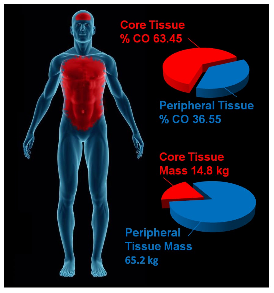
The obvious problems with this system are its bulk (Figure 1-13), likely high cost, lack of ease in field deployment (again related to its bulk and weight) and the intrinsic physiological problems associated with the induction of hypothermia via external cooling. As extensively discussed in Section Two, the mammalian body consists of multiple thermal compartments transiently ‘isolated’ from each other by differences in blood flow and heat conductivity.[89] Broadly, these compartments can be classified as strongly and weakly circulated (perfused); corresponding to the body core and periphery. The core tissues receive ~63% of the resting cardiac output (CO) but constitute only ~19% of the total body mass. By contrast, the peripheral tissues receive ~37% of the basal CO and constitute ~81% of the body’s mass (Figure 1-14).

 Figure 1-14: The parenchymatous organs that comprise the visceral core of the body receive an aggregate of ~63% of resting the cardiac output while comprising only ~19% of the body mass. By contrast, the peripheral tissue mass which accounts for ~81% of body mass receive only ~19% of the cardiac output. External cooling profoundly chills peripheral tissues before significantly reducing core temperature. Values for organ and tissue masses were obtained from: IAEA. Compilation of anatomical, physiological and metabolic characteristics for a Reference Asian Man. Volumes 1 and 2. Report IAEA-TECDOC-1005, (Vienna, Austria: International Atomic Energy Agency) (1998), Boecker, BB. References values for Basic Human Anatomical and physiological characteristics for use in radiation protection. Radiation Protection Dosimetry.105(1–4): 571–574;2003, de la Grandmaison GL, Clairand I, Durigon M. Organ weight in 684 adult autopsies: new tables for a Caucasoid population. Forensic Sci Int. 2001 Jun 15;119(2):149-54 and Heymsfield SB, Gallagher D, Mayer L, Beetsch J, Pietrobelli A. Scaling of human body composition to stature: new insights into body mass index. Am J Clin Nutr 2007;86:82–91.Values for organ and tissue blood flows were obtained from: Williams, LR, Leggett, RW. Reference values for resting blood flow to organs of man. Clin Physiol Meas. 10:187-212;1989. Graphic by M.G. Darwin
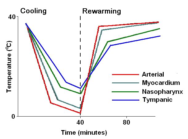
 The Pathophysiology and Biophysical Limitations of External Cooling

The objective of MTH is to provide protection against ischemia-reperfusion injury to the brain, heart, kidneys and liver; the visceral organs that constitute the strongly circulated core of the body. The peripheral tissues (skin, skeletal muscle, connective tissues and bone) are at once much more resistant to ischemia and less well perfused.  External cooling rapidly chills the ischemia-resistant peripheral tissues cooling them profoundly, while failing to provide protection to the vulnerable parenchymatous organs in the body core. This is not only undesirable in terms of its inefficiency; it also poses a number of hazards and risks.[90] Hypothermia is therapeutic in ischemia-reperfusion because it down-regulates the immune-inflammatory response; a response that is vital for host defense, wound healing and hemostasis. Hypothermia, like any major medical intervention that perturbs fundamental physiological processes, carries with it serious risks, as well as benefits. In both animals and humans, hypothermia is markedly immunosuppressive [91],[92] and interferes with the both the biochemistry of the clotting cascade and the production of platelets and clotting proteins.[93],[94]

In humans perioperative minimal hypothermia (MinH) (36oC) increases the rate of wound infections [95] and prolongs hospitalization. [96] These effects occur in part due to the regional thermoregulatory vasoconstriction MinH induces; which in turn leads to reduced oxygen delivery to injured  tissues, [97] inhibition of oxidative killing by neutrophils, [98] and reduced collagen deposition.[96]  MinH induces significant suppression of mitogenic responses to Concanavalin A (con A), phytohemagglutinin (PHA), and pokeweed mitogen (PWM) and these changes are known to persist for at least 48 h. The mitogens PHA and ConA activate T cells, whereas PWM stimulates both T and B cells, thus indicating that the suppressive effects of MinH involve a variety of lymphocyte subpopulations. Hypothermia of as little as 1oC significantly inhibits production of  interlukins (IL-1б, IL-2, IL-6) and TNFα in post-surgical patients, [99] and this suppression of cytokine production persists for least 24 hours after even a brief post-operative hypothermic interval.[96] The inhibition of pro-inflammatory cytokine production by IL-1б and TNFα induce tissue factor which is critical to angiogenesis, collagen elaboration and fibroblast activation; all essential processes in wound repair and hemostasis.[100],[101],[102] Significantly, many of these of adverse effects of post-operative MinH can be prevented by maintaining normothermia in the perioperative period.[96]

In the Hypothermia After Cardiac Arrest Study Group, patients treated with MTH experienced twice the incidence of sepsis. This finding is consistent with other studies where MinH and MTH were found to double the rate of post operative wound infection.[96]  A recent meta-analysis of MTH for traumatic brain injury (TBI) found that the incidence of pneumonia was also doubled for patients undergoing MTH.[103]

External cooling causes greater perturbation in hemodynamics than does central cooling, [104] resulting in increased systemic vascular resistance and decreased cardiac index; a phenomenon observed in all of the patients in the Bernard, et al., study that employed MTH for post-arrest cerebral resuscitation.[46]  This is particularly undesirable in the setting of MI, CHF and cardiogenic shock. For these reasons, as is the case with any potent therapy, careful attention must be paid to the dose-response curve, and overshoot or excessive regional cooling must be minimized or avoided.[105]

In patients cooled with ice packs and non-feedback controlled cooling blankets, there have been persistent and extensive problems with overshoot. In one study, 63% of patients overshot to <32oC, 28% to <31oC, and 13% were inadvertently cooled <30oC.[105] By contrast, patients in the European Resuscitation Council Hypothermia After Cardiac Arrest Registry who were cooled endovascularly had less overshoot (mean lowest temperature 32.9oC, IQR; 32.6oC to 33oC) when compared to patients cooled by other methods (mean lowest temperature 32.4oC, IQR: 31oC to 32.9oC).[106]

In part, the problem of overshoot in external cooling can be corrected by servo control of patient cooling. However, the fundamental problems of external cooling remain. Excessive cooling of poorly perfused peripheral tissues will inevitably result in ‘after-drop’ of core temperature as thermal equilibration occurs. This process is idiosyncratic and inherently difficult to model or predict. In large measure the speed and character of thermal equilibration between peripheral and central tissues will depend upon highly variable factors such as body morphology and composition, body surface area, cardiac output, regional blood flow distribution, and the administration of vasoactive medication such as ionotropes; with their attendant peripheral vasoconstriction.

When the tissues of non-hibernating (or unprepared hibernating mammals) are cooled to £20ºC a wide range of deleterious changes occur. The saturated fats which comprise cell membrane lipids undergo phase change, resulting in red and white blood cell rigidity; with accompanying inability to deform and pass through capillaries. Red cell aggregation also occurs and this, in association with reduced flow as a result of vasoconstriction, results in blood sludging and failure of the microcirculation.[107],[108]  Profound hypothermia, either local or systemic, results in hemoconcentration as a consequence of translocation of vascular water and electrolyte to the interstitial space.[109],[104],[110],[111],[112] This hemoconcentration further exacerbates regional ischemia in deeply chilled tissues.

Independent of injury from the freezing of water, moderate, profound or ultraprofound hypothermia is known to cause cellular damage which is referred to as ‘chilling injury.’ Chilling injury appears to be a multifactorial process in which alterations of membrane structure (reorganization of lamellar lipid sheets with lateral phase separation between regions of gel phase and regions of liquid crystal phase result in loss of membrane integrity), [113],[114] failure of ion pumping (with consequent disruption of cellular ionic homeostasis), [115],[116] depolymerisation of some elements of the cytoskeleton, [117],[118] generation of free radicals, [119],[120] and metabolic disruption due to selective inactivation of critical enzymes [121] all appear to play a role.

Cooling of tissues to 5ºC for as little as 1 hour has been shown to cause microvascular endothelial damage similar to that observed in ischemia-reperfusion injury; loss of endothelial cell tight junctions, infiltration of capillary and venule walls with leukocytes, and frank extravasation of red cells from injured vessels.[122] A possible reason for the similarity in the histological appearance of chilling injury with ischemia-reperfusion injury may be due to the fact that both types of injury appear to be caused, at least in part, by reactive oxygen species and by disruption of the cytoskeleton.

The molecular changes induced by moderate, profound and ultraprofound hypothermia may also directly compromise endothelial cell integrity. For example, chilling of several types of epithelial cells has been shown to result in disassembly (depolymerisation) of the intracellular microtubules resulting in compromise of the polarized membrane expression and function of some transport proteins in these cells.[123] These functions are slow to return to normal (@20 hr) and are associated with prolonged dysfunction of allografts that have undergone cold preservation storage.[124],[125]

Deep cooling of the peripheral tissues may also result in immunosuppresion in chilled limbs and skin, and possibly impaired hematopoiesis due to localized moderate hypothermia in bone marrow in the cranium, sternum, vertebrae, and to a lesser extent, in the pelvis. [In children, the long bones are the principal repository of hematocytoblasts, and this marrow would also be disproportionately chilled during external cooling.] In contrast to the anti-inflammatory effect of MTH, systemic hypothermia to £28ºC, either accidental or induced has been shown to increase levels of pro-inflammatory cytokines.

Core cooling is thus the gold standard for the induction of systemic hypothermia (mild, moderate, profound or ultraprofound) and external (peripheral) cooling should be used only where there is no other alternative for achieving truly rapid cooling, or maintaining it for the required 24-48 hours following induction.

 Consideration of Invasive Core Cooling Methods

Figure 1-15 (right): Typical (idealized) cooling and re-warming curve achievable with maximum extracorporeal (cardiopulmonary bypass) cooling.
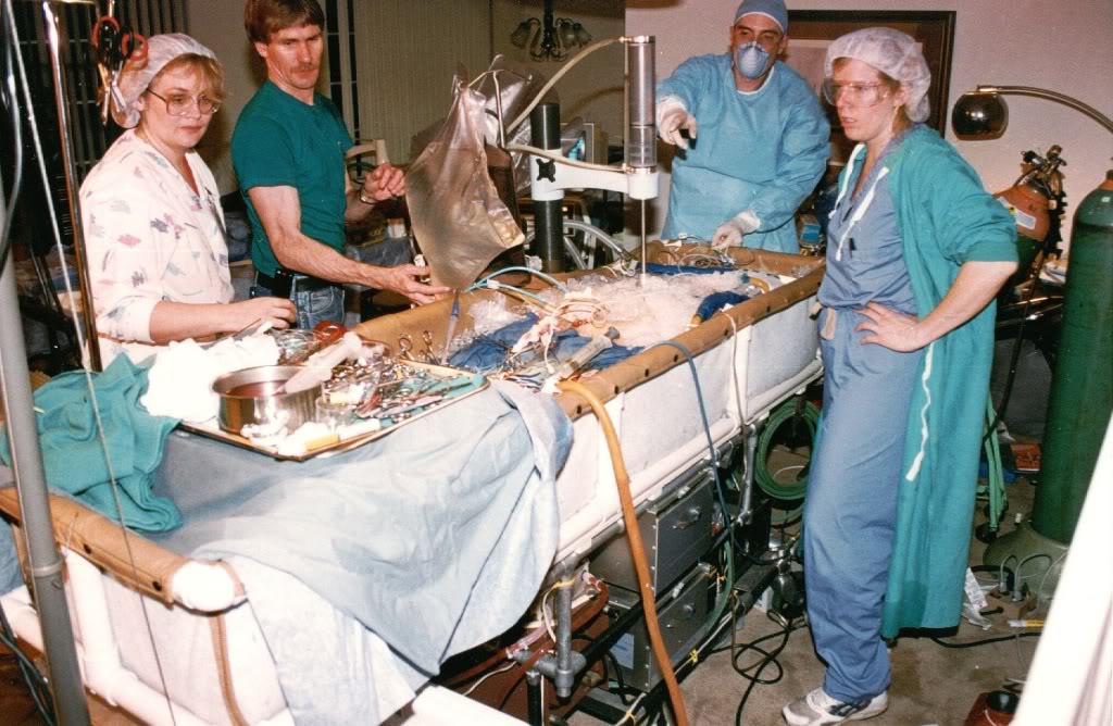
Extracorporeal cooling via cardiopulmonary bypass (CPB) [126] or high flow veno-venous heat exchange [127],[128]  allows for cooling rates of 0.8ºC/min to 1.0ºC/min (Figure 1-15) However, it cannot be applied rapidly enough given existing logistic and regulatory constraints. CPB requires complex hardware and highly skilled personnel who must maintain their clinical reflexes by practicing perfusion on a regular (preferably daily or at least weekly) basis. Even in centers of excellence, with a highly skilled, rapid-response CPB team at the ready (including a well-practiced surgeon or cardiologist), the soonest CPB can be initiated after cardiac arrest is typically 15-20 minutes.[129],[130]

The use of emergency CPB applied in this time frame is largely confined to patients undergoing cardiac catheterization and/or revascularization (i.e., angioplasty or stent placement) who arrest in the cardiac catheterization lab. When such patients experience cardiac arrest that is refractory to treatment with drugs and defibrillation – they are usually otherwise healthy – they have normally functioning lungs, normal fluid balance and fluid distribution (are neither dehydrated nor edematous from fluid overload), and have failure of a only a single organ –  the heart. Even under such ‘ideal’ conditions, CPR is often inadequate to maintain brain viability during the brief interval between cardiac arrest and the start of CPB.

While veno-venous extracorporeal cooling is less technically demanding, it still requires skill-intensive vascular access under field conditions, and reported rates of cooling are modest; ~0.012ºC/min [131],[87]. Both of these techniques may require anticoagulation and certainly require coagulation monitoring; which again is a barrier to in-field application.

Administration of large volumes (40mL/kg) of chilled intravenous fluid has also been used to reduce core temperature in the field in patients undergoing CPR. However this method is extremely slow (~0.058ºC/min) [132],[133],[134], and is sharply constrained by the maximum volume of fluid that can be administered.

 Figure 1-16 (left): Cryopatient undergoing ECMO and blood washout in the home. Even under ideal circumstances cardiopulmonary bypass takes in excess of 30 minutes to initiate.
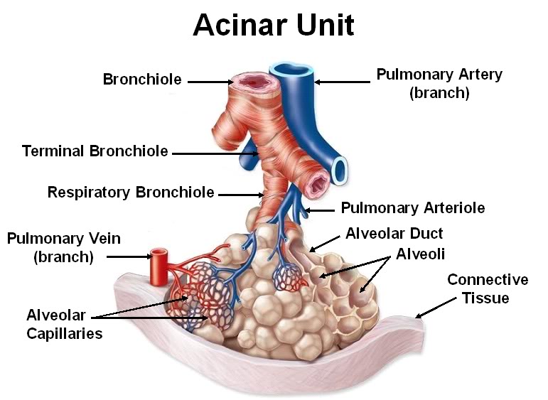
 The effectiveness of intravascular fluid administration in achieving durable core cooling is also a function of body composition. Obesity, which has become epidemic in the developed world, is now rapidly becoming a global problem with 1.1 billion adults worldwide classified as overweight,and 312 million of them as obese by the World Health Organization (WHO). [135] Vascular volume does not increase proportionally to increase in body weight; in fact, the vascular volume to body weight ratio falls toward an asymptotic value of approximately 45 ml/kg in the obese human.[136] Thus, the use of cold intravenous infusions will necessarily be even less effective in the obese than might be expected since the maximum volume of fluid that may be safely given does not increase linearly with body weight. Fat is also an extraordinarily good insulator, and obesity often unfavorably alters body surface to volume area. Both of these factors combine to dramatically reduce the speed and overall effectiveness of external cooling.

The limits of efficiency of core cooling that can be achieved by irrigating the peritoneal or pleural spaces with cold liquid are in the range of 0.05 to 0.3ºC/min [137],[138] and both of these techniques are invasive, require considerable technical skill, and carry with them risks of serious iatrogenic consequences.

As noted earlier, the ideal way to address these needs would be extracorporeal membrane oxygenation (ECMO) and cooling. However, the practicality of rapidly bringing this demanding technology to bear in the EMS setting is negligible. Surgery (or time-consuming percutaneous vascular access; at or beyond the optimum therapeutic window of |15 min post ROSC) is required to access the circulation and this cannot be accomplished under field conditions. Experience with emergent initiation of ECMO in patients presenting for experimental cryopreservation (Figure 1-16) are probably representative of the time required to achieve extracorporeal support under field conditions for patients with SCA. Such patients typically experience a delay of 60 to 80 minutes (and not infrequently longer) after the initiation of closed chest CPR before the application of CPB, even under optimum circumstances.[139],[140],[141]

Thus, what is needed for patients who have suffered prolonged (³5 min) cardiac arrest, or in whom ROSC cannot be effectively established, is the ability to rapidly induce hypothermia via core cooling simply and noninvasively.  In addition to moderating the injury cascade initiated by ischemia-reperfusion, mild (33-35oC) or moderate (28-32oC) hypothermia can indirectly act to improve gas exchange and hemodynamics by bringing the patient’s cerebral and systemic metabolic demands closer to those that can be delivered by CPR.

Exsanguinating Trauma Resulting in Cardiac Arrest

Closely related in pathophysiology to prolonged normothermic ischemia secondary to SCD is cardiac arrest secondary to exsanguinating trauma. It is estimated that ~20,000 US civilians a year die as result of hemorrhage from abdominal and thoracic injuries [36], or from poly-trauma. In developing nations this problem is even more severe as a disproportionate amount of trauma occurs in rural settings remote from tertiary care facilities; and with no helicopter or other airlift infrastructure available to shorten this interval.

Similarly, approximately 20% of the battlefield casualties who fail to reach tertiary care facilities die from intractable hemorrhage on the battlefield or during transport.[142],[143],[144],[145],[146] The US military, under the auspices of DARPA, has been funding a multimillion project to achieve ~30 minutes of battlefield ‘suspended animation’ using chilled, drug containing crystalloid solutions, to solve this major cause of war-related mortality [37] and preliminary studies have been positive.[147],[128],[147]

Cardiac arrest secondary to exsanguinating trauma offers a unique opportunity for intervention in the pathophysiological cascade of cardiac arrest. Because blood loss and deterioration of the patient to the agonal state occur over a time course of minutes, to an hour or longer, it is possible to begin induction of hypothermia before cardiac arrest occurs; potentially allowing for the opportunity to prevent many of the foreseeable irreversible pathological events which occur during ischemia as a result of cardiac arrest.

Beyond inhibition or moderation of the injury cascade attendant to ischemia-reperfusion injury, deep (15-27oC) profound (5-14oC) or ultra-profound (5-0oC) hypothermia offers the prospect of acting  as a bridge to definitive therapy in cases of uncontrolled hemorrhage in both the military and civilian settings, as well as in cases of cardiac arrest that are refractory to prompt defibrillation and that require prolonged CPR until CPB can be initiated and coronary revascularization or application of long-term circulatory support (i.e., left ventricular assist device (LVAD) or total artificial heart (TAH)) can be implemented. This was the vision of one of the fathers of CPR and the discoverer of MTH, Dr. Peter Safar. Safer envisioned a time in the near tomorrow when patients who were not salvageable in the field would be placed into what he termed a ‘metabolic lock-box’ and preserved until definitive therapy could be applied in hospital. His formal name for this paradigm was ‘emergency preservation and resuscitation’ (EPR). [148]

As is the case with truly effective implementation of MTH in cardiac arrest or neurotrauma the barrier to EPR is safe, rapid and easily implemented core cooling. One possible solution to this problem is to use the lungs as a heat exchanger.

The Lungs as Heat Exchangers

Possessing the surface area of a tennis court, and being obligated, as they are, to accept and thin film all of the cardiac output over much of that surface area, the lungs commend themselves as extraordinarily suited to serve as heat exchangers, as well as mass exchangers. The principal obstacle to utilizing the lungs for this purpose is the extreme poverty of gas as a heat exchange medium. While the specific heat of room air (gas) is 1.4 J g−1 K−1 cp at 23ºC [149] compared to 4.18 J g−1 K−1 cp for water (liquid) at 25ºC [150], this is not the rate-limiting factor for heat transfer using air or oxygen (0.918 J g−1 K−1 cp at 25ºC). While water possesses an extraordinarily high specific heat, this is atypical and is exceeded only by liquid ammonia (4.70 J g−1 K−1 cp at 25ºC) and hydrogen (gas) (14.30 J g−1 K−1 cp at 25ºC). The limitation of gases in facilitating heat exchange is their low density compared to liquids. Dry air has a density of ρSATP = 1.168 kg/m3 compared to 997.05 kg m-3 (25.0°C) for liquid water. This is roughly a 1,000 to 1 difference in density and is the primary reason why air, oxygen and gases in general transfer heat at rates too low to be useful for rapidly reducing body temperature.[151],[152]

Yet another constraint on the use of gases for heat exchange in the lungs of most vertebrates is their tidal, bi-directional scheme of ventilation which results in admixture of inspired and expired gases and sharply constrains the maximum flow rate of gas per unit of time (minute volume) ‘through’ (i.e., in and out of) the lungs. By contrast, the lungs of birds employ a unidirectional, flow-through approach to ventilation (circulatory lungs) which is more efficient at gas exchange and allows for higher peak minute volumes.[153]

The final constraint on using gases is the necessity of keeping the temperature of the gas (or other heat exchange medium) at or above 0ºC in order to avoid freezing damage to the tissues or provoking broncho-constriction which interferes with both gas and heat exchange.[154]   This sharply limits the ΔT, which will also decay steadily as the temperature of the patient decreases towards 0ºC.

The only solution to the heat exchange limitation imposed by gases is to replace the tidal gaseous breath, or some fraction of it, with an appropriate liquid. This is a formidable challenge because the respiratory systems of air breathing animals have evolved complex and delicate systems to facilitate gas exchange; and these systems are incompatible with the presence of bulk liquid in the airways.

Figure 1-17: Anatomy of the acinus. Anatomically, alveoli are not independent structures but rather have common walls and are organized around and attached to the terminal bronchiole. This unit of structural organization is the acinus. The acinus is reinforced with both comparatively rigid (collagen) and elastic (elastin) fibers and the arrangement of the alveoli in a honeycomb-like manner within the acinar unit provides added structural stability and give the acinus emergent properties similar to those of foam. The structural features of the acinus, acting in concert with surfactant (which lowers alveolar surface tension providing additional architectural stability), is termed interdependence. Interdependent structural stabilization of the alveoli maintains alveolar volume and surface area fairly constant during ventilation under normal conditions. In lung injury, even the collapse of a single alveolus in an acinus causes shear stress – not only in the walls of the collapsed alveolus, but also in the walls of the adjacent alveoli (see Figure 4-11). The shear stress that develops under such conditions may exceed 140 cmH20, and the stabilizing interdependence of the acinus is lost, with the alveoli increasingly behaving as independent units that change volume dramatically with each tidal cycle.
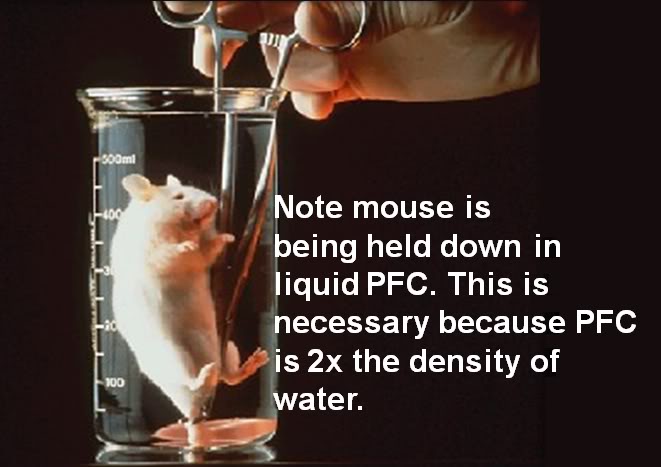
The smallest gas exchange units of the mammalian respiratory system, the alveoli, are a mere ~125 µ in diameter and, absent molecular reinforcement in the form of surfactant, are inherently unstable and collapse. Surfactant has evolved to function as a semi-aqueous thin film lining the alveolus and other small acinar structures (such as the terminal bronchioles and the alveolar ducts) (Figure 1-17) and it is dissolved, damaged, or inactivated when exposed to water, and to other molecules typically found in body fluids such as proteins and lipid particles. Surfactant has both hydrophilic and lipophylic components which impose the seemingly impossible constraint that any liquid introduced into the lungs must be both hydro- and lipophobic, as well as virtually completely chemically inert.

 A Brief Précis of the History and Development of Liquid assisted Pulmonary Cooling (LAPC) for Induction of Hypothermia during CPR

So far, only one class of compounds, the perfluorocarbons (PFCs), meets these requirements. With the demonstration in 1966 by Clark and Gollin that mice could survive extended periods of ‘liquid breathing’ with the PFC FX-80 (FC-75; perflurotetrahydrofuran) the feasibility of using liquids as gas exchange media began to be explored. Despite the (in retrospect) painfully obvious implication that PFCs could be used in the lungs for heat exchange as well as gas exchange, the idea did not occur to the author until sometime between the winter of 1993 and the spring of 1994. This happened while rereading Guyton’s Textbook of Medical Physiology during the course of several transcontinental flights across the United States (US). The author had been aware of Leland Clark’s ‘fluorocarbon breathing mice’ since shortly after these experiments were carried out in 1966; they were the subject of extensive media coverage, including dramatic footage shown on science-oriented television programs of the time (such CBS’ ‘The 21st Century’). Yet, it was not until re-reading Guyton’s chapter on respiratory physiology that the idea of using the lungs as a liquid-based heat exchanger became clear. Having spent literally 22-years obsessed with the problem of how to rapidly, non-invasively, and relatively simply, induce hypothermia in humans under field conditions; in hindsight it seems utterly obvious to use tidal liquid ventilation (TLV) employing a refrigerated PFC as a way of achieving rapid, homogenous, systemic cooling.

Figure 1-18: Mice can briefly tolerate breathing the PFC FC-77 but soon succumb to exhaustion from the work of breathing and hypothermia from the ambient temperature liquid.
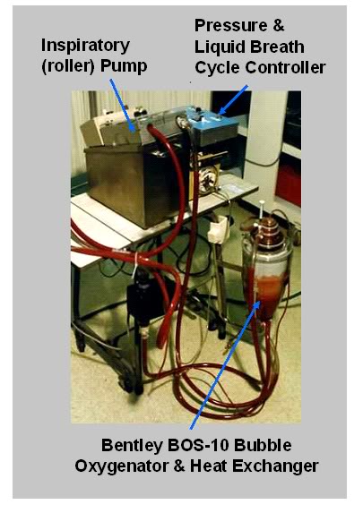
An investigation of the literature disclosed the feasibility of TLV based on the work of Shaffer and his colleagues. [155]  Unfortunately, acquiring a PFC suitable for liquid assisted ventilation was very problematic. The only commercially available PFCs at that time were the 3M Fluorinet™ fluids and Rimar-101 (the latter manufactured by the Miteni Corporation in Milan, Italy). Neither company distributed their products through the conventional chemical houses[6]. The reason for this was that both 3M and Miteni specifically embargoed the sale of their PFCs for any research application involving biological systems – and in particular any application that involved liquid assisted ventilation (LAV) or O2 carrying blood substitutes (oxygen therapeutics).

By January of 1995, the author and Respiratory Therapist Michael Fletcher had constructed the first TLV ventilator for research into inducing MTH during CPR, with the idea very much in mind that this technology had tremendous potential for broad medical application (post resuscitation syndrome, stroke, subarachnoid hemorrhage (SAH) and possibly traumatic brain and ischemic (and traumatic) spinal cord injury (TBI and SCI).

Figure 1-19: The first TLV apparatus for cardiopulmonary cerebral resuscitation-related research (left) used blood as the gas exchange medium. The device used a bubble oxygenator and heat exchanger and relied on gravity to provide ‘exhalation’ drainage from the lungs. The inspiratory roller pump and the timing and pressure controller functioned well, but blood proved to be too injurious to the lungs t serve as a gas exchange medium. These experiments made the need for an expiratory pump obvious. Photo: circa February, 1995.
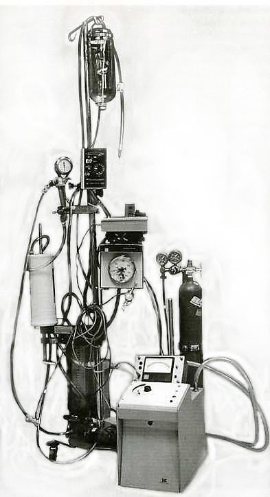
Because of the unavailability of PFCs, and because of the prior liquid breathing research by Johannes Klystra  [156],[157],[158] which demonstrated that mammals could survive after breathing a balanced salt solution under hyperbaric conditions (where gas solubility in water is dramatically improved), we decided to use bovine blood as the gas exchange medium in a non-survival canine model of TLV. Blood, unlike saline, has the proven capacity to deliver and exchange an adequate amount of O2 and CO2under normobaric conditions. A review the literature (then and now) indicated that this had not been attempted, or apparently even proposed. These two initial experiments using bovine blood in an acute canine model of TLV demonstrated that blood was not suitable for use, and that TLV was a demanding technique that required an exhalation pump in addition to an inspiration pump. The prototype device taught us a great deal, and in fact, the control unit from that first TLV ventilator was used until 2000 to conduct much of the early 21st Century Medicine, Inc., and Critical Care Research, Inc., work on liquid assisted pulmonary cooling (LAPC).

 Figure 1-16: Prototype tidal liquid ventilator using the ‘sweep flow’ (continuous liquid delivery/removal to the carina) technique circa 1995, and the active compression-decompression, high impulse Thumper™ mechanical CPR device (Figure 1-17) used to facilitate tidal movement of PFC between large and small airways (below). [Photo (left) courtesy of Charles Platt, 1995.]
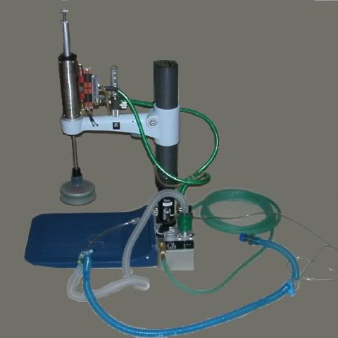
By March of 1995 several PFCs had become available in sufficient quantity for use in a canine model of TLV. A continuous flow approach was being used to deliver chilled PFC to the carina, with tidal movement of liquid in and out of the small airways being achieved by active compression and decompression of the chest wall using the suction cup of an Ambu CardioPump™ on a specially constructed, pneumatically driven Thumper™ CPR device. This technique was christened ‘sweep-flow liquid ventilation.’ [159]

Figure 1-17: Custom fabricated Michigan Instruments, Inc., Thumper™ active compression decompression cardiopulmonary resuscitator
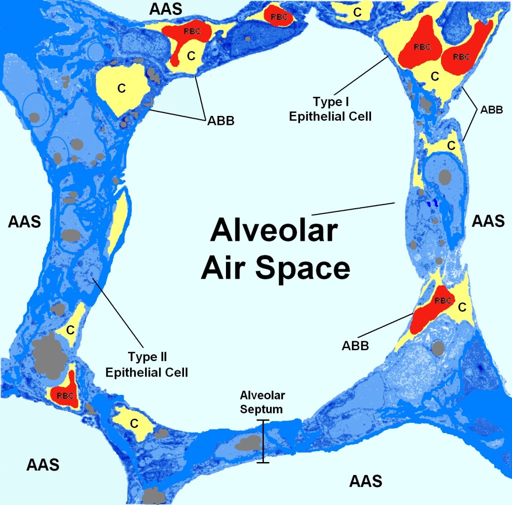
Dynamically sensing and controlling the amount of PFC in the animals lungs proved problematic with this system, and despite a very high predicted minute alveolar ventilation rate (with 80 to 100 liquid ‘breaths’ per minute from chest compression-decompressions), and minimal anatomical dead-space (the tip of the liquid delivery catheter was at the level of the carina), hypercarbia developed after the first 10 to 15 minutes of ventilation.

While survival of animals following TLV using PFCs was 100%, with a low incidence of adverse effects (providing TLV was discontinued prior to lethal levels of hypoxemia), the logistics of applying TLV to a field-setting under emergency conditions was deemed insurmountable. This was particularly true in the mid-1990s due to limitations on both portable computing power and the lack of adequately miniaturized and lightweight sensing and servo-control technology.

During the fall of 1996, the author learned of the development and initial clinical application of a new technique of liquid assisted ventilation (LAV) called partial liquid ventilation (PLV) which was being used to treat respiratory distress syndrome in (primarily) premature infants. This work was rapidly extended to include adults, and in the Spring of 1996 the author attended a three-day conference and workshop: the ‘Basics of Liquid Ventilation Management of Severe ARDS, ECMO, Liquid Breathing and PCIRV Conference’ in Ann Arbor, MI.[7]  In contrast to TLV, PLV was effected by simply adding PFC to the patient’s respiratory system until it was filled to functional residual capacity (FRC, ~30 ml/kg) while continuing conventional mechanical ventilation with gas; PLV was the essence of simplicity PFC was added to the endotracheal tube with a syringe until the desired volume was given or a meniscus of PFC appeared at the desired level in the ET tube. What is more, as the author had already observed while rounding at the University of Michigan Medical Center’s ICU, due to their unique physical properties, PFCs had the ability to rapidly reverse the hypoxemia and hypercarbia that accompany respiratory distress and its attendant pulmonary edema. Thus, PLV offered the added promise of restoring adequate gas exchange in fulminating pulmonary edema, as well as serving as a safe and effective heat exchange medium.

Early in 1997, due to the technical and logistic difficulties imposed by TLV, the author began to investigate the use of multiple lavages of the lungs in dogs using PFC chilled to ~4ºC. In this technique the animals’ lungs were filled with ~4 ºC PFC to vital capacity (VC, 60-70 ml/kg) following which the PFC was suctioned out and this process was repeated with additional loads of ~4ºC PFC until the desired core temperature was reached.[159]  While technically straightforward and very simple in application, this approach required large amounts of chilled PFC and had the obvious potential to interfere with gas exchange under conditions of hyper-metabolism, oxygen debt, or hypercarbia. Additionally, the time required for loading and suctioning out 60-70 ml/kg of PFC did not appear to be consonant with maximizing heat exchange, given that much of the PFC load would necessarily remain in the large and intermediate sized airways, where heat exchange is very slow compared to in the alveoli.

At this point it became apparent that a practical, field-applicable method of inducing hypothermia by PFC lung lavage would involve a hybrid approach, wherein both liquid and gas tidal ventilation would have to proceed simultaneously, or on a rapidly alternating basis. This work was largely set aside at that time (1997) due to a research commitment to develop a multi-modal method of dealing with ischemia-reperfusion injury attendant to prolonged cardiac arrest. This model involved the use of multiple drugs, as well as the rapid (~15 minutes) induction of mild post-insult hypothermia using the Safar, et al., [160] model canine model of cardiac arrest and resuscitation, which employed CPB for the induction of hypothermia.

 Figure 1-18 (above): The alveolar capillary unit is the fundamental repeating structural unit of the lung. Above is a schematic rendering of an alveolus as it would appear magnified ~3,000 times. The polyhedral alveolar air spaces (AAS) are separated from each other by the alveolar septa which are ~15µ thick. The Type I epithelial cells overlay the capillaries (C) and constitute the air side of the airway-blood barrier (ABB). The capillaries are the yellow spaces with the red blood cells (RBCs) inside them. The Type II epithelial cells are heavily vesiculated and secrete surfactant which maintains the structural integrity of the alveoli and prevents them from collapsing during exhalation. The total thickness of the ABB is only ~0.5µ which means that heat exchange between blood and PFC at the alveolar wall is extremely rapid – the rate limiting factor on heat exchange is, in fact, the rate at which PFC can be moved into and out of the alveolus.
With the success of this post-resuscitation hypothermia and multi-drug research cerebral resuscitation model in 1998, it became important to demonstrate that very rapid, in-field induction of hypothermia was technically and logistically possible. A decision was made to return to the LAPC research in order to validate the feasibility of this approach for immediate, simple, post-arrest induction of MTH (~34 ºC).The first results of the work were presented in1999 at the 28th Educational and Scientific Symposium of the Society of Critical care Medicine in San Francisco, CA. [161]  At the time, it was anticipated that completion of the research would take approximately 6 months.

The team that began this work consisted of the author, Steven B. Harris, M.D., Sandra Russell, BS, Michael Fletcher, R.T., Joan O’Farrell and Brian Wowk, Ph.D.  This project proved more formidable than initially anticipated and ultimately consumed over 2 years of full-time effort in order to develop the core elements of a workable technique, and achieve a basic understanding of the physiology and pathophysiology of fractional tidal liquid assisted pulmonary cooling (LAPC); the technique that was ultimately developed. This team worked together seamlessly to establish the effective volumes of PFC to be loaded and unloaded (i.e., fractional liquid tidal volumes), optimum dwell times for PFC, tolerable peak and mean airway pressures for ventilation during liquid cycling, workable gas ventilation strategies, and the basic mechanics of both heat and gas exchange using this technique.

 What LAPC Can Potentially Deliver

Properly implemented, LAPC would offer the following advantages:

  • Ease of application, requiring far less highly skilled personnel than are needed for CPB,
  • Technically less demanding, requiring fewer total personnel than CPB,
  • Effective at achieving a rate of heat exchange in the brain comparable to or better than that achievable with CPB,
  • Effective at achieving good gas exchange even with patients with severe lung disease/injury (pulmonary edema, acute lung injury (ALI) and acute respiratory distress syndromes (ARDS),
  • Relatively inexpensive to use,
  • Improves oxygenation and ventilation,
  • Reduces interfacial surface tensions (i.e., between liquid and gases and liquids, gases and alveolar membranes),
  • Reduces ventilation pressures,
  • Recruits alveoli,
  • Redistributes pulmonary blood flow and mitigates V/Q mismatch,
  • Mitigates barotraumas and volutrauma,
  • Lavages airway and alveolar debris,
  • Reduces inflammation and lung injury.
  • Allow CPR  to serve as a viable bridge to CPB and eventual definitive therapy to restore spontaneous circulation or supplement or replace it as necessary (i.e., LVAD or TAH)

The physiological considerations underlying LAPC are these:

1)    All of the blood that flows out from the heart to the body flows through the lungs first, where it is oxygenated and carbon dioxide (CO2) is removed. In order to facilitate gas exchange, the architecture of the lungs has evolved to provide an enormous surface area combined with an ultra-microscopic barrier between blood and air. The alveoli, the gas exchange units of the lungs, are polyhedral compartments ~250 µ in diameter which branch out from the terminal bronchioles (which are about twice the diameter of an alveolus; 250µ) (Figure 1-18, above). Each adult human lung contains ~300 x 106 alveoli with an aggregate surface area of 140 m2 [162] The alveolar epithelium is comprised of two types of cells; Type I epithelial cells that constitute the alveolar side of the air-blood barrier (ABB) and Type II epithelial cells which are intercalated in the epithelial lining and secrete surfactant. The Type I epithelial cells are so thinned out where they cover the capillary endothelial cells (which are even thinner) that the composite thickness of both cell layers and the basal lamina is ~0.5µ. These unique anatomical features of the lungs, combined with the physiological fact that they process all of the cardiac output, makes them ideal as a heat exchanger.

2) As previously noted, air, oxygen and other gases make extremely poor heat exchange media since they are roughly a thousand times less dense than water, they will remove heat at only roughly one thousandth the rate.

3) Mammals can survive briefly while spontaneously breathing perfluorochemicals (PFCs) and the usual cause of death when small mammals, such as mice, are left immersed in PFCs is hypothermia due to loss of heat from the large surface area of the lungs to the room temperature PFC in which they are immersed.[163] The severely injured lungs of human patients with pulmonary edema and/or acute lung injury (ALI) or acute respiratory distress syndrome (ARDS) not only tolerate extended (days to weeks) partial filling with PFCs of the appropriate vapor pressure, density, and viscosity, their ability to carry out gas exchange is improved, and there is evidence that long-term damage from ARDS may be reduced.[164],[165],[166],[167]

This technique of non-invasive in-field induction of hypothermia using the lungs is called liquid assisted pulmonary heat exchange (LAPHE), or liquid assisted pulmonary cooling (LAPC) when used for inducing hypothermia.

FOOTNOTES


[1] Perftoran is both a product and a company name; the company was formed in 1997 by the Ministry of Public Health and Medical Institute of the Russian Federation.  Perftoran is a perfluorocarbon-based oxygen carrier (PBOC), consisting of perflurodecalin, perfluoromethylcyclohexylpiperidine and proxanol and was approved for use by the

Pharmacologic State Committee on 24, June 1999.

[2] Oxygent has been licensed in the UK

[3] The author rejects the conventional designation of ‘sudden cardiac death’ because it is inaccurate; death is, by definition, the irreversible loss of life. Acute cardiac arrest is not death and the nomenclature used to describe it should reflect that fact.

[4] A few qualifying remark are in order here. This statement holds only when the new therapeutic modality fits well within the existing biomedical paradigms, and social and ethical milieus. Semmelweis and antisepsis in the 18th Century were vigorously resisted whereas the sulfanilamide and penicillin were rapidly embraced 60 years later. Few clinicians question the potential of hypothermia, or the biological basis of its therapeutic action, to the extent it is currently understood. What is questioned is, ‘is it worth it?’

[5]Data on cooling of humans during CPR is, of necessity, derived from the human cryopreservation program; implemented in the United States starting in 1967 as a controversial effort to achieve a form of ‘medical time travel’ for individuals experiencing medico-legal, but not biological death.

[6] e.g., Sigma, Fluka, Fischer, Mallinkrodt, Spectrum, Baker, etc.

[7] Basics of Liquid Ventilation Management of Severe ARDS, ECMO, Liquid Breathing and PCIRV Conference, Ann Arbor, Mi, March 8-9, 1996.

Posted in Cryonics Technology (General), Ischemia-Reperfusion Injury, Medicine | Tagged , , , , , , , , , , | Leave a comment

Cryonics “Castle”

By Mike Darwin

 Show: “Castle”

Season: 4

Episode: 3, “Head Case”

Air Date: 10/03/11

Series Creator: Andrew W. Marlowe

Writer: David Grae

Characters: Rick Castle, Kate Beckett

Location: Los Angeles, CA

Photos Credit: ABC/Adam Taylor

“Castle” Stars: Nathan Fillion as Richard Castle, Stana Katic as NYPD Detective Kate Beckett, Susan Sullivan as Martha Rodgers, Molly Quinn as Alexis Castle, Penny Johnson Jerald as NYPD Captain Victoria Gates, Tamala Jones as Medical Examiner Lanie Parish, Jon Huertas as NYPD Detective Javier Esposito, and Seamus Dever as NYPD Detective Kevin Ryan.

Guest Cast: William Atherton as Dr. Ari Weiss, Andy Umberger as Johnny Rosen, Judith Hoag as Cynthia Hamilton, Shaun Toub as Dr. Philip Boyd, Jordan Belfi as Beau Randolph, Jared Hillman as Eddie Peck.

NOTE: You can watch the full episode of “Castle” reviewed here on line at no charge: http://www.hulu.com/watch/282426/castle-head-case


ESSENTIAL BACKGROUND

 Figure 1: Stana Katic as NYPD Homicide Detective Kate Beckett enters the Passage cryonics facility with her gun drawn in search of the missing body of a homicide victim.

 I’m a regular viewer of “Castle,” so my take may be prejudiced. “Castle” is very light TV fare – it is a fanciful police procedural comedy/drama that centers on the adventures of a crime novelist, Rick Castle, who is on a perpetual “ride along” with an attractive female homicide detective named Kate Beckett. Beckett has served as the inspiration for one of Castle’s most successful characters, Detective Nikki Heat. Superficially, “Castle” is escapist fare that offers some respite from sadistic pornography of “Criminal Minds” or the now predictable, hard-boiled and increasingly preachy cynicism of the “Law and Order” franchises.“

Castle” is a throwback to the humorous, but morality and issue driven “detective” writing of Anthony BoucherHerbert Brean, and perhaps the most talented master of this genre, John Dickson Carr. As critic S. T. Karnick has aptly said of “Castle”and its predecessors:

“What these and their contemporaries excelled at was creating a sense of wonder, building a fantastic situation that has an inexorable logic of its own. In their way, they conveyed a sense of American life as a realm of astonishing possibilities ultimately grounded in common sense, logic and morality. It’s a form of fiction I enjoy greatly and which I think has much to recommend it.”

As TV fare it has more in common with “Ellergy Queen” or “Murder She Wrote” than CSI or “Blue Bloods.” Often I hear more of “Castle” episodes than I see of them, but it is good, non-traumatic, “wind down” entertainment before bed; nice to watch while reading book and preparing to doze off. However, a “good” episode will cause the book to be put down and will fully command my attention. This was a good episode of “Castle”, in fact I would argue that it was an extraordinary episode. I say this because “Castle” doesn’t usually explore ideas in any nuanced way, other than those surrounding romantic and family life, which are the core values the show seeks to explore, albeit 21st century style.

Castle is a highly successful author, divorced, more than a bit juvenile and a something of rake who finds his way into the beds of the occasional vixen who strays into the plotline. Detective Beckett is the serious, sober and grounded half of the duo, whose job it is to burst the bubble of most of Castle’s outrageous, and usually erroneously wild conspiracy theories of the crimes they encounter together.  The emotional subtext is that Castle is madly in love with Beckett, Beckett is arguably is in love with Castle and neither has the confidence in themselves, or their life choices to admit these feelings to each other, or to anyone else, for that matter.

Because “Castle” is, at least superficially not a serious TV drama, the idea of a cryonics-themed episode made me squirm more than a bit. The whole idea screamed “clichéd mockery.” As it turned out, this episode was some of the best cryonics-themed TV programming I’ve ever seen – at least in terms of thoughtfully exploring the multiple significant issues cryonics poses to the culture. Without as doubt this episode’s presentation of the emotional and value-driven reasons for why we cryonicists are doing what we are was the most accurate and moving of any I’ve seen  to date.

Figure 2: Nathan Fillion as the crime fiction writer Richard Castle exploring the cryonics facility. Passage Cryonics either has really bad Superinsulation, or they just finished filling every dewar in the facility.

CRYONICS AND HOMICIDE INTERSECT ON TV (AGAIN)

The plot line (warning, spoiler alert) is that a murder has occurred in a New York City street, but there is no body; just so much blood on the scene that the victim would have almost completely exsanguinated. Through various twists and turns, the victim is determined to be an academic who was pursuing promising research on a life extension technology that would add ~ 10 healthy years to a person’s life by causing the body’s dividing cells to produce young, rather old replacements for themselves. The identification of the likely victim leads Beckett, Castle and crew to a “self storage warehouse” where they discover an “under the radar” cryonics facility called “Passage.”

       Dr. Weiss: “He conducted cutting-edge research developing        life-extension techniques.”

       Castle: “Not that it did him any good.”

It was at this point that I started to grin.  This set-up precisely describes Alcor and its location from mid-1970s to the mid-1980s in Fullerton, CA. What’s more, the first man ever cryopreserved, James H. Bedford, was stored for a number of years by his family in a San Fernando Valley mini-warehouse that was part of franchise called “Self Storage;” something I found more than a bit of an irony at the time. Could the “Castle” writers have done their homework that well? Surely not; but, it was good for a grin, anyway.

Figure 3: Seamus Dever as NYPD Detective Kevin Ryan (left) Kate Beckett (center) and  Jon Huertas as NYPD Detective Javier Esposito draw a bead on the two cryonics technicians who are in the process of placing the missing homicide victim into long-term cryogenic storage.

Almost immediately after entering the cryonics facility, the homicide investigative crew encounters the Passage personnel sliding the missing murder victim into a dewar. Beckett informs them that the police are going to take custody and that the Medical Examiner (ME) will need to autopsy the body. Enter the smarmy, self-righteous and utterly self-assured President of Passage Cryonics, accompanied by his even more self assured, viperous and lawsuit threatening caricature of a lawyer. Remove the patient from cryopreservation (yeah, they actually use that word; we’re making progress) and Passage will sue the NYPD and the ME’s Office into financial oblivion! the attorney informs them.

       Castle: “You got any celebrities in here? Ted Williams? Jack Frost”

Beckett and crew phone the District Attorney for a warrant to seize the body, only to be told that, “the case law is murky on the issue of whether or not a coroner can autopsy a cryonics patient.” Incredible!  now the writers really have my attention, because the Dora Kent case was not a “recorded” case that definitively established precedent; the California Appellate Court let the lower (Superior) court’s ruling stand, but declined to grant the case “precedent setting status.”[1] Maybe these guys really did do their homework after all!

ART IMITATES LIFE

The researcher/patient’s wife is questioned by Detective Beckett (and Castle) and she comes across as a sympathetic person who wants, above all else, to defend her husband’s cryopreservation and ensure that he has another chance at an indefinitely extended life. In fact, she reminisces during her interview that, when she and her husband first met, he told her that he was so in love wit her that “one lifetime would never be enough” – he wanted to spend eternity with her – and life extension and cryonics were the tools to achieve that end.

Figure 4: At left, fictional pornographer Beau Randolph as portrayed by actor Jordan Belfi and at right, real pornographer Larry Flynt who did indeed at one time have a serious interest in cryonics. [2)

However, as it turns out, the ME may not need to do an autopsy after all. One of the victim’s associates, a famous pornographer who created the “College Girls Gone Wild” franchise has been bankrolling the life extension researchers academic’s work. And tellingly, they’ve just argued repeatedly over the “dead” man’s desire to make his life extending discovery “open source” for the entire world to further advance and benefit from. By now, I’m chuckling. Is this a reference to Hustler’s Larry Flynt? I’m beginning to think that I’m starting to see my life played out on a very B-list (but nevertheless amusing) TV show. [2]

 Figure 5: At left actor William Atherton as Dr. Ari Weiss, CEO of Passage Cryonics (shades of Avi Ben Abraham, center?) and at right, Dr. Max More, CEO of the Alcor Life Extension Foundation. [3]
Alas, the pornographer owns a gun, fired the very morning of the murder, that could possibly be the murder weapon. The cops need the slug, and the slug is in the cryopreserved body of the victim.  Cut to a testy conference between the ME, the cops, the CEO of Passage Cryonics and their oily lawyer. The ME insists on an autopsy of the body and it’s clear that she now has probable cause and will likely get the necessary court order. Suddenly, the Passage CEO stands up and announces that he has the answer; all cryonics requires is the brain, so why not give the ME the body for autopsy and allow the cryonics organization to keep the head? Now, I know the writers have done their homework. [3,4] One, two, or three coincidences? Maybe. But this many? Not a chance!

Figure 6: At right, Tamala Jones as Medical Examiner Lanie Parish discloses the results of her autopsy on the headless body of the murder victim. A plot lifted right from the Dora Kent case. [4,5]

A TANGLED STORYLINE

The head is removed, the body is autopsied, the slug is recovered, and, just like in real life (Larry Flynt), the wily pornographer is off the hook; they can’t pin the crime on him because he didn’t do it. He was, as he told the police, otherwise occupied murdering a noisy pigeon on his roof that morning. A compliant of animal cruelty is sworn out against him and he vanishes from the proceedings.

The once cooperative ME now demands the patient’s head, because, as it turns out,  he appears to have been serving as his own guinea pig by having the implants that cause tissue rejuvenation placed in his brain. The 0nly problem is that when the investigators go to retrieve the head (patient), surprise, he’s missing from the cryonics organization’s facilities!

       Beckett: Are you saying you lost his head?

So, who took him and why? It is soon discovered that the patient’s researcher friend has removed him from Passage in order to prevent his destruction by cranial autopsy. What was really going on was that the patient was dying of an inoperable malignant brain tumor (glioblastma multiforme) and this colleague was undertaking to try and save him with a highly experimental, and unfortunately, ineffective nanoparticle cancer treatment. Our cryonics patient was thus doomed to die of a brain tumor – a brain tumor that would, before it killed him, utterly destroy his brain, thus making any hope of recovery from cryopreservation impossible. So now, in addition to the Dora Kent case, the writers have folded in the Donaldson v. the Attorney General of the State of California case. [6]

The nano-cancer researcher colleague explains to the homicide investigators that even though the tumor was growing rapidly, the patient had decided to continue pursuing his life extension research and forgo being cryopreserved. He turns over the MRIs and other documentary evidence explaining why trace evidence of brain matter from the patient had been found in a secret lab, ending the need for further postmortem dissection.

Revelation of these facts also explained the seemingly anomalous download of a “cryopreservation cancellation document” for Passage Cryonics, recovered from the patient’s laptop. Finally, it dawns on Castle and Beckett that the shooting that ended this life cycle for the patient was the very thing that might be responsible for saving his life. They correctly reason that if he wasn’t cryopreserved while his brain was reasonably intact, then he would be lost forever.

THE 21ST CENTURY ROMEO & JULIET

Figure 7: 21st Century Romeos and Juliets use cryonics as a way to overcome the tragic circumstances of disease and death which threaten to separate them forever.

Bingo (!); the missing motive in the case in now apparent. If his wife was aware he was not only dying of brain cancer, but also that he planned to terminate his cryopreservation arrangements, then the only way she could hope to ensure their future together was to “kill” her husband now, while both his brain and his cryopreservation arrangements were still reasonably intact.

This was, in fact, exactly what she had done. As the show winds up there is a touching and very emotional monologue from the wife explaining that the tumor had warped her husband’s judgment and that he was no longer making decisions as he had when was well; she had no choice but to stop his heart with a gunshot, triggering his GPS-enabled bio-monitoring watch and summing the cryonics team.  The wife is placed in a holding cell and Castle, Beckett and the Passage President confer about the situation. Suddenly, the Passage CEO’s smartphone registers an alarm: a Passage client has experienced cardiac arrest, butit makesno sense since the GPS feature shows the location as right there in the jail.  It is quickly discovered that the wife has taken a cyanide tablet concealed in a ring she was wearing.[1] The wife lies lifeless on the floor of the cell and there is a moment of stunned silence, broken by the Passage CEO, who pleadingly asks if he can summon the cryonics team so that the wife can join her husband on the long journey into the future. Beckett says, “Yes,” having already expressed her sympathy with the wife for her act of “involuntary euthanasia” that put in him Passage cryonics with two bullets in his chest at the start of the story.

Whew! Every significant medico-legal issue in the public history of cryonics to date, all rolled into one ~ 45-minute long TV episode! That’s quite a feat! But a much more impressive one was that writer and the creator of “Castle” got all the important things right. No, they didn’t get much the technical side of cryonics right, and for that, we may arguably be thankful. The Passage cryonics patients, unlike the real ones, look like very startled solid-state versions of their living selves. This is the first time I’ve ever seen cryonics patients depicted with their eyes open – wide open, in fact.

But the shortcomings in the technical depictions of cryonics were more than compensated for by the fact that the show’s creative talents got the core messages of cryonics right. Medico-legal death is a process not a condition, and “irreversibility” is a function of brain structure and the sophistication (or lack thereof) of available medical science and technology. Life is a good thing, and the desire for indefinitely long and healthy lives, free from the burdens of aging, disease and death are reasonable goals being pursued by reasonable people. Indeed, they are romantic goals and they are technologies that offer everyday people the opportunity to continue expressing the best and brightest of their humanity; their love of each other, their pursuit of knowledge and growth, and their desire to transcend time.

Wow! That’s a lot, coming as it is from the principal engine of the popular culture: television. We cryonicists owe a sincere debt of gratitude and some heartfelt thanks to the writer, director and the  producers of this “Castle” episode.

Please, write them and communicate your appreciation:

http://beta.abc.go.com/shows/castle/discuss#linkIdAll

REFERENCES

1.     Alcor Life Extension Foundation, Inc. v. Mitchell (1992) 7 Cal. App. 4th 1287 [9 Cal.Rptr.2d 572]:http://law.justia.com/cases/california/caapp4th/7/1287.html. Retrieved 2011-09-05 .
2.    Green, M. Her death ends the improbable love match of porn merchants Althea and Larry Flynt.  People Magazine, 28(3);1987: http://www.people.com/people/archive/article/0,,20096764,00.html.  Retrieved 2011-09-05 .
3.    Cieply, M. Iraquis ask firm about cloning Saddam Hussein. Los Angeles Times, 09 September, 1990: http://articles.latimes.com/1990-09-09/business/fi-718_1_saddam-hussein. Retrieved: 06 October, 2011.
4.    Babwin, D. Coroner says lethal dose of drugs killed cryonics case figure. The Press Enterprise, Riverside County, CA, 28 February, 1988, start page: A-1.
5.    Perry, R.M., our finest hours: notes on the Dora Kent case. http://www.alcor.org/Library/html/DoraKentCase.html. Retrieved: 06 October, 2011.
6.    Donaldson v. Lungren (1992) 2 Cal. App. 4th 1614 [4 Cal.Rptr.2d 59]: http://law.justia.com/cases/california/caapp4th/2/1614.html. Retrieved: 06 October, 2011.

FOOTNOTE

[1] A great deal of suspension of disbelief is required in watching in CASTLE; as anyone who has ever been arrested, or who is familiar with police procedure knows, all jewelry and other possessions, right down to hairpieces (but generally excluding corrective eyeglasses and dentures) are removed from any subject taken into custody.

Posted in Cryonics Philosophy, Culture & Propaganda | 14 Comments

Doing the Time Warp

By Mike Darwin


It’s astounding;
Time is fleeting;
Madness takes its toll.
But listen closely…

Not for very much longer.

I’ve got to keep control.

I remember doing the time-warp
Drinking those moments when
The Blackness would hit me

And the void would be calling…

Let’s do the time-warp again.

             — “The Time Warp,” Rocky Horror Picture Show by Richard O’Brien.

Yesterday, on Cryonet2, a post caught my eye and ended up having a special resonance for me. The subject under discussion was a media story about a man who had been in prison for ~20 years and who, upon his release, found it so difficult to adapt to the technological and social change that occurred during his time in confinement, that he set fire to an abandoned building in order to be returned to prison for a good long time. The person commenting on this article wrote, “I find the story is bit hard to swallow, having some familiarity with the general issue. Prisons have televisions. Cell phones are commonly smuggled into prisons, and computers are common in prisons, including some for prisoner use.”

At first blush, his incredulity seems justified, and even with deeper consideration his skepticism may seem appropriate, because people from Bronze Age cultures can and do adjust just to cultural change and displacement far more massive than that prisoner experienced. For example, some of the Hmong People from Vietnam successfully made the jump to the US at the end of the Vietnam War (by 1978 some 30,000 were living in the US) and many actually out-competed their new compatriots in the US.

Figure 1: The Hmong people of South Vietnam lived a culture suspended in time between the Stone and Bronze and ages in the 1970s. Above is a typical Hmong village from that time period.

Figure 2: A Hmong village in southern Vietnam in 2004.
Nevertheless, the phenomenon of the culturally and temporally displaced prisoner who is unable to adjust to a changed world is, in fact, a commonplace that has been observed for many years in US prisoners who’ve served long sentences – certainly, since the 1940s. It is very real, and television and conversation with other recently interned inmates does little to relieve it, at least in some people. There is a huge and material difference between seeing a novel technology in use, and experiencing the transformative effect it has, not only on your life in general terms, but on your way of thinking and behaving. The science fiction author Larry Niven captured something of this when he posited “flash crowds” as an unforeseen social effect of teleportation technology. As it turn out, Niven’s “flash crowds” didn’t have to wait teleportation: the development and widespread application of smartphone and social networking technologies have not only created flash crowds, but enabled flash mobs that propel revolution forward (the Arab Spring) or coordinate riotous looting (the London Riots of the Summer of 2011).

This phenomenon should arguably be of special interest to cryonicists , because we propose to cross many decades or even centuries or longer, without even the advantage of network television programming, or tales of the outside world told by the newer inmates on our ward, or cell block. Because the average person is inescapably enveloped in and carried along by the time stream of the culture he inhabits, it is unlikely that he will have any experience of what it means to be cut off from technological advance and the enormous cultural change that accompanies it.

It’s so dreamy, oh fantasy free me.
So you can’t see me, no, not at all.
In another dimension, with
voyeuristic intention,
Well secluded, I see all.

With a bit of a mind flip

You’re into the time slip.

And nothing can ever be the same.

You’re spaced out on sensation.

Like you’re under sedation.

Let’s do the time-warp again.

                    – “The Time Warp,” Rocky Horror Picture Show by Richard O’Brien.

You can get a potentially deadly taste of this by traveling back in time by the expedient of “geographical atavism,” which is what I call going to places on earth where the technological level is decades, centuries or millennia earlier than the present, and then living there. This is getting difficult to do, since even the lepers in India now have cell phones.

You can also do it by virtue of living off the street, either in the US, or preferably in a “foreign” country. At first, you try to reach for all sorts of technology that isn’t there, and only gradually do you stop doing this and realize that you are now fundamentally different from them – e.g., all the other people in the world. You can no longer communicate effortlessly any further than you can shout. There is no medical care beyond basic first aid, and every step you take is taken with the knowledge that a misstep could be lethal. You are hot when it is hot, and you are cold if you haven’t prepared well, and you are still cold often even if you have prepared. If you can’t find food to eat you are hungry, or you have to ask (beg) for food.

Figure 3: Marks and Spencer was originally a moderately upscale department store chain in the United Kingdom. In recent years they have branched out into selling high quality foods, including “luxury” sandwiches and prepared meals under the brand name of M&S Simply Food. M&S adheres to a rigid policy of discarding most unsold prepared foods at the end of each business day, as well as to discarding baked goods, chocolates, flowers and most other food items at, or just beyond their peak of freshness. This creates the opportunity for “no cash outlay” gourmet meals for those with a dustbin key, a thick hide and a near total lack of “normal” social inhibition.
Figure 4: In the UK, as opposed to the US, dustbins (aka dumpsters in the US) are typically locked with a mechanism that looks a bit like a 3-sided Allen wrench (inset photo at left).  It seems deceptively simple to open, say with needle nose pliers, but this is not the case. The triangular post that operates the mechanism has rounded edges, is recessed and requires substantial force to turn. It is thus highly desirable, if not necessary, to have a “dustbin key;” an item which can be procured at some £ (dollar) stores for  £ 1 (about $1.60 US). This locking mechanism on UK dumpsters appears to be universal, and is considered in the same light as other keyed utility mechanisms, such as water, gas and electricity.

Figure 5: Kitchen, or food waste, must be segregated from other waste in the UK. Typically, the chain merchants such as the Co-Op, M&S Simply Food, and Pret a Manger (another upscale ready-to-eat sandwich shop) conveniently over-bag their various types of food waste in clean, unused plastic bags – often double-bagging it. Food is also discarded on a fairly predictable schedule, so it is possible to retrieve refrigerated and frozen goods in pristine condition, whilst still safely chilled or frozen. The letters “KP” stand for “kitchen policing” wherein the word “police” is used as a verb to mean “to clean” or “to restore to order.”

 
Figure 6: A not untypical haul from an hour or so of foraging in the dustbin at M&S, the local fruit monger and one of the high-end green grocers. One broken egg in a carton of 6 or 12 eggs means the entire lot is discarded. Since I don’t eat land vertebrates, my flat mate had a steady supply of choice cuts of meats – fresh and frozen.
 Figure 7: Living “off the street” as a latter day hunter-gatherer carries with it wholly unexpected difficulties in readjusting to the more typical existence of the technologically sophisticated work-a-day world. I had not previously understood why people would pay good money for something as trivial and ephemeral as fresh flowers. When I returned to the US this past July, after experiencing months of beautiful, sweet smelling flowers in the flat every day, I was disconcerted to find that I had acquired a costly and wholly unsupportable new taste.

Figure 8: Sadly, my difficulty in readjusting to “normal” life was not confined to missing the presence of the severed reproductive organs of plants. I found I had grown accustomed to what were, to me, gourmet meals: good bread and fresh fruit whenever I felt like having them. Above, cold potato leek vichyssoise, smoked salmon with organic string beans, an organic free range egg and Italian tomatoes with a vinaigrette dressing. I have never before (or since) eaten so well so consistently.
 Figure 9: Dishes, cutlery, pots, pans and household appliances were available in dustbins for the taking and with very little competition. The books, clothing, duvet, TV and 3-shelf stand in the photo at bottom right were all acquired from the street within a matter of ~2 weeks. In the UK a TV license is required to watch television – something that, fortunately, both of my flat mates possessed. Failing this, the advent of Blue Ray technology has caused conventional DVD players (and DVDs) to be treated as barely better than rubbish in many large cities in the US & UK.
If you live this way for quite awhile you become transformed. If you return to a world of truly enormous choices and possibilities, even if it is one you formerly inhabited with ease, it is very fatiguing, and it can be confusing and stressful, as well. Since time immemorial men have set on journeys of transformation and enlightenment. From the travels of Gilgamesh to Jesus Christ to Buddha, to the mythical travels of Swift’s Gulliver, all such journeys have in common the individual removing himself more or less completely from his normal environment and thus from his accustomed, culturally imposed way of experiencing and seeing his own life. And there is one more thing; they involve danger and some degree of hardship. The important lesson in this latter element for cryonicists is that any environment different from that in which you have grown to maturity in is a dangerous one. The nuances of other languages and cultures, and the even more subtle nuances of the myriad unspoken but vital cues for survival are necessarily inaccessible to the stranger in a strange land.

Many men who take such a journey return transformed – and sadly – inarticulate and unable to communicate what they have experienced. It is not uncommon for them to repeatedly return to such sojourns, or to attempt to rework the “mundane” lives they have returned to, in an effort to mirror the transcendence they have experienced during their journey(ies). This transcendence, so elusive and so impossible to put into words, consists mostly of the radical change in perspective that occurs when a person is removed from his “time stream.” If we grow up in a reasonably stable culture and remain there throughout our lives with our cohorts, our perception of reality necessarily becomes circumscribed.

The exigences of daily life act to preclude our living in the world that exists beyond our moment-to-moment experience of it. The time we spend in conscious contemplation of the distant past and the far future becomes negligible – we become both confined and defined by the time stream we inhabit; the unfolding of events that are largely determined by our culture and our cohorts. And so it is all over the world – different peoples in different lands, all existing at the same time, but in different currents, eddies and streams that are largely isolated from each other. This is a crippling state of affairs, because we either lose, or altogether fail grasp the larger perspective of the universe as a vast, complex place which is unfolding not only in myriad ways, but over myriad different timescales, as well. That reality has important implications for our survival, both as individuals, and as a species.

Well I was walking down the street
just a-having a think
When a snake of a guy gave me an
evil wink.
He shook-a me up, he took me by surprise.
He had a pickup truck, and the
devil’s eyes.
He stared at me and I felt a change.
Time meant nothing, never would again.

Let’s do the time-warp again.

       — “The Time Warp,” Rocky Horror Picture Show by Richard O’Brien.
 

Several years ago, I was digging through a dustbin in back of a charity shop in London. They were usually a good source for classical CDs (they toss any CD without a jewel case, and any that don’t sell in a fortnight). I found this device in the dustbin (Figure 10).

Figure 10: My first MP3 player was mistakenly acquired because I thought it was a jump drive. I was stunned when I puzzled out that this tiny device could store up to 100 popular songs and index and “shuffle” them! The carrying lanyard was a promotional giveaway handed out on the street in Soho during London’s Gay Pride celebration.

I pulled the end off of it and saw it had a male USB plug. I figured I’d found myself a jump drive – a brand new one, too, since it still had the adhesive protective plastic covering the little screen on the front of it. I took it home and hooked it up to my computer. It took me a fair bit of time before I could understand what it was; an MP3 player. In fact, I didn’t know what an MP3 player was. I did know about iPODs, but only from TV; because they are small, they are also inconspicuous, and I’d not really seen them up close, nor did I know anyone who had one. I had noticed that people no longer used CD players in public and that they, along with CDs, were now a commonplace in the charity shop dustbins (just as perfectly “good” Sony Trinitron, and other nice color CRT TVs are now a commonplace outside thrift stores in the US and the UK (they can’t throw then in the trash because of the heavy metal content, so they set them out to be carted away by people who can’t afford flat screen technology).

 The MP3 player I had found had something like a 100 songs on it! Imagine that! I had no idea that you could carry around a hundred songs, let alone hundreds, or thousands, on such a tiny thing! What was even more astonishing to me was when I realized that MOST of the volume of the device was consumed by the primitive mid-20th century AAA alkaline batteries that powered it. Gradually, I realized that I could get most of the music I liked for free on-line, or from my UK CD collection, and organize it such that I could have the music “match” my travels around London. I could go to the Design Museum, the V&A, or anywhere else I liked and create a perfect soundtrack of music, period or otherwise, to accompany me! In effect, I could make a personalized soundtrack for my life! I quickly realized that if I had several MP3 players, I could select from a nearly endless variety of “collections” to suit my mood – Edith Piaf, The Beatles, Louis Armstrong, torch singers from Dietrich to Sara Vaughn, or the Goldberg Variations (while dozing on a long train or bus ride).

The dustbins of charity shops (thrift stores in the US) are a seemingly inexhaustible source of all manner goods. Many urban charity shops have no laundry facilities and do not find it profitable to carry bedding, linens, or items of clothing such as underwear, socks, and the like – even if they are new and still in the packaging. These things are thus often discarded outright. Items not sold within a fortnight are also typically discarded, as are items that the shop chooses not to sell; medical supplies and equipment, some kinds of music or art, many types of books (most confine themselves to the trendy, bestselling authors and “coffee table” books; the rest are discarded, often still in the boxes). Often,. Whole households of goods flow into the shop as a result of the death of an elderly person whose relatives live far away, or who are uninterested. In such cases the overflow of goods (beyond the capacity of the shop’s shelves) passes immediately into the waste stream.  Furniture, dishes, every kind of household appliance and gadget imaginable, and “obsolete” technology such as CD players, low megapixel digital cameras, flatbed scanners, cordless and mobile (cellular) telephones are present in abundance, as are all manner of toys and child-related items (car seats, cribs).  Finally, and very importantly, anything that the staff who work or volunteer in the shops do not recognize, understand or value, is also discarded.

The shop where I found the MP3 player was staffed by elderly female volunteers. I quickly learned to seek out shops staffed in this manner, because they were almost a guaranteed source of the most exotic technological goods.  In a few short weeks I had accumulated 3 MP3 players, an ASUS EAH6670/DIS/1GD5 Radeon HD 6670 video card[1], two “tiny” digital recorders on neck lanyards and ~ 5 gigs of add-on memory for my laptop and my desktop computers, as well as half a dozen jump drives.

Interestingly, just as the charity store staff was blind to things of value they did not understand (and thus they discarded them), I soon discovered that the same phenomenon applied to me, and others like me, but in reverse. It was impossible for us to see things of value, sometimes of considerable value, even when they were right in front of our eyes, unless we knew what to look for! The ASUS video card was a prime example. There are countless electronic bits and boards in dustbins, and in this case, it took the savvy of a young man who played computer games to recognize the manufacturer, thus saving the card from being salvaged for its muffin fan and instead allowing it to make its way into my desktop computer.

Figure 11: An ASUS EAH6670/DIS/1GD5 Radeon HD 6670 video card. I picked it out of the dustbin for the muffin fan, only to be told it was likely a working video card.

 The technology embodied in the MP3 players was transformative in ways I had not even begun to understand from watching television – and I watch/listen to a lot of television. I had no idea that there were competing brands of tiny, non-hard drive, digital music machines – let alone that they had gotten so small. The people on the street that I interacted with surely had them, but I paid no mind, because I assumed that the earbuds they often sported were connected to radios. Radios had gotten very small; I knew that, because I found AM/FM radios the size of matchboxes in the dustbins frequently. But that was the limit of my understanding; even though I was immersed in a culture where such devices had become commonplace.

I don’t like social media, like Facebook and Linkedin (please, stop sending me spam for Linkedin!), but I do understand them, and I know how powerful they are. A guy in prison hasn’t a clue, and he can’t get a clue from TV, or from hearing about it from another inmate. In fact, his position is much like me and the MP3 player; no one saw fit to explain to me that such technology had evolved, let alone that it was so inexpensive and commonplace that MP3 players were given away as promotional items and might easily be so little valued as to be tossed into the waste stream.

Figure 11: In the UK, homeless people on the street are a rarity, compared to the US. Both the UK and most of Western Europe maintain social welfare programs that are readily available to almost all residents who want them. Three of the most common reasons that people refuse this safety net are substance abuse, having a dog, or some other unwillingness or inability, such as mental illness, to comply with the rules of Council housing or other government social welfare requirements (including criminal activities).

And, why would they? Such technology is a commonplace for those in the mainstream of the culture, something they take for granted, and it is not likely to be a topic of conversation except amongst peers. When you cease to be a peer, and you step outside of your cohort, you have exited one of the time streams that the rest of the world inhabits. If you have children, they will help keep you oriented and in sync with the culture. However, if you are isolated by prison (or by choice) in a rapidly technologically evolving world, you are in for some major surprises – and for no small amount of cognitive dissonance.

I am a technophile who became involved in cryonics as a child. I’ve lived my whole life in expectation (and largely in welcoming anticipation) of technological advance. Statistically, people who are in prisons, or who gather at watering holes such as urban dustbins, are very different from most of the rest of Western, “civilized” humanity. Many are emotionally or intellectually damaged, and most tend to “live in the moment.” Their event horizon extends only so far as the next cigarette, the next hit of spliff or Tina, and maybe to some consideration of where and how they will spend that coming night. They know almost nothing about the past, and are constitutionally unable to see beyond a few days, or weeks into the future. Many will start to forage for a tarp or a piece of plastic to protect them from the rain only when the sky clouds over, or it actually begins to rain.  This, as it turns out, is a critically important observation, because it points up the powerful leverage to be had by living in longer timescales.

The capitalist philosopher A. J. Galambos divided the timescales we humans inhabit in the following way: [2]

Trivial Timescale: Moment to activities and thoughts which dominate most of our daily awareness time; I need to make a phone call, check my mail, brush my teeth, get something to eat, go to the loo.

Personal Timescale: What kind of training should I take, whom should I marry, how should I plan for my retirement, how should I apportion my estate, how can I provide from my children and grandchildren?

Species Timescale: Concern and involvement with history, the environment, the future of mankind. The Species Timescale lasts as long as man himself.

Cosmic Timescale: How does the universe work, what causes the stars to shine, how long will the universe last, can we live forever?

Galambos correctly pointed out that while almost all of us (of necessity) spend most of our time in the Trivial Timescale, preoccupied with things of the moment, to the extent we transcend the trivial we gain power and control over the world around. Newton and Einstein may have spent only a brief moment of their total conscious time in the Cosmic Timescale, but the benefits in terms of technological advance were enormous and gave us not just the laws of Classical Physics and of Relativity, but the calculus, the tools for spaceflight and the capability for self annihilation with the hydrogen bomb.

So, in the sense that people inhabit prisons, or who are squatters or otherwise homeless, are so often condemned to live only in the moment, and thus exclusively in the Trivial Timescale, they are fundamentally different.They are “transtemporally crippled,” and in many ways have less foresight than a really clever dog or cat. It is not uncommon for such people to be unable to retain even very basic possessions, such as a sleeping bag, a CD player and essential toiletries such as a toothbrush, deodorant and a razor. “Now” pretty much encompasses their sphere of action with respect to the past and the future. To expect such people to stay technologically and culturally integrated with a rapidly changing world – especially when imprisoned away from it for decades – is akin to expecting your dog or cat to discourse learnedly on the nuances of Shakespeare, or to explain to your the excitement experienced whilst listening to Justin Bieber.

Of course, slipping out of the time stream is not confined to the sphere of technology, or to prisoners behind bars. It is the fate of every kind of exile everywhere. I have been long exiled from cryonics, and much longer still isolated from the social wellsprings (scant that they may be) that constitute cryonics organizations. Thus, I have no idea if many of the people with whom I once worked and socialized with are still alive, and if they are, where, what and who they are now. I wonder, often, at the anonymous case reports that appear on-line, and try to fathom if it is one of the many people I once knew so well, but that exile for over 20 years has left me isolated from? And those are just the people from my past

Figure 12: Frank Cole crossing the Sahara desert and the whole of the African continent, from the Atlantic Ocean to the Red Sea on camel in 1990. Cole was murdered by Tuareg bandits near Timbuktu, Mali, in late October 2000.
 Very recently, someone made a derogatory remark about a man I had not previously heard of. He was a cryonicist and a filmmaker by the name of Frank Cole (1954-2000). The commentator remarked on his stupidity for being “killed by bandits in Africa.” I was struck by this remark, because, even more astonishing to me than the discovery that MP3 players existed in 2007, was the discovery of even the possibility that a man like this Frank Cole, could have been a cryonicist. The idea was incomprehensible to me.

It took me quite awhile to find out something of who Frank Cole was, and it was not until I saw his searing final film, Life Without Death, that I think I began to grasp what he was about. But, truth to tell, it was not until I read his former lover, Anne Milligan’s reminiscence of him (see below), that I felt I fully understood him. And that made me very sorry that I had missed the opportunity to know him, because in his work I believe I see the same, almost otherworldly ability to see the culture and the world “we” inhabit from outside, above, below, or beyond it. And in his work I see the exact same vision of the loss of those we love as the penultimate evil, and of death as what it is; the ultimate horror and the ultimate evil.

Figure 13: Frank Cole as a young man.
 I think Cole would have understood when I say that that ability, or practice, if you will, is the perhaps the best psychological preparation possible for recovery from cryopreservation. It is not a place you can ever get to by watching TV, reading books, or otherwise attempting to escape the time stream you inhabit by being distracted from it.

To understand that kind of alienation and isolation, and to taste of its absolute irreversibility requires that you step completely out of the world you inhabit and go to another one that is embedded not just in a different time, but in a different era, and in a different place in space. To do that is, necessarily, to take a horrific risk, because where you will be is not a simulation, and there is no recall from error or mischance, and no opportunity for “a reboot.”

In writing this, I am reminded of one of the songs that was on that discarded MP3 player from the dustbin in London, when it came into my possession. It was by a British pop group called ‘The Enemy.’ It’s lyrics come to mind now, as I think of Frank Cole, the nature, fragility and arbitrariness of life, and how absolutely essential it is that we continue to transcend our accepted experience of it, forever and ever, even for trillions and trillions of years, as the Ancient Egyptians liked to say.

We’ll Live And Die In These Towns

Lyrics by The Enemy

You spend your time in smokey rooms
where haggled old women
with cheap perfume say,
“It never happens for people
like us you know.”
Well nothing ever happened on its own and well,

the toilets smell of desperation
the streets all echo of aggregation
and you wonder
why you can’t get no sleep
when you’ve got nothing to do,
and you’ve had nothing to eat.
Your life’s slipping
and sliding right out of view
and there’s absolutely nothing
that you can do well

We’ll live and die,
we’ll live and die in these towns
don’t let it drag you down
don’t let it drag you down now
we’ll live and die,
we’ll live and die in these towns
don`t let it drag you down
don`t let it drag you down now

Dirty dishes from a TV meal
that went cold from the wind
through a smashed up window
You can’t go out if anybody calls ya
cause you can’t have a bath
when there`s no hot water
and your friends are out
on the town again
and you ask yourself if it will ever end
and it`s all too much for your head to take
just a matter of time
before you break, well

We’ll live and die,
we’ll live and die in these towns
don’t let it drag you down
don’t let it drag you down now
we’ll live and die,
we’ll live and die in these towns
don`t let it drag you down
don`t let it drag you down now

now…
now…

we’ll live and die,
we’ll live and die in these towns
don’t let it drag you down
don’t let it drag you down now
we`ll live and die,
we’ll live and die in these towns
don’t let it drag you down
don’t let it drag you down now

Our critics often say that practical immortality will result in a world of boredom – in a world of eternal sameness inhabited by people making the same choices over and over again. There is merit to this criticism because success, a prerequisite for indefinite survival, breeds complacency. Even with lives as short and turbulent as ours in the developed West are today, it is easily possible to become anesthetized by the time stream we are embedded in. When this happens, we lose all consciousness of the bigger picture, indeed the true picture of reality and we risk losing our ability to transcend the Trivial Timescale and inhabit the Cosmic one, however briefly. Lose that and we lose our ability to survive. Men like Frank Cole remind us that while there is great peril in journeys of transcendence which allow us to step out of our given time stream and cultural imperative. However without them, we face the even greater peril of forgetting, or failing even to understand the complex, challenging and utterly alien nature of the universe as it really exists.

My Life with Frank Cole

October 4, 2009

I delivered this tribute at the Book Launch “Life Beyond Death: The Cinema of Frank Cole” & Film Retrospective sponsored by the Canadian Film Institute at the National Library, Ottawa Oct 3rd, 2009. Frank Cole was a Canadian Documentary Filmmaker who was killed in 2000 by bandits near Mali while crossing the Sahara Desert. Rick Taylor is a Professor at Carleton University, Author and Frank’s best friend.

Dear Rick,

I just finished Life Without Death. I read it in one sitting and was sucked down the rabbit hole. It’s a beautiful book and I especially wanted you to know how much I loved your memoir, Saltwater Road to the Sahara. Your lovingly recreated details brought everything back so vividly It was poignant and bittersweet

And thank you for portraying me with such kindness and especially saying that I loved him whole heartedly because I did, though in truth, I don’t often revisit those memories now, weighed down as they are, with the silent echo of words never spoken, with youth’s uncertainty and unbending pride.

I enjoyed the book immensely but I was sorry that no one had written about Frank from a woman’s perspective because that dynamic informed both his art and life. I don’t think a man, even you Rick, could fully comprehend what it was like to be Frank’s Eve, to be the snake, the seductive field of sleeping poppies. To inherit the complicated push pull of his relationship with his mother. To stand innocent against the charge that intimacy leads to complacency, loss of purpose, and ultimately loss of self.

And so I hope you’ll indulge me while I revisit the piece of Frank’s story that was also mine, through the lens of my sensibilities.

I met Frank in the late 70’s in response to a laundromat ad for a roommate. To say that Frank was different is, of course, an understatement. While it’s true that he seemed remarkably serious and mature for his age, there was something more. His clipped words were punctured with unnerving silences and delivered with an enigmatic assuredness that seemed to announce that he had not only cornered Truth but had it up against the wall by the throat.

The disarming combination of animal magnetism, a rejection of society’s conventions, and a driving intensity body-slammed those he met through their comfort zone. People either loved or hated Frank, they were never indifferent.

In those days he was the enfant terrible in the Algonquin Film program and our apartment became the meeting place of a never-ending parade of Ottawa’s counter culture, drawn by Frank’s aura. It was palpable – Life seemed to be to be more meaningful, more vibrant, and more exciting in his presence. I was captivated and determined. I set out to impress Frank Cole.

Though Frank was not traditionally handsome, there were plenty of women vying for his attention. With his love of the outrageous and the absurd, I sensed that he would be won over by nothing less than a grand gesture.

So one night I placed a small table outside his bedroom and covered it with linen cloth and formal place setting for one. Wearing only a fedora and boots, I perched on the plate and knocked. I can still hear him roar with appreciative laughter as he opened the door… In the morning he took photographs and wrote a terse and clinical account of the night.

That was my introduction to Frank as the outsider. At 24 he had already adopted the practice of precisely and unflinchingly documenting his life with his uncanny ability to be both the observer and the observed,

It isn’t easy to pinpoint the various trajectories that coalesced into Frank’s view of the world. No doubt accompanying his parents to war torn countries, long separations at boarding school, his beloved brother Peter‘s open heart surgery at 8, and later the death of his grandparents, all contributed to his lifelong fear of becoming dependent on anyone or any thing including his own basic needs.

Being in relationship with Frank meant that I too, was expected to engage in this struggle. Work always came first and often our dates started around midnight after Frank had completed a long day of disciplined writing. (Canada Council really got its money’s worth)

He refused to play the role of boyfriend – he wouldn’t meet my parents or socialize with my friends. When we went on trips, he kept a notebook where he meticulously divided expenses down to the last cent. He allowed me to move in with him several times only to kick me out when, as he put in, things got a little too cozy. It was a joke among his friends that when I got sick he would move back home to his mother, and leave me to fend for myself

But just when I’d think that I had his nihilistic angst-ridden, intellectual little ass pigeon-holed, a new aspect of his personality would emerge. Like when he took me to visit the Mountenays. .

The whole family would surround the car whooping “Mamma, it’s Frankie, Mamma, come on out and see Frankie” and he’d smile warmly, laughing wholeheartedly at their childlike jokes and shyly acquiescing to their boisterous wranglings to get him to join this or that team for baseball or cards.

There was nothing patronizing, or condescending in either his personal dealings with them or his affectionate tribute “The Mountenays”. He loved being sucked into the vortex of their exuberance and vitality. And perhaps he also envied their complete lack of self consciousness, and their ability to dissolve into the collective, two things that were totally foreign to his nature.

He was both drawn and repulsed by the rawness of life, the primal power of sex and fascinated by those who clashed with it full on, unencumbered by society and Hallmark sentiments.

After the Mountenays, he considered making a film about the sex trade and we spent endless nights in New York City strip clubs and on Rue St Laurent in Montreal peering into shadowy doorways, and talking to prostitutes.

In light of what was to happen, I still feel a twinge of remorse for telling him that he could never inhabit the desperate world of these people because he had a safety net. Unlike them, he could go home.

Frank wrote his life like a well scripted film. All the action was subservient to the main theme. He told me that he couldn’t marry me or make a home with me and he didn’t want our child to be born.

But in unguarded moments, the dispossessed part of him that yearned for intimacy seeped around the corner of his resolve. One time after being ill with a high fever, he told me that in his delirium he had imagined his body was divided into tiny squares, all of which I had lovingly cared for in turn. “It was great” he admitted wistfully.

Night after night, under the blanket of darkness, I witnessed his agonizing and repeated struggle to conquer his need for connection, and love. No matter that he failed more often than he succeeded.

He told me that I would commit suicide in his film. Sacrifice myself to set him free to force him to be independent, strong and alone. Catching the mirrored reflection of his eyes , I realized he wished it were true.

In the end, the very things that attracted me to Frank made life with him hell. Once, in a heated argument, hurled the most vitriolic insult in his arsenal, predicting with disdain that I would end up a housewife”

keeper of the home, keeper of the heart….

In his world of no compromise, my only option was to disown myself as a woman, to devalue the gifts of the feminine. It broke my heart to choose …me. I ended the relationship.

In October 1988, I received a call from Frank asking to see me. Over the years our paths had crossed mostly at his parents’ home. After dinner we would retreat to the basement, where, drawing up battle lines, we would spar with feigned indifference over such topics as cryogenics and the locus of the self, (Frank insisting on the head, while I argued for the heart), the campaigns of Alexander the Great, the merits of Juan Butler and always his safety in the desert.

But this night was different. His usual bravado was absent- he was drawn and pale. After some time he answered the question I could not ask. He was going to the Sahara in the morning . “Annie “, he said quietly,” don’t leave me alone tonight, don’t go. I’m afraid ”.

That night I held faith on Frank’s behalf, folding him into my body like a child, words spilling like beads of blood, dropping into the confessional of night. In the morning he was gone and when he returned months later, the wall was back . We never spoke of it again.

And now I sit with Frank’s account of that trip open on my knee. I notice the date, Oct 28, 1988, a week since his arrival, a week since the last night we had spent together. He writes that he despairs over how he will ever manage to cross the Sahara. Then he adds “ I rode behind Sid Ahmed toward a bed of sand. He chose it because of it’s softness and because it had a bush that provided shelter from the wind. He checked it for scorpions and snakes and then covered any fallen thorns with sand. He laid down a groundsheet for my sleeping bag… like a father putting a child to bed. This was how,. This….was how. ..with these people’s hearts…, nothing would be impossible.

I read that paragraph again and again, – grief and gratitude flooding me in equal measure.

Frank Cole lived his life with courage. He called it the strength to be free. As for me, I like to remember that in October 1988, in the Sahara Desert, he came to recognize the true meaning of the word. Courage – Avec Coeur- with heart. Because Frank Cole lived his life with heart.

And that is a life well lived

With much affection

Annie

Copyright E. Anne Milligan 2009


Footnotes


[1] The ASUS card features 1480 stream processors, a 810MHz core clock, 1000MHz (4.0Gbps) effective memory clock, 1GB GDDR5 128-bit memory, supports Direct X 11, Bus Standard PCIE 2.1, one DVI output, one HDMI output, one Mini DisplayPort output.

[2] Galambos, Andrew (1998), Sic itur ad astra: This is the way to the stars, Volume One – The Theory of Volition, San Diego, California: The Universal Scientific Publications Company, Inc., ISBN 0-88078-004-5.

Posted in Cryonics Biography, Cryonics Philosophy, Culture & Propaganda, Philosophy | 19 Comments

Interventive Gerontology 1.0.02: First, Try to Make it to the Mean: Diet as a life extending tool, Part 3

The Adventist Health Studies

Figure 1: Survival of California Adventist men (1980-1988) and other California men (1985) beyond the age of 30 years. The difference between the 2 groups was significant (P,.001). These were non-Hispanic white subjects. Hazards for 1989 are used for non-Adventist Californians older than 94 years (see the “Subjects and Methods” section of the text). AHS indicates Adventist Health Study; CI, confidence interval.
The Seventh-day Adventist Church (SDA) is a Christian denomination that was founded in 1963 as an offshoot of the Millerite movement in the US during the middle part of the 19th century. Ellen White, the principal founder of SDA, advocated a lifestyle incorporating the following five behaviors: not smoking, eating a plant based diet, eating nuts several times per week, engaging in regular exercise and maintaining normal body weight throughout the individual’s lifetime[1] Adventists also typically eschew alcohol (~8% drink), tobacco (~1.8% smoke), butter, strong seasonings (including pepper), caffeine (coffee, tea, cola) and consider the eating of pork, shellfish, and other foods proscribed as “unclean” in Leviticus as especially unwholesome.[2]

Beginning in 1960, two studies were conducted to determine the effects of the SDA lifestyle on all-cause mortality, as well as on disease-specific mortality and morbidity. The first study was conducted in the interval from 1960 to 1965. The Adventist Mortality Study, also known as the Adventist Health Study-1 (AHS-1) was comprised of 22,940 California Adventists and consisted of an intensive 5-year follow-up, and a more informal 25-year follow-up.[3] The AHS-1 found that the mean lifespan for California Adventist men was 6.2 years longer than for non-Adventist California men. The mean lifespan extension achieved by SDA women was more modest; a 3.7-year advantage over their non-SDA counterparts. These statistics were based on life table analyses.[4]

The reduction in disease specific mortality was impressive, with the overall death rate from neoplasms being 60% lower for SDA men and 76% lower for SDA women.[3, 5] The incidence of breast and colorectal cancer were dramatically lower than in the control population with SDA women experiencing 85% less breast cancer [6-8]and SDA men and women experiencing 62% less colorectal cancer.[3, 9, 10]The incidence of coronary heart disease (CHD) was 66% lower for SDA men and 98% lower for SDA women.[11-13] On average Adventist men live 7.3 years longer and Adventist women live 4.4 years longer than other Californians.

The second Adventist Health Study (AHS-2) took place in the time period between 1974 and 1988 and involved approximately 34,000 Californian Adventists over the age of 25. AHS-2 was designed to try to determine which components of the SDA lifestyle provided protection against specific types of disease. The AHS-2 found that the consumption of red and white meat was associated with an increase of colon cancer and that, independent of meat consumption, eating legumes was protective against the disease.[5, 10, 14] The consumption of nuts was found to be inversely related to the incidence of myocardial infarction, and regular consumption of nuts several times a week reduced the incidence of coronary heard disease CHD by ~50%.[15-17] A strong inverse relationship was found between the risk of CHD and the consumption whole grain wheat bread, as opposed to white bread (~45% reduction in CAD).[16] In men, the frequent consumption of tomatoes and of soy milk was associated with a ~60% reduction in the incidence of prostate cancer.[16, 18, 19]

Figure 2: Survival of California Adventist women (1980-1988) and other California women (1985) beyond the age of 30 years. The difference between the 2 groups was significant (P,.001). These were non-Hispanic white subjects. Hazards for 1989 are used for non-Adventist Californians older than 94 years (see the “Subjects and Methods” section of the text). AHS indicates Adventist Health Study; CI, confidence interval.
Unlike the Cretan diet, the dietary practices of the SDAs are less homogenous and typically incorporate foods commonly consumed by Americans (although with more moderation), including many associated with degenerative disease, such as refined sugar and snack foods. Similarly, the SDA diet typically strives to replace traditional American foods with healthier alternatives, while maintaining the flavor, texture and appearance of the original dishes.[20] One way this is done is by using a range of proprietary textured vegetable protein products (TVP) derived from wheat or soy (with corn or soy oil providing the calories from fat) as meat substitutes. There is also a heavy emphasis on the consumption of vegetables, nuts, whole grains and fruits.[21, 22]

Figure 3: Examples of textured vegetable protein products made to resemble commonly eaten meat dishes in the US.

These products have historically been manufactured by companies owned by or closely associated with the SDA church[23] and this was an added factor in their widespread use. Lentils are also often substituted for meat in traditional American recipes, such as meatloaf and soup. The use of TVP meat substitutes increase compliance by making products that allow for the preparation of foods that fill the cultural niche of beef, chicken and turkey. There are even faux-meat hot dogs available (Figure 3). Nuts are also commonly used as an ingredient in TVP dishes to provide added flavor and a more meat-like mouth feel.[20] Examples of commonly used SDA “meatless meat products” (Figure 3) along with their ingredients and nutritional content are available at http://fatsecret.com/calories-nutrition/worthington-loma-linda#Meatless_Foods.

The primary sources of lipids in the SDA diet have historically been from corn and soy oils, and to a lesser extent oils from nuts (corn oil has partly been replaced by canola oil in the contemporary SDA diet). In examining the commonalities between the SDA and the Cretan diet, the following components seem the most likely candidates to explain the reduction in morbidity and mortality observed in both populations:

  • No or very low consumption of red meat
  • No or low consumption of meat (excluding fish) in general
  • Large consumption of fresh fruits and vegetables
  • Use of free range hens’ eggs
  • No or low consumption of butter
  • No or low consumption of unfermented milk products
  • Emphasis on legumes in the diet
  • Emphasis on the regular consumption of nuts
  • Fat intake primarily in the form of polyunsaturated or monounsaturated fats of vegetable origin
  • Regular exercise
  • Maintenance of near ideal body weight over the lifespan
  • Abstention from smoking

Which Diet for a New Lifestyle?

Figure 4: The Greek Food Column and the three critically important lifestyle elements that accompany it; balance, proportionality and regular exercise.

The Lyon Heart Study clearly showed that the diet of Crete can be adhered to over a period of 5 years. Figure 4 is the Greek Column Food Guide based on the diet of Crete. The visualization of this food guide in the form of a Greek column includes the concepts of genetic variation and nutrition and balanced energy intake and energy expenditure; it is based on foods, not food groups. Although it excludes certain foods made with hydrogenated oils, it does not restrict the intake of naturally occurring foods. It also takes into consideration moderation, variety and proportionality. Dietary guidelines shown in Table 1 provide further information on how to implement the diet of Crete.

Table 1.
The seven dietary guidelines of The Cretan Diet
1. Eat foods rich in (n-3) fatty acids such as fatty fish (salmon, tuna,
trout, herring, mackerel), walnuts, canola oil, flaxseeds and green
leafy vegetables. Or, if you prefer, take (n-3) supplements.
2. Use monounsaturated oils such as olive oil and canola oil as your
primary fat.
3. Eat seven or more servings of fruits and vegetables every day.
4. Eat more vegetable protein, including peas, beans and nuts.
5. Avoid saturated fat by choosing lean meat over fatty meat (if you
eat meat) and low fat over full fat milk products.
6. Avoid oils that are high in (n-6) fatty acids, including corn,
safflower, sunflower, soybean, and cottonseed oils.
7. Reduce your intake of trans fatty acids by cutting back on
margarine; vegetable shortening; commercial pastries; deep-fat
fried food; and most prepared snacks, mixes and convenience
food.

Studies on the diets of hunter-gatherers suggest that (n-3) fatty acids were present in practically all foods that humans ate, and present in equal amounts with (n-6) fatty acids (i.e., 1:1 ratio). The depletion of the (n-3) fatty acids in Western diets is the result of the industrialization of farming, and to a lesser extent, the recent emergence of aquaculture. The high ratio of (n-6) to (n-3) fatty acids (16.74:1 instead of 1:1) is a consequence of the inexpensive mass production of vegetable oils and their substitution in much of the diet for saturated fats as a consequence of economic considerations, government policy (corn and soy subsidies) and erroneous health advice by the “experts.” The latter, led by Ancel Keyes,  recommended the indiscriminate substitution of saturated fat and butter with oils high in (n-6) fatty acids to lower serum cholesterol. This effort was successful in reducing the incidence of CVD, however it has not reduced the incidence of other pro-inflammatory diseases, and the mean lifespan has not increased fully commensurate with the decrease in CVD mortality.

The results of the Seven Countries Studies and the Lyon Heart Study based on a modified Cretan diet that is balanced in (n-6) and (n-3) fatty acids, rich in antioxidant micronutrients, and in chemoprotective trace minerals  from fruits, vegetables, wild growing herbs and greens is associated with decreased rates of heart disease and cancer; more so than any other diet, drug intervention, or technique. Indeed, all attempts to date to administer nutrients believed to be protective against disease as supplements have been unsuccessful. Attempts to reduce the incidence of CVD with vitamin C, vitamin E and with folic acid and vitamin B-6 (the latter to achieve reduction in elevated serum homocyeteine levels) have failed, suggesting that the biochemical protection these molecules provide in vitro, and in laboratory animal settings, requires the presence of other molecular species in order to act in vivo.

What appears to be unique about the Cretan (and to a lesser extent the SDA diet) is the content of bioprotective nutrients with a broad range of action, specifically the following: 1) a more balanced intake of essemtial fatty acids (EFAs) from vegetable, animal and marine sources; a ratio of (n-6) to (n-3) fatty acids of ;2:1 instead of the 15:1 in most Western diets (it is 16.74:1 in the US); and 2) a diet rich in antioxidants, i.e., high in vitamin C, vitamin E, b-carotene, glutathione, resveratrol, selenium, phytoestrogens, folate, and other phytochemicals from green leafy vegetables; phenolic compounds from wine and olive oil; high intakes of tomatoes, onions, garlic and herbs, especially oregano, mint, rosemary, parsley and dill, which contain  lycopene, allyl thiosulfinates, salicylates, carotenoids, indoles, onoterpenes, polyphenols, flavonoids and other phytochemicals used in cooking vegetables, meat and fish.

Some Serious Caveats Regarding the Applicability of Historical Data

In asking people about how long they expect to live, I’m often surprised by the high degree of confidence they exhibit based on the longevity of relatives. If you challenge the assumption that because their aunts, uncles or parents lived into their 80s or 90s that they will too, you will likely be met with the vehement assertion that this fact pretty much guarantees a similar outcome for the respondents. This assertion would be more credible if their long lived 1st or 3rd degree kin were reared under identical, or at least under similar conditions. And therein lies the rub, because this is usually not the case.

Figure 5: Average weekly hours spent on home production from 1900 to 2000 for two aggregates of the population; those in their productive prime, and those in their declining years.

It must be remembered in making historical comparisons with contemporary Westerners in terms of both life expectancy, and dietary or other interventional lifespan studies, that 20th century Cretans and Adventists were, of necessity, far less sedentary than is the average 21st century Westerner today. In this cohort of people housework (household production) involved a considerable amount of exercise, and often no small amount of hard physical labor. Until the middle of the 20th century in the US, laundry was done by hand, in whole or in part, and clothing was hung up to dry, taken down and ironed. Even operating automobiles involved clutching, shifting gears and manual operation of windows – small things by themselves, but cumulatively important.

Figure 6: Between 1950 and 2000 there was a ~ 20% reduction in the types of work classified as “high activity.” What is neither shown nor known is the degree to which both high and low activity jobs have become less strenuous. [24]
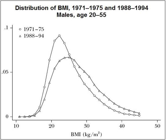
Meal preparation in 1965 required ~ 16.5 hours per week and the total numbers of hours spent in home production was on the order of 51.8 hours at that time. [25] As can be seen in Figure 5, time spent on home production decreased significantly beginning around 1960. Beyond the decrease in total hours spent on housework, there was a much larger decrease in the amount of physical effort required. Washing machines and clothes dryers, prepared meal components and entire prepared meals, as well as countless other “labor saving” devices, goods and services have markedly decreased fitness. The same has been true of strenuous physical activity in the work place where the overall number of high activity jobs have decreased by ~ 20% from 1970-2000.[26, 27]  There has also been a large shift in the workplace demographic since the mid-2oth century. Life expectancy increased from 47.3[28] years in 1900 to 77.8 years today, a consequence of which (in part) was the exodus of teens from the workforce. In 1920, ~20% of the US labor force was comprised of males aged 15 to 18 years of age.[28] Today, very few teenagers work full time jobs, and the number of teens employed in summer jobs has decreased from ~60% in 1994, to ~40% in 2008.[29] Of those teens who do find summer employment very few are in physically demanding (and consequently usually hazardous) areas of work, such as construction or agriculture. This change, coupled with increased TV viewing and other sedentary activities, translates into reduced fitness in the age 15-30 demographic.

Figure 7: The graph above shows the distribution of the Body Mass Index between the 1971–1975 and 1988–1994 surveys. Over this time, median BMI increased by 0.9; the 75th percentile increased by 1.5; and the 95th percentile increased by 2.7.[238]
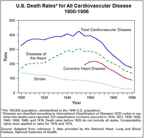
In their article, “Why Have Americans Become More Obese?” Cutler, et al., take the contrary position and argue that it is not reduced energy expenditure (or fitness) in the the population, but rather, the reduced investment required in terms of time per calorie consumed, that has been the primary cause of the change in US, and increasingly Western European eating habits (and thus is responsible for the current epidemic of obesity and type II diabetes).[30] Superior food packaging and preservation have cut not just meal preparation time dramatically, but also cleanup time. The mess generated in the preparation of multiple elements of a meal is now confined to the factory and the cleanup is included in the price of the food. It is also no longer necessary to spend as much time cooking, or even heating food, because it can be rapidly prepared and be made ready to eat in a matter of minutes from refrigerator or cupboard by the use of the microwave oven. These technological changes have thus reduced the threshold for eating formerly time consuming and messy to prepare dishes to the point of almost no effort or expenditure of time at all. It is now almost as easy to eat a piece of cake or pie, a brownie, or complex entree as it once was to eat an apple. All the mess and time involved in baking a cake or a pie from scratch is gone.

Regardless of the cause, we are most certainly not our parents or our grandparents, and as the current epidemic of obesity and type II diabetes attests, we are not likely to age or die as they did, either. Any doubts about the difference between “us” and “them” (or even “us then” and “us now”) should be laid to rest by a careful perusal of Figure 7.

The generations who participated in the AHS and Seven Countries Study were also fed differently. In Europe, they were subjected to nearly a decade of reduced calorie consumption, and even in the US, the relatively high cost of calories (in time, if nothing else) combined with less leisure time and fewer options for sedentary work, no doubt acted to limit calorie consumption, compared to today. This reduced calorie consumption may have been protective, and might have served to add years to life even in the presence or the absence of a more optimal diet. These generations of people were also fed on agricultural products derived mostly from small farms where crops and livestock had the opportunity to acquire a broad range of micro-nutrients and phytochemicals that are now less abundant in the food supply.

How Square is Curve Already?

Figure 8: The death rate from cardiovascular disease in the US has plummeted since the turn of century in part due to the replacement of saturated fats with of polyunsaturated fats in the diet.[31]
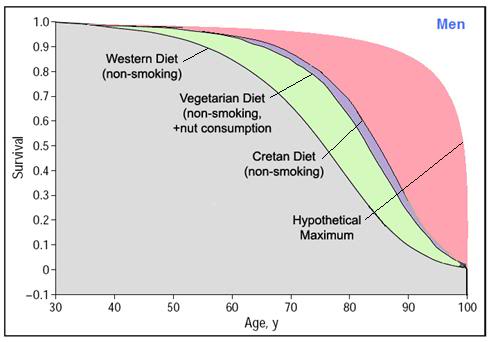
It should also be pointed out that data from longitudinal studies like the AHS-1&2 and the Seven Countries Study reach us as light does from a distant star. When we point and look at the star in the crook of the handle of the Big Dipper we say, “Look, there’s Alcor!” But of course that isn’t the Alcors we are looking at, but rather the light that shows what they looked like 83 years ago. Similarly, all of the data in AHS-1&2 and Seven Countries Study is a generation or two (or three!) old by the time we have it. The participants in those studies are mostly dead now, as indeed they would have to be in order for us to be able to plot lifespan curves for them. Thus, it is easy to make the mistake of saying, “If I adopt this diet I can expect 7 additional years of life, or 10 additional years of life, because that’s what the study participants experienced.

At least one problem with that assumption is that some of the benefits from both studies have very likely already been realized in the form of the switch from saturated to poly- and monounsaturated fats in the diet, which began in the early 1960s and continues through the present. The most significant benefit from both the Seven Countries Cretan diet and the Adventist Vegetarian diet has been the reduction in mortality (and morbidity) from CVD that has been ongoing since ~1968 in the US. The death rate from CVD has been halved since 1960 when both of these studies were undertaken (Figure 8). To those who vilify Ancel Keys for not getting it just right, I can only say, “Look at (Figure 8) and try to tell me that you could have done better.” So, we’ve undoubtedly used up some of benefit from these dietary interventions in terms of mean lifespan extension.

Figure 9: These curves show the best case extension of mean lifespan that can be anticipated with the Adventist Vegetarian diet or the Cretan Diet.
Finally, it is critically important to understand that both the Cretan and the Adventist Vegetarian diets are really not “diets” at all, but rather lifestyles. Both lifestyles have in common a strong emphasis on low impact exercise and a non-sedentary way of life. Both lifestyles were a product of a time without televisions or computers, and both lifestyles required then, and will require now, considerably greater time for food preparation and cleanup. The upside of that is that we should also eat less, if Cutler et al., are correct. That is important to consider as well, because, leaving aside whether fats, carbohydrates or protein should comprise X- percentage of a given diet’s calories, one thing both these diets have in common is modest to moderate calorie restriction.  Five, or possibly even 10 extra years of healthy, productive life should hopefully make the practical costs worthwhile.

The Caveman Diet, or Just How Credulous Are You?

“There are races of people who are all slim, who are stronger and faster than us. They all have straight teeth and perfect eyesight. Arthritis, diabetes, hypertension, heart disease, stroke, depression, schizophrenia and cancer are absolute rarities for them. These people are the last 84 tribes of hunter-gatherers in the world. They share a secret that is over 2 million years old. Their secret is their diet- a diet that has changed little from that of the first humans 2 million years ago, and their predecessors up to 7 million years ago. Theirs is the diet that man evolved on, the diet that is coded for in our genes. It has some major differences to the diet of “civilization”. You are in for a few big surprises.
The basic principles of the Paleolithic Diet are so simple that most high school students can understand them. Within 15 minutes from now you will grasp the major elements. At the technical level, Paleolithic Diet Theory has a depth and breadth that is unmatched by all other dietary theories.” – Dr. Ben Balzer, M.D.

The ideas underlying the Cretan Diet and the SDA Vegetarian diet are complex and do not admit of easy reduction to a catchphrase. The actual foods permitted and consumed in both diets differ markedly – one proscribing all meat, the other urging fish consumption, one obtaining most of the dietary fat calories from PUFAs, and the other from monounsaturated olive oil… It is these differences in the face of the common outcome of greatly improved health and moderate extension of the mean lifespan that are, in fact, key, because they tell us about the likely underlying common mechanisms and thus possibly of their action. They also offer us the opportunity for more choice, and therefore for more flexibility and the likelihood of greater compliance.

The Paleo Diet: A diet so unscientific, only a caveman would do it.

That is not, however, how people make a quick buck. Neither diet is particularly ‘sexy.’ And both diets require an understanding of the underlying biology that makes them work in order to be credible. It’s not possible, or at least not as easy to offer up a one sentence explanation for the feeble minded, such as, “This is a healthy diet that will extend lifespan because it is the natural human diet that our ancestors were evolved to eat.”[32-34] That sounds great because it is simple, easy to understand and “seems right” to a lot of uninformed, ignorant and fearful people. It also speaks to that deep and abiding suspicion that our health (and our other woes) is an artifact of our having lost our way – either from the primordial Garden of Eden, or from our biologically appropriate evolutionary ground state (i.e., before we embarked on agriculture). In fact, the emphasis on a 1:1 or 2:1 ratio of (n-6) to (n-3) fatty acids was derived from observations of contemporary hunter gatherer populations who have a low incidence of inflammatory and age-associated degenerative disease compared to that seen in post-agricultural populations. That was a useful insight that was subsequently validated in many human studies, the best of which extended over a period of decades. That’s the heart and soul of Level 1, Evidence Based Medicine.

In 1988, S. Boyd Eaton, Marjorie Shostak and Melvin Konner published a book entitled The Paleolithic Prescription: A Program of Diet & Exercise and a Design for Living[32] advocating a diet based on what the authors hypothesized the primordial pre-agricultural human diet was like. Subsequently, well over a dozen books have been published advocating variations on this diet based on arm chair hypothesizing from findings in the scientific and ethnographic literature.  The diet (depending upon the version you come across) is low (10-15% energy) or moderate in fat , low in carbohydrate (20–40% energy), and  high in protein diet (19–35% energy) which provides 55–65% of total calories from meat, 35–45% of calories from non-grain and low glycemic index vegetable sources with a primarily saturated fat intake (10%–58% energy) similar to or higher than that found in Western diets.[35-37]

The first problem with this approach is that the diet is not validated; the AHS and the Seven Counties studies had the considerable advantage of being able to study actual, living human beings under real world conditions, and then apply those insights to other populations, including populations already suffering from CVD. Indeed, that is where so many of the insights, as well as so many of the unresolved questions regarding these diets/lifestyles come from (i.e., the data are complex and robust). Late Paleolithic people are not only long dead and gone, they are really long dead and gone, and contemporary hunter gatherers – the few that remain – cannot be considered equivalent. Ironically, most of the data cited on the relationship between CVD and diet by the originators of the Paleolithic diet are from the Seven Countries Study![32, 37]

Even more to the point, there is present in the hypothesis of Eaton, Konner et al.,[32, 33] the notion that 10,000 years of agriculture is evolutionarily insignificant. In essence, they posit that human evolution with respect to diet stopped 10,000 years ago.[32, 35] At first glance this might seem to be credible, because human evolution has occurred over a period of millions years and it would seem that any changes that would occur in population genetics over a mere 10,000 years must be trivial. However, this is not the case for several reasons. First, the rate of evolution is a function of a complex interplay of multiple factors, including environmental change and selection pressure. It is only necessary to look at the various breeds of dogs, or pigeons created by artificial selection to understand that evolutionary change can be swift.

The introduction of agriculture was a watershed event and it would be astonishing if it was not accompanied by significant evolutionary adaptations to the dietary changes that resulted.  To understand that this is so it is only necessary to examine the strong natural selection for the gene that controls lactase production.[38] Human populations with a long history of cattle herding and milk consumption can metabolize lactose present in cow’s milk throughout adulthood, whereas populations that did not domesticate cattle cannot. Natural selection for the heterozygous carriers of the sickle-cell gene to maintain sickle-cell trait in populations exposed to malaria is another post-advent of agriculture evolutionary adaptation. This adaptation was selected for as a direct result of an agriculture-induced alteration to the environment; the clearance of the tropical forests in central Africa, which in turn led to the explosion in the population of the Anopheles mosquitoes that are the vectors for the Plasmodium parasite that causes malaria.

Recently developed techniques for measuring genetic variability now allow for the determination of selection operating in the human genome.[39] Directional selection has been identified in the glucose-6-phosphate dehydrogenase (G6PD) gene, which confers resistance to malaria.[40] What is more, G6PD resistance has evolved not once, but twice in humans, in both Africa and in the Americas.[41] Similarly, the genes expressing chemokine receptor 5 (CCR5) among Europeans, which confers resistance to the human immunodeficiency virus (HIV) are likely to have been selected for within this population over a period of several hundred years in response to Yersinia pestis (bubonic plague) and tuberculosis, both of which use the CCR5 receptor as an entry portal into the host.[42]  Numerous other studies have also provided evidence for the recent operation of natural selection on the human genome as a result of very recently developed techniques that allow for comparisons over long sections of DNA.[43-46]

In addition to the conservation of lactase production into adulthood, there is substantial evidence of evolutionary adaptation to the high carbohydrate diet that was a product of agriculture. The incidence of obesity that occurs upon exposure to high calorie “affluence diets” is known to vary greatly by ethnicity. The Pima people (or Akimel O’odham) are a racial group of Amerindians living in central and southern Arizona. One-half of adult Pima Indians have diabetes and 95% of those with diabetes are overweight or obese.
Obesity is thought to be 50-90% heritable. Genome scans in obesity studies are highly reproducible and, despite ethnic and environmental differences, the loci at chromosomes 2 and 10 are generally confirmed as the source of the phenotype. Obesity is “oligogenic,” with expression modulated by “polygenic modifier genes” interacting with the environment in food choices, physical activity, and smoking.[38] Prior to their introduction to the “American” diet after WWII the Pima were not obese and diabetes was extremely rare.[39-41] The diet of the Pima was a very low fat, high carbohydrate diet consistent with the subsistence agriculture of the desert southwest.[42, 43]  Some variations in the ectonucleotide pyrophosphatase phosphodiesterase gene-1 (ENPP1) are associated with a 50% increase in the risk of morbid obesity in adults and a 69% increased risk of childhood obesity. An ENPP1 mutation, for example, which is known to protect against obesity and type II diabetes, is present in about 90 percent of non-Africans, but nearly absent in Africans and, not coincidentally, in the Pima. Human evolution in response to environmental change and in response to dietary change is both ongoing and dynamic.[47][44]

Of course, the Paleolithic diet may be the best diet yet conceived. I could give many reasons why I believe this is not so, but absent hard data gleaned from human trials, I can’t prove much. And that is my final and most important point. I did a Pubmed search using the keywords “Paleolithic diet” and I got 67 hits. Of those 67 hits only 9 were papers that involved actual human or animal application of the diet, or even discussion of same. I’ve copied all of the cites for these studies below:

1: Konner M, Eaton SB. Paleolithic nutrition: twenty-five years later. Nutr Clin Pract. 2010 Dec;25(6):594-602. PubMed PMID: 21139123.

2: Jönsson T, Granfeldt Y, Erlanson-Albertsson C, Ahrén B, Lindeberg S. Apaleolithic diet is more satiating per calorie than a Mediterranean-like diet in  individuals with ischemic heart disease. Nutr Metab (Lond). 2010 Nov 30;7:85. PubMed PMID: 21118562; PubMed Central PMCID: PMC3009971.

3: Klonoff DC. The beneficial effects of a Paleolithic diet on type 2 diabetes and other risk factors for cardiovascular disease. J Diabetes Sci Technol. 2009 Nov 1;3(6):1229-32. PubMed PMID: 20144375; PubMed Central PMCID: PMC2787021.

4: Eaton SB, Konner MJ, Cordain L. Diet-dependent acid load, Paleolithic[corrected] nutrition, and evolutionary health promotion. Am J Clin Nutr. 2010 Feb;91(2):295-7. Epub 2009 Dec 30. Erratum in: Am J Clin Nutr. 2010 Apr;91(4):1072. PubMed PMID: 20042522.

5: Jönsson T, Granfeldt Y, Ahrén B, Branell UC, Pålsson G, Hansson A, Söderström  M, Lindeberg S. Beneficial effects of a Paleolithic diet on cardiovascular risk factors in type 2 diabetes: a randomized cross-over pilot study. Cardiovasc Diabetol. 2009 Jul 16;8:35. PubMed PMID: 19604407; PubMed Central PMCID: PMC2724493.

6: Frassetto LA, Schloetter M, Mietus-Synder M, Morris RC Jr, Sebastian A. Metabolic and physiologic improvements from consuming a paleolithic, hunter-gatherer type diet. Eur J Clin Nutr. 2009 Aug;63(8):947-55. Epub 2009 Feb  11. PubMed PMID: 19209185.

7: Osterdahl M, Kocturk T, Koochek A, Wändell PE. Effects of a short-term intervention with a paleolithic diet in healthy volunteers. Eur J Clin Nutr. 2008 May;62(5):682-5. Epub 2007 May 16. PubMed PMID: 17522610.

8: Jönsson T, Ahrén B, Pacini G, Sundler F, Wierup N, Steen S, Sjöberg T, Ugander M, Frostegård J, Göransson L, Lindeberg S. A Paleolithic diet confers higher insulin sensitivity, lower C-reactive protein and lower blood pressure than a cereal-based diet in domestic pigs. Nutr Metab (Lond). 2006 Nov 2;3:39. PubMed PMID: 17081292; PubMed Central PMCID: PMC1635051.

9: Eaton SB, Eaton SB 3rd. Paleolithic vs. modern diets—selected pathophysiological implications. Eur J Nutr. 2000 Apr;39(2):67-70. PubMed PMID: 10918987.

If I enter the keywords “Mediterranean diet” I get 2,269 hits, of which 225 are reports of clinical trials. I will not copy those here!

That’s it. Nine papers of poor quality and not a single clinical trial demonstrating reduced morbidity or mortality – even in CHD or type II diabetes.  Sixty-seven papers of hypothesizing 25 years after this diet was put forth. That is dismal science and it is inexcusable to take a position advocating such an intervention in the complete absence of any evidence that it will actually extend the human (or the laboratory animal) lifespan when there is a large body of high quality data that supports far less extreme, and far more practical dietary and lifestyle interventions that will accomplish those ends.

I have no problem with people coming up with a hypothesis, however kooky or sane, and then proceeding to try it out – even on people – as long as those people have informed consent and the data they are given is accurate. In looking over the various books and the countless media articles on the Paleolithic diet, I was struck by how much the Paleolithic Diet’s hype reminded me of the Pritikin diet hype, and even more so of the Pearson & Shaw Life Extension Revolution circus from 30 years ago. “Live to be 100!” “Feel great! Experience all day energy every day!” “Lose Weight!” Well, at least one of those is very likely true, and that is that most people who undertake any version of the Paleo diets I’ve reviewed will likely lose weight. But as to the other claims? Right now they are preposterous. The sad thing is that for first the time in history we have one diet/lifestyle choice that satisfies EBM-1 criteria, and another that satisfies EBM-2 criteria. Both are “proven” to reduce morbidity from a range of degenerative diseases, and both have been proven to significantly extend mean lifespan…

Max More, CEO Alcor Life Extension Foundation
As I so often say, “You pays your money and you takes your chances.” Still,  it is embarrassing to see cryonicists buy into yet another quick fix cure all, with no appropriate science to back it up. In his article “The Cryo-Paelo Solution”[48] Alcor President Max More advocates the Paleolithic Diet as a life extending add-on to cryonics. This recommendation is supplemented by a web interview.[49] His citations consist these of these popular books on the subject: Loren Cordain, The Paleo Diet; Nora T. Gedgaudas, Primal Body, Primal Mind; Mark Sisson, The Primal Blueprint; Gary Taubes, Why We Get Fat;Gary Taubes, Good Calories, Bad Calories; Arthur de Vany, The New Evolution Diet. The expert More cites as the one to consult for an introduction to Paleo-dieting is Loren Cordain, author of  The Paleo Diet. The quote that open this section on the Paleolithic diet is by Dr. Ben Balzer, M.D., and is from the “Introduction” to Cordrain’s book. Need I say more?

End of Part 3

References

1.            White E: Ministry of Healing: http://books.google.com/books/about/The_Ministry_of_Healing.html?id=2bu2ry223JAC: Kessinger Publishing, LLC; 1905.

2.            Rucker C: The Seventh-Day Diet: A Practical Plan to Apply the Adventist Lifestyle to Live Longer, Healthier, and Slimmer in the 21st Century: Pacific Press Publishing Association 2002.

3.            Mills PK, Beeson WL, Phillips RL, Fraser GE: Cancer incidence among California Seventh-Day Adventists, 1976-1982. Am J Clin Nutr 1994, 59(5 Suppl):1136S-1142S.

4.            Kahn HA, Phillips RL, Snowdon DA, Choi W: Association between reported diet and all-cause mortality. Twenty-one-year follow-up on 27,530 adult Seventh-Day Adventists. Am J Epidemiol 1984, 119(5):775-787.

5.            Grundmann E: Cancer morbidity and mortality in USA Mormons and Seventh-day Adventists. Arch Anat Cytol Pathol 1992, 40(2-3):73-78.

6.            Zollinger TW, Phillips RL, Kuzma JW: Breast cancer survival rates among Seventh-day Adventists and non-Seventh-day Adventists. Am J Epidemiol 1984, 119(4):503-509.

7.            Fraser GE: Diet as primordial prevention in Seventh-Day Adventists. Prev Med 1999, 29(6 Pt 2):S18-23.

8.            Mills PK, Annegers JF, Phillips RL: Animal product consumption and subsequent fatal breast cancer risk among Seventh-day Adventists. Am J Epidemiol 1988, 127(3):440-453.

9.            Mills PK, Beeson WL, Phillips RL, Fraser GE: Bladder cancer in a low risk population: results from the Adventist Health Study. Am J Epidemiol 1991, 133(3):230-239.

10.          Phillips RL, Snowdon DA: Dietary relationships with fatal colorectal cancer among Seventh-Day Adventists. J Natl Cancer Inst 1985, 74(2):307-317.

11.          Fraser GE, Dysinger W, Best C, Chan R: Ischemic heart disease risk factors in middle-aged Seventh-day Adventist men and their neighbors. Am J Epidemiol 1987, 126(4):638-646.

12.          Fraser GE, Lindsted KD, Beeson WL: Effect of risk factor values on lifetime risk of and age at first coronary event. The Adventist Health Study. Am J Epidemiol 1995, 142(7):746-758.

13.          Fraser GE, Shavlik DJ: Risk factors for all-cause and coronary heart disease mortality in the oldest-old. The Adventist Health Study. Arch Intern Med 1997, 157(19):2249-2258.

14.          Giem P, Beeson WL, Fraser GE: The incidence of dementia and intake of animal products: preliminary findings from the Adventist Health Study. Neuroepidemiology 1993, 12(1):28-36.

15.          Fraser GE, Sabate J, Beeson WL, Strahan TM: A possible protective effect of nut consumption on risk of coronary heart disease. The Adventist Health Study. Arch Intern Med 1992, 152(7):1416-1424.

16.          Fraser GE: Associations between diet and cancer, ischemic heart disease, and all-cause mortality in non-Hispanic white California Seventh-day Adventists. Am J Clin Nutr 1999, 70(3 Suppl):532S-538S.

17.          Sabate J: Nut consumption, vegetarian diets, ischemic heart disease risk, and all-cause mortality: evidence from epidemiologic studies. Am J Clin Nutr 1999, 70(3 Suppl):500S-503S.

18.          Willett W: Lessons from dietary studies in Adventists and questions for the future. Am J Clin Nutr 2003, 78(3 Suppl):539S-543S.

19.          Phillips RL, Snowdon DA: Association of meat and coffee use with cancers of the large bowel, breast, and prostate among Seventh-Day Adventists: preliminary results. Cancer Res 1983, 43(5 Suppl):2403s-2408s.

20.          Beck J, Beck, JJ, Jarnes, K.: Adventist Sabbath Dinner Cookbook: Pacific Press Publishing Association; 2001.

21.          Council GCoS-dAN: The Seventh-day Adventist Position Statement on Vegetarian Diets: http://www.sdada.org/position.htm. In.; 2010.

22.          Health LLUSoP: The Vegetarian Food Pyramid: http://www.vegetariannutrition.org/food-pyramid.pdf. In. Loma Linda: Loma Linda University; 2008.

23.          Center S: History of Loma Linda Foods: http://www.soyinfocenter.com/HSS/loma_linda_foods.php. In. Lafayette: Soyinfo Center; 2004.

24.          King G, Fitzhugh, EC, Bassett, DR Jr, McLaughlin, JE, Strath SJ, et al.: Relationship of leisure-time physical activity and occupational activity to the prevalence of obesity. Int J Obes Relat Metab Disord 2001, 25:606-612.

25.          Ramey V: Time Spent in Home Production in the Twentieth-Century United States: New Estimates from Old Data :http://weber.ucsd.edu/~vramey/research/Home_Production_published.pdf. The Journal of Economic History 2009, 59(1).

26.          Borodulin K, Laatikainen, T, Juolevi, A, Jousilahti, P. : Thirty-year trends of physical activity in relation to age, calendar time and birth cohort in Finnish adults. Eur J Public Health 2008, 18(3):339-344.

27.          Brownson R, Boehmer, TK, Luke, DA.: Declining rates of physical activity in the United States: what are the contributors. Annu Rev Public Health 2005, 26(421-43).

28.          Census USBot: U.S. Bureau of the Census, Historical Statistics of the United States, Colonial Times to 1970. 1971.

29.          Camarota S, Jensenius, K. : A Drought of Summer Jobs: Immigration and the Long-Term Decline in Employment Among U.S.-Born Teenagers: http://www.cis.org/articles/2010/teen-study.pdf. In: Backgrounder. Center for Immigration Studies; 2010.

30.          Cutler D, Glaeser, EL, Shapiro, JM.: Why have americans become more obese? Journal of Economic Perspectives 2003 17(3):93-118.

31.          National Heart LaBI: Morbidity & mortality: 1998 chartbook on cardiovascular, lung, and blood diseases. In. Edited by Health UDoHaHSNIo. Rockville, Maryland: US Government Printing Office; 1998.

32.          Eaton S, Shostak, M, Konner, M.: The Paleolithic Prescription: A Program of Diet & Exercise and a Design for Living. New York:: Harper & Row; 1988.

33.          Konner M, Eaton SB: Paleolithic nutrition: twenty-five years later. Nutr Clin Pract, 25(6):594-602.

34.          Lindeberg S: [Paleolithic diet and evolution medicine: the key to diseases of the western world]. Lakartidningen 2005, 102(26-27):1976-1978.

35.          O’Keefe J, Cordain L.: Cardiovascular Disease Resulting From a Diet and Lifestyle at Odds With Our Paleolithic Genome: How to Become a 21st-Century Hunter-Gatherer. Mayo Clin Proc 2004;, 79:101-108.

36.          Cordain L, Eaton SB, Miller JB, Mann N, Hill K: The paradoxical nature of hunter-gatherer diets: meat-based, yet non-atherogenic. Eur J Clin Nutr 2002, 56 Suppl 1:S42-52.

37.          Marlowe F: Hunter-gatherers and human evolution. Evolutionary Anthropology 2005, 14:54 -67.

38.          Froguel P, Boutin P: Genetics of pathways regulating body weight in the development of obesity in humans. Exp Biol Med (Maywood) 2001, 226(11):991-996.

39.          Bennett PH, Burch TA, Miller M: Diabetes mellitus in American (Pima) Indians. Lancet 1971, 2(7716):125-128.

40.          Bennett PH, Rushforth NB, Miller M, LeCompte PM: Epidemiologic studies of diabetes in the Pima Indians. Recent Prog Horm Res 1976, 32:333-376.

41.          Zimmet P, Arblaster M, Thoma K: The effect of westernization on native populations. Studies on a Micronesian community with a high diabetes prevalence. Aust N Z J Med 1978, 8(2):141-146.

42.          Ravussin E, Valencia ME, Esparza J, Bennett PH, Schulz LO: Effects of a traditional lifestyle on obesity in Pima Indians. Diabetes Care 1994, 17(9):1067-1074.

43.          Boyce VL, Swinburn BA: The traditional Pima Indian diet. Composition and adaptation for use in a dietary intervention study. Diabetes Care 1993, 16(1):369-371.

44.          Malhotra A, Kobes S, Knowler WC, Baier LJ, Bogardus C, Hanson RL: A Genome-Wide Association Study of BMI in American Indians. Obesity (Silver Spring).

45.          More M: The cryo-paelo solution: http://www.alcor.org/magazine/2011/03/07/the-cryo-paleo-solution/. Cryonics (on line edition) 2011.

46.          Snyder S: Alcor CEO Max More and the paleo diet: http://samsnyder.com/2011/05/22/alcor-ceo-max-more-and-the-paleo-diet/. In: A Blog by Sam Snyder. Sam Snyder; 2011.


Posted in Gerontology | 50 Comments

Interventive Gerontology 1.0.02: First, Try to Make it to the Mean: Diet as a life extending tool, Part 2

Figure 1: Ancel Keys (January 26, 1904 – November 20, 2004) was the American physiologist and epidemiology of cardiovascular disease (CVD). He was responsible for two famous diets: K-rations formulated as balanced meals with a long shelf life for combat soldiers in World War II and the Mediterranean (Cretan) diet. Keys is shown (at right, above) two months before his 101st birthday.
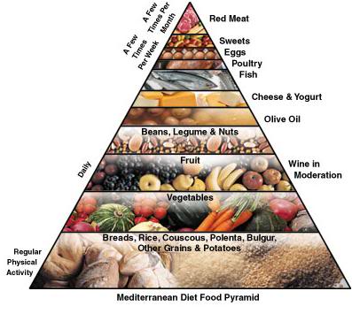
The Seven Countries and Adventist Health Studies

Nathan Pritikin drew his initial conclusions about the effect of dietary change from classified data he was privy to during World War II (WWII) on the patterns of age-associated disease in Europe as a consequence wartime calorie restriction and severe reduction of fat intake due to the severely reduced availability of meat and dairy products.  He also observed that the incidence of age-associated degenerative diseases was  very low in human populations where the diet was very low in fat (~10% of the total caloric intake), contained no refined sugars and consisted mostly of fresh vegetables and fruits  with very little meat being consumed. Similarly, the physiologist Ancel Keys (Figure 1), who was working with the Army Quartermaster Corps in developing K-Rations,[1] became involved in the Army’s program to create scientifically informed re-feeding programs for POWs and civilians suffering from starvation, saw the same kind of data. Unlike Pritikin, Keys had the opportunity to do human experimentation afforded by wartime conditions.[2, 3] Keys working hypothesis was different than Pritkin’s, namely that it was primarily saturated fats in the diet that were responsible for the high incidence of cardiovascular disease (CVD) in the affluent and well-fed West.

This epidemiological approach to identifying patterns of food consumption associated with increased or reduced risk of degenerative disease was also being pursued around this time in the US, by physicians at the Loma Linda Medical Center in Loma Linda, CA. Loma Linda was an almost exclusively Seventh Day Adventist community at that time, and these physicians believed that their patients, who practiced a vegetarian diet in conjunction with abstention from tobacco and alcohol, were considerably healthier than the non-Adventist population in California. They began a study of diet, lifestyle and the incidence of disease and all-cause mortality in 1958; the Adventist Health Study-1 (AHS-1) [4-26]

Keys returned to Europe after the war and began a study of six European countries, which later became the Seven Countries Study. [27-72] The dietary recommendations which emerged from the Seven Countries Study are commonly referred to as the “Mediterranean diet.” However, the use of the words “Mediterranean diet” to describe these recommendations is a misnomer. The countries of the Mediterranean basin have large differences in diet, lifestyle and in their corresponding rates of morbidly and mortality. The country with the lowest death rate  (14.0 – 18.0 per 1000 persons), is Crete, whose death rate has been at this level since at least 1930. [73] The diet of Crete is archetypical of the ‘traditional’ Greek diet before the introduction of continental European and American foods into Greece after ~ 1960.

The Seven Countries Study was the first to generate robust data on the incidence of cardiovascular disease in a range of populations (US, Finland, The Netherlands, Italy, the former Yugoslavia, Japan and Greece) with a fairly broad spectrum of dietary patterns. The study showed differences on the order of 5 to 10-fold in coronary artery disease (CAD) between the populations studied. [36, 50, 74]

Figure 2: The Cretan diet food pyramid.

Both the Seven Countries Study and the AHS-1 demonstrated large reductions in disease-specific morbidly and mortality, as well as in all-cause mortality; primarily as a result of diet and lifestyle differences. In the case of the Seven Countries Study, extensive follow-on research using well designed prospective studies, resulted in the development of a set of dietary guidelines which became known as the “Mediterranean” or more correctly, the “Cretan diet.” The guidelines which constitute the Cretan diet satisfy Level-1 EBM criteria for extension of the mean lifespan by ~10 years, reduction in all cause mortality, high levels of compliance, and very importantly, titratability. In other words, the degree of compliance with the diet is roughly commensurate with the benefit that results. [75-77]

What About Cholesterol?

For thirty years an acrimonious debate has raged in the scientific and medical communities over whether cholesterol, or any molecular species of lipoprotein, “causes heart disease” or other CVD. The causes of the inflammatory events that underlie the start of arterial plaque formations are complex, possibly multifactorial (e.g., genetic, viral, microbial, environmental, etc.) and by no means fully elucidated. Keys, Pritikin and many others mistakenly believed that “elevated” serum cholesterol was the primary cause of CVD. This hypothesis is well supported by the epidemiological data. However, there are many people who have normal or even slightly low levels of total cholesterol, or of the low density lipoprotein (LDL) species whose oxidation is usually cited as the motor of atherogenesis.

However, the observations of Keys and Pritkin extended beyond a cause and effect relationship between cholesterol and CVD. In different ways, both men demonstrated that altering the total serum cholesterol level and/or the ratio between the LDL and high density lipoprotein (HDL) species, they could reduce the incidence of the disease. In Pritkin’s case, he even demonstrated that the disease could be reversed by the expedient of a very low fat diet.[78-80] Pritkin demonstrated his theory on a very small population of people; principally those who read his book, or otherwise followed his dietary advice. Keys, on the other hand, conducted an experiment on a grand scale.
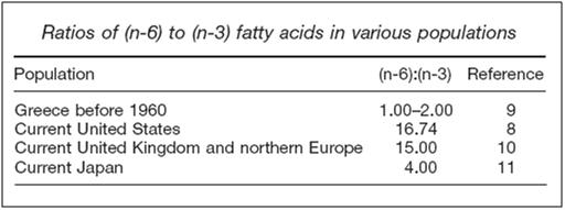
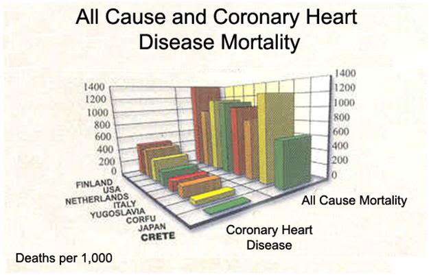
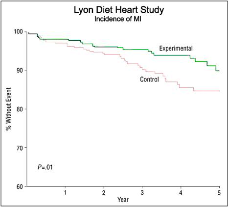
Throughout the 1960s Keys campaigned relentlessly to persuade physicians, public health authorities and the public themselves (directly) to replace the bulk of the calories they consumed in (saturated) fat with polyunsaturated fat. The purpose of this international dietary intervention was to reduce the serum cholesterol of the population, and thus the incidence of CVD. This effort enjoyed unprecedented success and it has resulted a doubling of the proportion of the unsaturated fatty acid, linoleic acid, in the tissues of Americans between 1960 and 1975.[81]  The mortality rate from coronary heart disease (CAD) in the US began to fall, starting in 1968, and it has continued to decline since then. It has been estimated that approximately 50% of the decline in CVD is as a result of dietary and lifestyle changes, exclusive of the reduction in tobacco abuse.[82]

As a scientist, I am acutely interested to understand the details of the pathophysiology of atherosclerosis. As someone who wants to avoid CVD, I am much more concerned with what works, even if the biomechanics are incomplete, or even contradictory. In this case, what works is that on a population basis, blood lipids are highly predictive of the risk of disease. Similarly, for most patients, raising HDL and lowering LDL are protective against both the onset of CVD, and to a lesser extent, its progression. Lipid status is thus a useful screening tool, as well as an instructive guide to the individual patient’s likely response to treatment. It is not necessary to believe that “cholesterol,” or any particular species of lipoprotein “causes” CVD. It is only necessary to understand that they are, at the least, useful biomarkers on a population wide basis and that they are often useful in the intelligent management of individual patients.

 Table 1: Fatty Acids Ratios in Different Diets

From the inception of the AHS-1 in 1958, and the Six Countries Study around the same time,[68, 83] the primary focus of the research, as was the case with Pritikin, was on the possible relationship of the diet to the etiology of CVD, with special emphasis on the fatty acid composition of the diet. The 5-year follow-up in the Seven Countries Study found favorable all-cause death rates in Greece, Italy and Japan, as compared with the other countries, including dramatically lower rates of CVD in Crete, and to a lesser extent in Japan.[84]The diet of Crete has in common with the diet of hunter-gatherers similar quantities of antioxidants, saturated fat, fiber, monounsaturated fat and, very importantly, the ratio of (n-6) to (n-3) fatty acids. [75-77]

One the basis of insights gained from the Seven Countries Study a wide range of epidemiologic investigations,  controlled clinical trials and relevant animal experiments have confirmed the hypotriglyceridemic, anti-inflammatory and antithrombotic aspects of (n-3) fatty acids (28 –35) as well as the criticality of (n-3) fatty acids, particularly Docosahexaenoic (n-3) (DHA) acid in the diet for the normal development of the retina and brain in the human infant. As a consequence of these insights, a study of the (n-3) fatty acid composition of diets that were known to be associated with reduced rates of CVD and cancer was undertaken.[75] The initial conclusion was that the high olive oil intake, which accounts for ~35% of the calories consumed in the Cretan diet, was likely responsible for the low rates of CVD and cancer. However, the Japanese have a similarly low incidence of these diseases and yet only ~11% of their calorie intake is from fat, none of which is from olive oil.

Figure 3: Mortality and morbidity difference between populations of patients with coronary artery disease (CAD) eating the Cretan diet and those on the low fat American Heart Association Step 1 diet[151]

The common factors between the populations of Crete and Japan were that they both consumed large amounts of vegetables (including wild plants), fruits, nuts and legumes, all of which are rich sources of folic acid, glutathione and vitamins E, C and other antioxidants. Wild plants are rich sources of (n-3) fatty acids and antioxidants and their consumption is not confined to humans. Both in Crete and in rural Japan, chickens and other livestock, such as goats and cows, are free ranging and consume wild vegetation in abundance (and in the case of chickens, insects, arachnids and worms) which are rich in (n-3) fatty acids and antioxidants, as well as cytoprotective and vasculoprotective trace minerals which are concentrated in the food chain. The result is poultry, eggs and milk which contain radically different ratios of (n-3) to (n-6) fatty acids and are enriched with selenium. For example, eggs from Crete have a ratio of (n-6) to (n-3) of 1:3, whereas the US ‘battery egg’ has a ratio of 19:4. Of course, the presence of this favorable ratio of lipids in eggs is also reflected in prepared foods which contain eggs (such as noodles, some breads and soups, etc.) and in the flesh of the animals that are slaughtered and eaten.

Analysis of the serum (n-3) fatty acid levels of the populations in Crete and Japan demonstrated that they had had higher concentrations of (n-3) fatty acids than did the other populations in the study, all of whom had a far high incidences of age associated degenerative diseases. The two populations with the lowest CAD in the Seven Countries Study consumed the highest amount of α-linoleic acid (α-LNA) the major sources of which were the wild herb purslane, walnuts and figs. By contrast, the Japanese obtained their α-LNA from canola and soybean oils. Interestingly, the Seventh Day Adventists, who experience an increase in mean lifespan of 7.28 years (95% confidence interval, 6.59-7.97 years) in men and by 4.42 years (95% confidence interval, 3.96-4.88 years) in women over that of their non-Adventist cohorts in the US[10], consume a vegetarian diet that is rich in nuts and oils containing α-LNA. The SDAs, like the people of Crete, have not only higher levels of α-LNA, but also lower levels of linoleic acid[9, 85, 86]


Figure 4: The Lyon Diet Heart Study demonstrated a 50 to 70% reduction of the risk of recurrence of myocardial infarction (MI) after four years of follow-up in coronary heart disease (CHD patients. The Lyon diet employed α-LNA as 0.6 to 1% of total daily energy or about 2 g per day in patients who follow a traditional Mediterranean diet. Supplementation with very long chain omega-3 fatty acids (c.1g per day) in patients following a Mediterranean type of diet was shown to decrease the risk of cardiac death by 30% and of sudden cardiac death by 45% in the GISSI trial. [87]

In 1994 de Lorgeril and Renaud published the results of a prospective study to evaluate a diet which contained the types and ratios of essential fatty acids (EFAs) found to be effective at reducing CVD in the Seven Countries Studies. The Lyon Heart Study (LHS), as it came to be known, was a randomized, single-blind secondary prevention trial that combined a modified Cretan diet enriched with α-LNA with that of the Step I American Heart Association diet.[87] The LHS demonstrated a reduction in all-cause mortality of 70% in the experimental population which consumed a diet low in butter and processed meats, but high in fish, nuts, fruits and vegetables (Figure 5).[87] The LHS followed subjects for 5 years after the start of the intervention and examined the reduction of risk for coronary artery disease (CAD) as well as in mortality from all cancers. The reduction in CAD was 56% (P 5 0.03) over that of control subjects, and in cancer mortality it was 61% (P 5 0.05)!

Figure 5: Cardiac morbidity and mortality in the Lyon Diet Heart Study. Of particular importance to cryonicists is the reduction in mortality from sudden cardiac death (SCD). [87]

Olive Oil, or Something Else?

Figure 6: The author has serious questions about whether experiments conducted using industrially prepared laboratory animal chows (right) are representative of the results obtained when fresh fruits and vegetable as well as foods consumed in their native state are used (left).
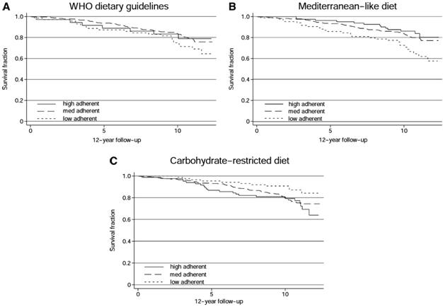
An initial and obvious conclusion from The Seven Countries Study was that a significant part of the reduction in morbidity and the extension of lifespan due to the Cretan diet was a consequence of the consumption of a large fraction of the calories in the diet in the form of monounsaturated  fats (MUFAs), principally as a result of olive oil consumption. However, recent animal studies have yielded paradoxical results. For instance, experiments in green monkeys have shown that a diet high in MUFAs (olive oil source) causes atherosclerosis equivalent to that observed in animals fed a diet high in saturated fats (SFAs).[88] This effect appears to result from an increased secretion of cholesteryl oleate enriched lipoproteins, as well as due to an increase in the circulating blood levels of chylomicron remnants, which are highly atherogenic lipoproteins. [89-91]

These paradoxical animal results have raised questions amongst epidemiologists and nutritionists about whether MUFAs really have beneficial effects in humans. Green monkeys are metabolically and genetically different than humans and the human data indicate that dietary MUFAs have favorable effects on CHD risk. There is also a significant amount of mechanistic data that indicate that there are molecular species in olive oil that have potent antioxidant and anti-inflammatory properties. In particular, the polyphenolic compounds hydroxytyrosol and oleuropein have been shown to possess these properties both in vitro and in vivo.[92-96] There is also the issue of the way in which olive oil is incorporated into the chow for experimental animals (Figure 6). Olive oil incorporated into a manufactured chow along with other dietary ingredients is not the way in which humans consume it, and this factor should be taken into consideration in future studies.

Figure 7: The titratability of the beneficial effects of the Cretan diet are nicely illustrated in this series of graphs showing all cause mortality over a ten year period with three variations of the Cretan diet; the world Health Organization recommended diet base on the Seven Countries Study, a broadly similar diet, and a carbohydrate restricted version of the Cretan Diet. Kaplan-Meier survival curves for individuals considered adequate reporters of dietary intake, grouped as low-, medium-, or high-adherent individuals to the dietary patterns investigated. Crude hazard ratios (HRs) and 95% CIs were calculated from Cox proportional hazards regression analyses with the use of low-adherent individuals as the reference group for each dietary pattern. A: World Health Organization (WHO) dietary guidelines, according to the Healthy Diet Indicator: medium adherent (HR: 0.70; 95% CI: 0.43, 1.15), high adherent (HR: 0.97; 95% CI: 0.45, 2.07). B: Mediterranean-like diet, according to the Mediterranean Diet Score: medium adherent (HR: 0.68; 95% CI: 0.44, 1.04), high adherent (HR: 0.29; 95% CI: 0.12, 0.70. C: Carbohydrate-restricted (CR) diet, according to the CR diet score: medium adherent (HR: 1.92; 95% CI: 1.02, 3.62), high adherent (HR: 2.17; 95% CI: 1.05, 4.45).[148]

Anti-inflammatory and Cytoregulatory Lipids in the Cretan Diet

One proposed resolution to the paradoxical animal findings regarding the atherogenicity of olive oil in the laboratory is the observation that both the high fat MFA diet of the Cretans, and the low fat PFA diet of the Japanese, are rich in (n-3) fatty acids and antioxidants, in particular resveratrol, glutathione, vitamin C, vitamin E, lycopene, b-carotene, polyphenols and polyamines obtained from fruits, vegetables, wild plants, and olive oil. [97] [98-101] Additionally, both diets are enriched in α-LNA and eicosapentaenoic acid [EPA, 20:5 (n-3)] from the consumption of large amounts of fish, relative to the control countries.[159, 171, 174] Because olive oil is high in the monounsaturated fatty acid oleic acid [18:1, (n-9)] and low in saturated (n-6) fatty acids it cannot compete with the endogenous desaturation and elongation of α-LNA, or with the incorporation of α-LNA into the constituent phospholipids of cell membranes. This is particularly important in the case of red blood cell (RBC) and platelet membranes, where they act to increase the deformability of RBCs and decrease the aggreability and adhesions of platelets. [102-108]

The ratio of (n-6) to (n-3) lipids in the Cretan diet is between 2:1 and 1:1, which is very close to the dietary ratio of the Japanese, as that of hunter-gatherer societies. The beneficial effects of such a ratio and their importance in normal growth and development [109, 110] as well as in the reduction of risk for CVD, hypertension, type II diabetes, osteoarthritis and, to a lesser extent cancer, are voluminously documented in the literature.[111] [112-116] The traditional Greek diet is very low in animal fat and thus the saturated fat content is quite low (7–8%). This low intake of SFAs is complemented by the high intake of  (n-6) and (n-3) EFAs which are also rich in phytoestrogens and other phytochemicals  as well α-LNA, vitamin C, vitamin E and glutathione.[117, 118] These molecules have been shown to have hypoglycemic, hypocholesterolemic and antitumor properties in animal experiments.[119-124] Consistent with these findings is the fact that the mortality from breast, prostate, bladder and colorectal cancer is lower in both the Cretan and the AHS populations than is the case for controls. [14, 20, 86, 125-129]

The principal EFA in the US diet is LA, an (n-6) fatty acid which is the precursor to the eicosanoids – molecules which have proinflammatory and cytoproliferative effects. The EFAs are converted to prostaglandins by the cyclooxygenases and to leukotrienes (LT) by the lipoxygenases. Arachidonicacid [(AA); 20:4(n-6)] and EPA, an (n-3) fatty acid, compete for cyclooxygenases and lipoxygenases, resulting in the production of eicosanoids with opposing effects. In general, AA-derived eicosanoids, such as the 2-series prostanoids and 4-series LTs, have pro-inflammatory effects, whereas EPA-derived eicosanoids, such as the 3-series prostanoids and5-series LTs, have anti-inflammatory effects. A focus of recent research has been to understand the importance of the (n-6) to (n-3) ratio, rather than the absolute level of either species of PUFA in cancer prevention.[102, 130]

Figure 8: The major active product of the omega-6 fatty acids is arachadonic acid which is converted to the 2-series prostaglandins and 4-series leukotrienes by the action of cyclooxygenase. The 2-series prostaglandins are pro-inflammatory.  In addition to the AA produced endogenously there are vast supplies available from the diet, most notably in meat, eggs and peanut oil.  In the Western diet there are comparatively few products of omega-3 metabolism to moderate the pro-inflammatory action of excessive dietary omega-6consumption. If the amount of omega-3 fatty acids in the diet is increased, their metabolites (primarily EPA and DHA) compete with arachidonic acid for access to cyclooxygenase resulting increased production of anti-inflammatory mediators as well as a decrease in the pro-inflammatory mediators, thereby significantly reducing the ratio of pro-inflammatory to anti-inflammatory mediators.
In animal studies (rats) LA increases the size and number of tumors, whereas fish oil [containing the (n-3) fatty acids EPA and DHA] decreases the incidence of tumor formation, as well as tumor size.[131] This finding is consistent with other studies in rats that indicate that the potent inhibitors of prostaglandin synthesis, the NSAIDs indomethacin and flurbiprofen are effective at reducing the incidence of spontaneously occurring breast cancer. Epidemiological studies in humans have also indicated a potentially chemoprotective effect as result of long term consumption of NSAID drugs.[132-136] Fish oils have been used to adjust systemic levels of  (n-3) fatty acids in animals models of colon, lung, breast, pancreatic and prostate cancers to reduce prostaglandin synthesis, with resulting chemoprevrention and/or slowed growth and metastases in neoplastic disease in the laboratory setting.[131]

These studies, together with the epidemiologic evidence, appear to confirm the importance of a (n-6) to (n-3) ratio of 2:1 as being chemoprotective in cancer, and raise the possibility that (n-3) fatty acids might be used as adjuvant therapy to reduce the risk of recurrence and metastases of breast cancer in humans following surgery and chemotherapy.[132-136] Epidemiological studies have also consistently shown that fish oil consumption protects against the development of a broad range of cancers, but especially breast and prostate cancer. [137-144] Thus, it is not the absolute level of either (n-3) or (n-6) lipids, but rather their presence in a ratio of 1:1 or 2:1 that chemoprotective against a number of cancers[128, 129, 145, 146] Western diets have a ratio of 10–20:1.[147]

Figure 9: The Cretan diet provides significant protection against Alzheimer’s disease (AD) in patients who have been diagnosed with mild cognitive impairment (MCI). Survival curves based on Cox analysis comparing cumulative AD incidence in subjects with MCI at the first evaluation by Mediterranean diet (MeDi) adherence tertile (P for trend = .02). The figure is derived from a model that is adjusted for cohort, age, sex, ethnicity, education, APOE genotype, caloric intake, body mass index, and time between the first dietary assessment and the first cognitive assessment. Duration of follow-up is truncated at 10 years. Results of log-rank tests for pairwise comparisons are as follows: middle vs low tertile, 2 = 4.26, P = .03; low vs high tertile, 2 = 1.39, P = .23; and middle vs high tertile, 2 = 0.12, P = .72.[148, 149]

Both the Cretan and the Adventist vegetarian diets confer substantial protection against the mild cognitive impairment (MCI) of aging and against Alzheimer’s disease (AD)(Figure 9)[148, 149] Interestingly, the AHS-2 results demonstrate a link between the incidence of dementia and the consumption of all meat products, including fish and poultry. This may account for added benefit of Adventist Vegetarian diet over what would be expected on the basis of its lipid constituents and the presence of some adverse foodstuffs, such as refined sugar. Perhaps meat consumption is associated with adverse effects, per se? The literature is open to interpretation on this point.[150, 151]

The two highest quality studies examining the effect of vegetarian diets on lifespan, as well as morbidity were conducted by Key, et al., and were published in 2009.[152, 153] These studies found no significant difference in lifespan between the control and the vegetarian populations in the study. However, as so often happens in studies of this kind, both the control and the vegetarian group experienced statistically significant lower rates of mortality than the general population (UK). This kind of confounding result may be due to self-selection of on average healthier people within the general population to serve as controls. Another limitation on these studies is that they were barely powered adequately to detect small to moderate differences in mortality. The vegetarian group in this study had a lower body mass index (BMI) and consequently less obesity. The incidence of CVD and cancer were not statistically significant between the groups.

End of Part 2

References

1.            Gottlieb SH: “K-ration”. Diabetes and cardiovascular risk. Diabetes Forecast 2002, 55(2):44-46.

2.            Kalm LM, Semba RD: They starved so that others be better fed: remembering Ancel Keys and the Minnesota experiment. J Nutr 2005, 135(6):1347-1352.

3.            Keys A, Taylor HL, Blackburn H, Brozek J, Anderson JT, Simonson E: Coronary Heart Disease among Minnesota Business and Professional Men Followed Fifteen Years. Circulation 1963, 28:381-395.

4.            Skinner JD, Carruth BR, Pope J, Varner L, Goldberg D: Food and nutrient intake of white, pregnant adolescents. J Am Diet Assoc 1992, 92(9):1127-1129.

5.            Grundmann E: Cancer morbidity and mortality in USA Mormons and Seventh-day Adventists. Arch Anat Cytol Pathol 1992, 40(2-3):73-78.

6.            Fraser GE: Associations between diet and cancer, ischemic heart disease, and all-cause mortality in non-Hispanic white California Seventh-day Adventists. Am J Clin Nutr 1999, 70(3 Suppl):532S-538S.

7.            Fraser GE: Determinants of ischemic heart disease in Seventh-day Adventists: a review. Am J Clin Nutr 1988, 48(3 Suppl):833-836.

8.            Fraser GE, Lindsted KD, Beeson WL: Effect of risk factor values on lifetime risk of and age at first coronary event. The Adventist Health Study. Am J Epidemiol 1995, 142(7):746-758.

9.            Fraser GE, Sabate J, Beeson WL, Strahan TM: A possible protective effect of nut consumption on risk of coronary heart disease. The Adventist Health Study. Arch Intern Med 1992, 152(7):1416-1424.

10.          Fraser GE, Shavlik DJ: Ten years of life: Is it a matter of choice? Arch Intern Med 2001, 161(13):1645-1652.

11.          Fraser GE, Shavlik DJ: Risk factors for all-cause and coronary heart disease mortality in the oldest-old. The Adventist Health Study. Arch Intern Med 1997, 157(19):2249-2258.

12.          Giem P, Beeson WL, Fraser GE: The incidence of dementia and intake of animal products: preliminary findings from the Adventist Health Study. Neuroepidemiology 1993, 12(1):28-36.

13.          Mills PK, Beeson WL, Phillips RL, Fraser GE: Cancer incidence among California Seventh-Day Adventists, 1976-1982. Am J Clin Nutr 1994, 59(5 Suppl):1136S-1142S.

14.          Singh PN, Sabate J, Fraser GE: Does low meat consumption increase life expectancy in humans? Am J Clin Nutr 2003, 78(3 Suppl):526S-532S.

15.          Snowdon DA, Phillips RL, Fraser GE: Meat consumption and fatal ischemic heart disease. Prev Med 1984, 13(5):490-500.

16.          Mills PK, Preston-Martin S, Annegers JF, Beeson WL, Phillips RL, Fraser GE: Risk factors for tumors of the brain and cranial meninges in Seventh-Day Adventists. Neuroepidemiology 1989, 8(5):266-275.

17.          Singh N, Graves J, Taylor PD, MacAllister RJ, Singer DR: Effects of a ‘healthy’ diet and of acute and long-term vitamin C on vascular function in healthy older subjects. Cardiovasc Res 2002, 56(1):118-125.

18.          Taylor CB, Allen ES, Mikkelson B, Kang-Jey H: Serum cholesterol levels of Seventh-day Adventists. Paroi Arterielle 1976, 3(4):175-179.

19.          Kahn HA, Phillips RL, Snowdon DA, Choi W: Association between reported diet and all-cause mortality. Twenty-one-year follow-up on 27,530 adult Seventh-Day Adventists. Am J Epidemiol 1984, 119(5):775-787.

20.          Beeson WL, Mills PK, Phillips RL, Andress M, Fraser GE: Chronic disease among Seventh-day Adventists, a low-risk group. Rationale, methodology, and description of the population. Cancer 1989, 64(3):570-581.

21.          Masley SC, Weaver W, Peri G, Phillips SE: Efficacy of lifestyle changes in modifying practical markers of wellness and aging. Altern Ther Health Med 2008, 14(2):24-29.

22.          Mills PK, Annegers JF, Phillips RL: Animal product consumption and subsequent fatal breast cancer risk among Seventh-day Adventists. Am J Epidemiol 1988, 127(3):440-453.

23.          Phillips RL, Garfinkel L, Kuzma JW, Beeson WL, Lotz T, Brin B: Mortality among California Seventh-Day Adventists for selected cancer sites. J Natl Cancer Inst 1980, 65(5):1097-1107.

24.          Phillips RL, Snowdon DA: Dietary relationships with fatal colorectal cancer among Seventh-Day Adventists. J Natl Cancer Inst 1985, 74(2):307-317.

25.          Vang A, Singh PN, Lee JW, Haddad EH, Brinegar CH: Meats, processed meats, obesity, weight gain and occurrence of diabetes among adults: findings from Adventist Health Studies. Ann Nutr Metab 2008, 52(2):96-104.

26.          Fonnebo V: The Tromso Heart Study: coronary risk factors in Seventh-Day Adventists. Am J Epidemiol 1985, 122(5):789-793.

27.          Hertog MG, Kromhout D, Aravanis C, Blackburn H, Buzina R, Fidanza F, Giampaoli S, Jansen A, Menotti A, Nedeljkovic S et al: Flavonoid intake and long-term risk of coronary heart disease and cancer in the seven countries study. Arch Intern Med 1995, 155(4):381-386.

28.          Coronary heart disease in seven countries. I. The study program and objectives. Circulation 1970, 41(4 Suppl):I1-8.

29.          Twenty-five year incidence and prediction of coronary heart disease in two Italian rural population samples. The Italian Research Group of the Seven Countries Study. Acta Cardiol 1986, 41(4):283-299.

30.          The diet and all-causes death rate in the Seven Countries Study. Lancet 1981, 2(8237):58-61.

31.          Alberti-Fidanza A, Fidanza F, Chiuchiu MP, Verducci G, Fruttini D: Dietary studies on two rural italian population groups of the Seven Countries Study. 3. Trend Of food and nutrient intake from 1960 to 1991. Eur J Clin Nutr 1999, 53(11):854-860.

32.          Alberti-Fidanza A, Paolacci CA, Chiuchiu MP, Coli R, Fruttini D, Verducci G, Fidanza F: Dietary studies on two rural Italian population groups of the Seven Countries Study. 1. Food and nutrient intake at the thirty-first year follow-up in 1991. Eur J Clin Nutr 1994, 48(2):85-91.

33.          Alberti-Fidanza A, Paolacci CA, Chiuchiu MP, Coli R, Parretta MG, Verducci G, Fidanza F: Dietary studies on two rural Italian population groups of the Seven Countries Study. 2. Concurrent validation of protein, fat and carbohydrate intake. Eur J Clin Nutr 1994, 48(2):92-96.

34.          Alonso A, Jacobs DR, Jr., Menotti A, Nissinen A, Dontas A, Kafatos A, Kromhout D: Cardiovascular risk factors and dementia mortality: 40 years of follow-up in the Seven Countries Study. J Neurol Sci 2009, 280(1-2):79-83.

35.          Alvarez Guisasola F, Mavros P, Nocea G, Alemao E, Alexander CM, Yin D: Glycaemic control among patients with type 2 diabetes mellitus in seven European countries: findings from the Real-Life Effectiveness and Care Patterns of Diabetes Management (RECAP-DM) study. Diabetes Obes Metab 2008, 10 Suppl 1:8-15.

36.          Cheng TO: Ancel Keys, seven countries study, China connections, and K rations. J Am Diet Assoc 2005, 105(3):349.

37.          de Lorgeril M, Salen P: Mediterranean type of diet for the prevention of coronary heart disease. A global perspective from the seven countries study to the most recent dietary trials. Int J Vitam Nutr Res 2001, 71(3):166-172.

38.          Ducimetiere P, Richard JL, Cambien F, Rakotovao R, Claude JR: Coronary heart disease in middle-aged Frenchmen. Comparisons between Paris Prospective Study, Seven Countries Study, and Pooling Project. Lancet 1980, 1(8182):1346-1350.

39.          Farchi G, Fidanza F, Giampaoli S, Mariotti S, Menotti A: Alcohol and survival in the Italian rural cohorts of the Seven Countries Study. Int J Epidemiol 2000, 29(4):667-671.

40.          Feskens EJ, Tuomilehto J, Stengard JH, Pekkanen J, Nissinen A, Kromhout D: Hypertension and overweight associated with hyperinsulinaemia and glucose tolerance: a longitudinal study of the Finnish and Dutch cohorts of the Seven Countries Study. Diabetologia 1995, 38(7):839-847.

41.          Feskens EJ, Virtanen SM, Rasanen L, Tuomilehto J, Stengard J, Pekkanen J, Nissinen A, Kromhout D: Dietary factors determining diabetes and impaired glucose tolerance. A 20-year follow-up of the Finnish and Dutch cohorts of the Seven Countries Study. Diabetes Care 1995, 18(8):1104-1112.

42.          Fidanza F, Alberti A, Lanti M, Menotti A: Mediterranean diet score: correlation with 25-year mortality from coronary heart disease in the Seven Countries Study. Nutr Metab Cardiovasc Dis 2004, 14(6):397.

43.          Alberti A, Fruttini D, Fidanza F: The Mediterranean Adequacy Index: further confirming results of validity. Nutr Metab Cardiovasc Dis 2009, 19(1):61-66.

44.          Jacobs DR, Jr., Adachi H, Mulder I, Kromhout D, Menotti A, Nissinen A, Blackburn H: Cigarette smoking and mortality risk: twenty-five-year follow-up of the Seven Countries Study. Arch Intern Med 1999, 159(7):733-740.

45.          Jansen DF, Nedeljkovic S, Feskens EJ, Ostojic MC, Grujic MZ, Bloemberg BP, Kromhout D: Coffee consumption, alcohol use, and cigarette smoking as determinants of serum total and HDL cholesterol in two Serbian cohorts of the Seven Countries Study. Arterioscler Thromb Vasc Biol 1995, 15(11):1793-1797.

46.          Jansen MC, Bueno-de-Mesquita HB, Buzina R, Fidanza F, Menotti A, Blackburn H, Nissinen AM, Kok FJ, Kromhout D: Dietary fiber and plant foods in relation to colorectal cancer mortality: the Seven Countries Study. Int J Cancer 1999, 81(2):174-179.

47.          Jansen MC, Bueno-de-Mesquita HB, Rasanen L, Fidanza F, Menotti A, Nissinen A, Feskens EJ, Kok FJ, Kromhout D: Consumption of plant foods and stomach cancer mortality in the seven countries study. Is grain consumption a risk factor? Seven Countries Study Research Group. Nutr Cancer 1999, 34(1):49-55.

48.          Kafatos A, Diacatou A, Voukiklaris G, Nikolakakis N, Vlachonikolis J, Kounali D, Mamalakis G, Dontas AS: Heart disease risk-factor status and dietary changes in the Cretan population over the past 30 y: the Seven Countries Study. Am J Clin Nutr 1997, 65(6):1882-1886.

49.          Karon JM, Parker RA: Re: “The diet and 15-year death rate in the Seven Countries Study”. Am J Epidemiol 1988, 128(1):238-241.

50.          Keys A, Menotti A, Aravanis C, Blackburn H, Djordevic BS, Buzina R, Dontas AS, Fidanza F, Karvonen MJ, Kimura N et al: The seven countries study: 2,289 deaths in 15 years. Prev Med 1984, 13(2):141-154.

51.          Kromhout D: [The 'Seven Countries Study': 40 years of research in coronary heart diseases in 7 countries]. Ned Tijdschr Geneeskd 1997, 141(1):7-9.

52.          Kromhout D, Bloemberg B, Feskens E, Menotti A, Nissinen A: Saturated fat, vitamin C and smoking predict long-term population all-cause mortality rates in the Seven Countries Study. Int J Epidemiol 2000, 29(2):260-265.

53.          Kromhout D, Bloemberg B, Seidell JC, Nissinen A, Menotti A: Physical activity and dietary fiber determine population body fat levels: the Seven Countries Study. Int J Obes Relat Metab Disord 2001, 25(3):301-306.

54.          Kromhout D, Katan MB, Menotti A, Keys A, Bloemberg B: Serum cholesterol and long-term death rates from suicide, accidents, or violence. Seven Countries Study Group. Lancet 1992, 340(8814):317.

55.          Kromhout D, Menotti A, Bloemberg B, Aravanis C, Blackburn H, Buzina R, Dontas AS, Fidanza F, Giampaoli S, Jansen A et al: Dietary saturated and trans fatty acids and cholesterol and 25-year mortality from coronary heart disease: the Seven Countries Study. Prev Med 1995, 24(3):308-315.

56.          Kromhout D, Nedeljkovic SI, Grujic MZ, Ostojic MC, Keys A, Menotti A, Katan MB, van Oostrom MA, Bloemberg BP: Changes in major risk factors for cardiovascular diseases over 25 years in the Serbian cohorts of the Seven Countries Study. Int J Epidemiol 1994, 23(1):5-11.

57.          Lanti M, Menotti A, Nedeljkovic S, Nissinen A, Kafatos A, Kromhout D: Long-term trends in major cardiovascular risk factors in cohorts of aging men in the European cohorts of the Seven Countries Study. Aging Clin Exp Res 2005, 17(4):306-315.

58.          Mamalakis G, Jansen E, Cremers H, Kiriakakis M, Tsibinos G, Kafatos A: Depression and adipose and serum cholesteryl ester polyunsaturated fatty acids in the survivors of the seven countries study population of Crete. Eur J Clin Nutr 2006, 60(8):1016-1023.

59.          Mamalakis G, Kiriakakis M, Tsibinos G, Kafatos A: Depression and adipose polyunsaturated fatty acids in the survivors of the Seven Countries Study population of Crete. Prostaglandins Leukot Essent Fatty Acids 2004, 70(6):495-501.

60.          Mariotti S, Capocaccia R, Farchi G, Menotti A, Verdecchia A, Keys A: Differences in the incidence rate of coronary heart disease between north and south European cohorts of the Seven Countries Study as partially explained by risk factors. Eur Heart J 1982, 3(5):481-487.

61.          Mariotti S, Capocaccia R, Farchi G, Menotti A, Verdecchia A, Keys A: Age, period, cohort and geographical area effects on the relationship between risk factors and coronary heart disease mortality. 15-year follow-up of the European cohorts of the Seven Countries study. J Chronic Dis 1986, 39(3):229-242.

62.          Menotti A: The relationship of total serum cholesterol to coronary heart disease in older men. The Italian rural areas of the Seven Countries Study. Ann Epidemiol 1992, 2(1-2):107-111.

63.          Menotti A, Blackburn H, Kromhout D, Nissinen A, Adachi H, Lanti M: Cardiovascular risk factors as determinants of 25-year all-cause mortality in the seven countries study. Eur J Epidemiol 2001, 17(4):337-346.

64.          Menotti A, Blackburn H, Kromhout D, Nissinen A, Karvonen M, Aravanis C, Dontas A, Fidanza F, Giampaoli S: The inverse relation of average population blood pressure and stroke mortality rates in the seven countries study: a paradox. Eur J Epidemiol 1997, 13(4):379-386.

65.          Menotti A, Blackburn H, Seccareccia F, Kromhout D, Nissinen A, Aravanis C, Giampaoli S, Mohacek I, Nedeljkovic S, Toshima H: The relation of chronic diseases to all-cause mortality risk–the Seven Countries Study. Ann Med 1997, 29(2):135-141.

66.          Menotti A, Conti S, Corradini P, Giampaoli S, Rumi A, Signoretti P: [Incidence and prediction of coronary heart disease in the Italian cohorts of the Seven Countries Study. 10 year experience (author's transl)]. G Ital Cardiol 1980, 10(7):792-806.

67.          Menotti A, Jacobs DR, Jr., Blackburn H, Kromhout D, Nissinen A, Nedeljkovic S, Buzina R, Mohacek I, Seccareccia F, Giampaoli S et al: Twenty-five-year prediction of stroke deaths in the seven countries study: the role of blood pressure and its changes. Stroke 1996, 27(3):381-387.

68.          Menotti A, Keys A, Aravanis C, Blackburn H, Dontas A, Fidanza F, Karvonen MJ, Kromhout D, Nedeljkovic S, Nissinen A et al: Seven Countries Study. First 20-year mortality data in 12 cohorts of six countries. Ann Med 1989, 21(3):175-179.

69.          Menotti A, Keys A, Blackburn H, Aravanis C, Dontas A, Fidanza F, Giampaoli S, Karvonen M, Kromhout D, Nedeljkovic S et al: Twenty-year stroke mortality and prediction in twelve cohorts of the Seven Countries Study. Int J Epidemiol 1990, 19(2):309-315.

70.          Menotti A, Keys A, Blackburn H, Karvonen M, Punsar S, Nissinen A, Pekkanen J, Kromhout D, Giampaoli S, Seccareccia F et al: Blood pressure changes as predictors of future mortality in the seven countries study. J Hum Hypertens 1991, 5(3):137-144.

71.          Menotti A, Keys A, Blackburn H, Kromhout D, Karvonen M, Nissinen A, Pekkanen J, Punsar S, Fidanza F, Giampaoli S et al: Comparison of multivariate predictive power of major risk factors for coronary heart diseases in different countries: results from eight nations of the Seven Countries Study, 25-year follow-up. J Cardiovasc Risk 1996, 3(1):69-75.

72.          Menotti A, Keys A, Kromhout D, Blackburn H, Aravanis C, Bloemberg B, Buzina R, Dontas A, Fidanza F, Giampaoli S et al: Inter-cohort differences in coronary heart disease mortality in the 25-year follow-up of the seven countries study. Eur J Epidemiol 1993, 9(5):527-536.

73.          Allbaugh L: Crete: A Case Study of an Underdeveloped Area. Princeton, N.J: Princeton University Press; 1953.

74.          Keys A, Menotti A, Karvonen MJ, Aravanis C, Blackburn H, Buzina R, Djordjevic BS, Dontas AS, Fidanza F, Keys MH et al: The diet and 15-year death rate in the seven countries study. Am J Epidemiol 1986, 124(6):903-915.

75.          Simopoulos A, Robinson, J.: The Omega Diet. The Lifesaving Nutritional Program Based on the Diet of the Island of Crete. New York: HarperCollins; 1999.

76.          Simopoulos AP: The traditional diet of Greece and cancer. Eur J Cancer Prev 2004, 13(3):219-230.

77.          Simopoulos AP: What is so special about the diet of Greece? The scientific evidence. World Rev Nutr Diet 2005, 95:80-92.

78.          Ornish D: Can lifestyle changes reverse coronary heart disease? World Rev Nutr Diet 1993, 72:38-48.

79.          Hubbard J, Inkeles, S, Barnard, RJ.: Nathan Pritikin’s Heart. N Engl J Med 1985, 313:52.

80.          Barnard R, Pritikin, R,  Rosenthal, R, et al.: Pritikin Approach to Cardiac Rehabilitation; Rehabilitation Medicine. St. Louis: Mosby Company, ; 1988.

81.          Katan M, Beynen, AC.: Linoleic acid consumption and coronary heart disease  in U.S.A. and U.K. Lancet 1981, 15(2(8242)):371.

82.          Goldman L, Cook, EF.: The decline in ischemic heart disease mortality rates: An analysis of the comparative effects of medical interventions and changes in lifestyle. . Ann Intern Med 1984 101(6)::825-836.

83.          Keys A: Coronary heart disease in seven countries. Circulation

 1970, 41: (suppl.): 1-211


84.          Keyes A: Coronary heart disease in seven countries. Circulation

 1970, 41: (suppl.): 1-211


85.          Sandker GW, Kromhout, D, Aravanis, C, Bloemberg, BP. Mensink, R P, Karalias, N, Katan, MB.: Serum lipids in elderly men in Crete and The Netherlands. . Eur J Clin Nutr 1993, 47:201-208.

86.          Willett W: Lessons from dietary studies in Adventists and questions for the future. Am J Clin Nutr 2003, 78(3 Suppl):539S-543S.

87.          de Lorgeril M, Salen P: The Mediterranean diet in secondary prevention of coronary heart disease. Clin Invest Med 2006, 29(3):154-158.

88.          Rudel L, Parks, JS,  Sawyer, JK.: Compared with dietary monounsaturated and saturated fat, polyunsaturated fat protects african green monkeys from coronary artery atherosclerosis Arteriosclerosis, Thrombosis, and Vascular Biology 1995, 15:2101-2110

89.          Higashi K, Ishikawa, T, Shige H, Tomiyasu, K, Yoshida, H, Ito, T, Nakajima, K, Yonemura, A, Sawada, S, Nakamura, H.: Olive oil increases the magnitude of postprandial chylomicron remnants compared to milk fat and safflower oil. J Am Coll Nutr 1997, 16::429-434.

90.          de Bruin T, Brouwer, CB, van Linde-Sibenius, TM, Erkelens,DW. : Different postprandial metabolism of olive oil and soybean oil: a possible mechanism of the high-density lipoprotein conserving effect of olive oil. Am J Clin Nutr 1993, 58:477-483.

91.          Bray G, Popkin, BM. : Dietary fat intake does affect obesity. Am J Clin Nutr 1998, 68:1157-1173.

92.          Briante R, Febbraio F, Nucci R: Antioxidant properties of low molecular weight phenols present in the mediterranean diet. J Agric Food Chem 2003, 51(24):6975-6981.

93.          Boskou D: Olive oil. World Rev Nutr Diet 2007, 97:180-210.

94.          Konstantinidou V, Covas MI, Munoz-Aguayo D, Khymenets O, de la Torre R, Saez G, Tormos Mdel C, Toledo E, Marti A, Ruiz-Gutierrez V et al: In vivo nutrigenomic effects of virgin olive oil polyphenols within the frame of the Mediterranean diet: a randomized controlled trial. FASEB J, 24(7):2546-2557.

95.          Wahle KW, Caruso D, Ochoa JJ, Quiles JL: Olive oil and modulation of cell signaling in disease prevention. Lipids 2004, 39(12):1223-1231.

96.          Battino M, Ferreiro MS: Ageing and the Mediterranean diet: a review of the role of dietary fats. Public Health Nutr 2004, 7(7):953-958.

97.          Manios Y, Detopoulou V, Visioli F, Galli C: Mediterranean diet as a nutrition education and dietary guide: misconceptions and the neglected role of locally consumed foods and wild green plants. Forum Nutr 2006, 59:154-170.

98.          Ferro-Luzzi A, James WP, Kafatos A: The high-fat Greek diet: a recipe for all? Eur J Clin Nutr 2002, 56(9):796-809.

99.          Renaud S, de Lorgeril, M., Delaye, J., Guidollet, J., Jacquard, F., Mamelle, N., Martin, J. L., Monjaud, I., Salen, P, Touboul, P.: Cretan

Mediterranean diet for prevention of coronary heart disease. Am J Clin Nutr 1995, 61((suppl.)):1360S-1367S.

100.        Mamelle N: Mediterranean dietary pattern in a randomized trial. Prolonged survival and possible reduced cancer rate. Arch Intern Med 1998, 158:1181-1187.

101.        de Lorgeril M, Salen P: Modified cretan Mediterranean diet in the prevention of coronary heart disease and cancer: An update. World Rev Nutr Diet 2007, 97:1-32.

102.        Simopoulos A: Omega-3 fatty acids in health and disease and in growth and development. m J Clin Nutr 1991(54):438-463.

103.        Freese R, Mutanen M, Valsta LM, Salminen I: Comparison of the effects of two diets rich in monounsaturated fatty acids differing in their linoleic/alpha-linolenic acid ratio on platelet aggregation. Thromb Haemost 1994, 71(1):73-77.

104.        Valsta LM, Salminen I, Aro A, Mutanen M: Alpha-linolenic acid in rapeseed oil partly compensates for the effect of fish restriction on plasma long chain n-3 fatty acids. Eur J Clin Nutr 1996, 50(4):229-235.

105.        Sebedio JL, Vermunt SH, Chardigny JM, Beaufrere B, Mensink RP, Armstrong RA, Christie WW, Niemela J, Henon G, Riemersma RA: The effect of dietary trans alpha-linolenic acid on plasma lipids and platelet fatty acid composition: the TransLinE study. Eur J Clin Nutr 2000, 54(2):104-113.

106.        Garcia-Marcos L, Canflanca IM, Garrido JB, Varela AL, Garcia-Hernandez G, Guillen Grima F, Gonzalez-Diaz C, Carvajal-Uruena I, Arnedo-Pena A, Busquets-Monge RM et al: Relationship of asthma and rhinoconjunctivitis with obesity, exercise and Mediterranean diet in Spanish schoolchildren. Thorax 2007, 62(6):503-508.

107.        Burri BJ, Dougherty RM, Kelley DS, Iacono JM: Platelet aggregation in humans is affected by replacement of dietary linoleic acid with oleic acid. Am J Clin Nutr 1991, 54(2):359-362.

108.        Chan JK, McDonald BE, Gerrard JM, Bruce VM, Weaver BJ, Holub BJ: Effect of dietary alpha-linolenic acid and its ratio to linoleic acid on platelet and plasma fatty acids and thrombogenesis. Lipids 1993, 28(9):811-817.

109.        Birch E, Hoffman, DR, Uauy, R, Birch, DG, Prestidge, C.: Visual acuity and the essentiality of docosahexaenoic acid and arachidonic acid in the diet of term infants. Pediatr Res 1998, 44: 201-209.

110.        Birch E, Birch, D., Hoffman, D., Hale, L., Everett, M, Uauy, R.: Breast-feeding and optimal visual development Strabismus

  J Pediatr Ophthalmol 1993, 30: 33-38.

111.        Simopoulos A, Kifer, RR, Martin, RE.: Health  effects of polyunsaturated fatty acids in seafoods. Orlando: Academic Press; 1986.

112.        Hamazaki T, Okuyama, H. (ed.): Fatty Acids and Lipids-New Findings: http://content.karger.com/ProdukteDB/produkte.asp?Aktion=showproducts&searchWhat=books&searchParm=toc&ProduktNr=224409. Basel: Karger; 2001.

113.        Leaf A, Weber, PC.: Cardiovascular effects of n-3 fatty acids. N Engl J Med 1988, 318:549-557.

114.        Simopoulos A: Fatty acids in the prevention-management of cardiovascular disease. Can J Physiol Pharmacol 1997, 75:234-239.

115.        Simopoulos A: Essential fatty acids in health and chronic disease. Am J Clin Nutr 1999, 70 ( (suppl.)):560S-569S.

116.        Simopoulos A, Leaf, A, Salem, N., Jr.: Essentiality of and recommended dietary intakes for omega-6 and omega-3 fatty acids. Ann Nutr Metabolism 1999, 43:27-130.

117.        Weisburger J: Mechanisms of action in antioxidants as exemplified in vegetables, tomatoes and tea. Food Chem Toxicol 1999, 37(943-948).

118.        Hertog M, Hollman,  PCH, Katan, MB.: Content of potentially anticarcinogenic flavonoids of 28 vegetables and 9 fruits commonly consumed in The Netherlands. J Agric Food Chem 1992, 40:2379-2383.

119.        Slater T, Block, G,  eds.: Antioxidant vitamins and b-carotene in disease prevention. Am J Clin Nutr 1991, 53( (suppl.)):189S-396S.

120.        Agradi E, Vegeto E, Sozzi A, Fico G, Regondi S, Tome F: Traditional healthy Mediterranean diet: estrogenic activity of plants used as food and flavoring agents. Phytother Res 2006, 20(8):670-675.

121.        Gerber M: Biofactors in the Mediterranean diet. Clin Chem Lab Med 2003, 41(8):999-1004.

122.        Heinrich M, Leonti M, Nebel S, Peschel W: “Local Food – Nutraceuticals”: an example of a multidisciplinary research project on local knowledge. J Physiol Pharmacol 2005, 56 Suppl 1:5-22.

123.        Visioli F, Galli C: Biological properties of olive oil phytochemicals. Crit Rev Food Sci Nutr 2002, 42(3):209-221.

124.        Cristina F: Mediterranean diet health benefits may be due to a synergistic combination of phytochemicals and fatty-acids. BMJ 2005, 331(7508):E366.

125.        Mills PK, Beeson WL, Phillips RL, Fraser GE: Bladder cancer in a low risk population: results from the Adventist Health Study. Am J Epidemiol 1991, 133(3):230-239.

126.        Hirayama T: Mortality in Japanese with life-styles similar to Seventh-Day Adventists: strategy for risk reduction by life-style modification. Natl Cancer Inst Monogr 1985, 69:143-153.

127.        Zollinger TW, Phillips RL, Kuzma JW: Breast cancer survival rates among Seventh-day Adventists and non-Seventh-day Adventists. Am J Epidemiol 1984, 119(4):503-509.

128.        Levi F, La Vecchia, C, Lucchini, F, Negri, E.: Cancer mortalityin Europe. Eur J Cancer Prev 1995, 1990-1992(4):389-417.

129.        Stamatiou K, Alevizos A, Mariolis A, Sofras F: [Mediterranean diet and prostate cancer]. Actas Urol Esp 2006, 30(3):340.

130.        Fernandez E, Gallus S, La Vecchia C: Nutrition and cancer risk: an overview. J Br Menopause Soc 2006, 12(4):139-142.

131.        Cave W, Jr.: Fatty acid diet effects on tumorigenesis in experimental animals. . World Rev Nutr Diet 1991, 3(66):462-476.

132.        Cowing B, Saker, KE.: Polyunsaturated fatty acids and epidermal growth factor receptor/mitogen-activated protein kinase signaling in

mammary cancer. J Nutr 2001, 131::1125-1128.

133.        Schley PD, Brindley DN, Field CJ: (n-3) PUFA alter raft lipid composition and decrease epidermal growth factor receptor levels in lipid rafts of human breast cancer cells. J Nutr 2007, 137(3):548-553.

134.        Ge Y, Chen Z, Kang ZB, Cluette-Brown J, Laposata M, Kang JX: Effects of adenoviral gene transfer of C. elegans n-3 fatty acid desaturase on the lipid profile and growth of human breast cancer cells. Anticancer Res 2002, 22(2A):537-543.

135.        Bardon S, Le MT, Alessandri JM: Metabolic conversion and growth effects of n-6 and n-3 polyunsaturated fatty acids in the T47D breast cancer cell line. Cancer Lett 1996, 99(1):51-58.

136.        Chajes V, Sattler W, Stranzl A, Kostner GM: Influence of n-3 fatty acids on the growth of human breast cancer cells in vitro: relationship to peroxides and vitamin-E. Breast Cancer Res Treat 1995, 34(3):199-212.

137.        Nielsen N, Hansen, JPH.: Breast cancer in Greenland-selected epidemiological, clinical and histological features. J Cancer Res Clin Oncol 1980, 98:287-299.

138.        Terry P, Lichtenstein, P, Feychting, M, Ahlbom, A, Wolk, A.: Fatty fish consumption and risk of prostate cancer. Lancet 2001, 357:1764-1766.

139.        Wargovich MJ: Fish oil and colon cancer. Gastroenterology 1992, 103(3):1096-1098.

140.        Caygill CP, Charlett A, Hill MJ: Fat, fish, fish oil and cancer. Br J Cancer 1996, 74(1):159-164.

141.        Lindner MA: A fish oil diet inhibits colon cancer in mice. Nutr Cancer 1991, 15(1):1-11.

142.        Mandal CC, Ghosh-Choudhury T, Yoneda T, Choudhury GG, Ghosh-Choudhury N: Fish oil prevents breast cancer cell metastasis to bone. Biochem Biophys Res Commun, 402(4):602-607.

143.        Moreira AP, Sabarense CM, Dias CM, Lunz W, Natali AJ, Gloria MB, Peluzio MC: Fish oil ingestion reduces the number of aberrant crypt foci and adenoma in 1,2-dimethylhydrazine-induced colon cancer in rats. Braz J Med Biol Res 2009, 42(12):1167-1172.

144.        Chiang KC, Persons KS, Istfan NW, Holick MF, Chen TC: Fish oil enhances the antiproliferative effect of 1alpha,25-dihydroxyvitamin D3 on liver cancer cells. Anticancer Res 2009, 29(9):3591-3596.

145.        Miano L: [Mediterranean diet, micronutrients and prostate carcinoma: a rationale approach to primary prevention of prostate cancer]. Arch Ital Urol Androl 2003, 75(3):166-178.

146.        Yee LD, Young DC, Rosol TJ, Vanbuskirk AM, Clinton SK: Dietary (n-3) polyunsaturated fatty acids inhibit HER-2/neu-induced breast cancer in mice independently of the PPARgamma ligand rosiglitazone. J Nutr 2005, 135(5):983-988.

147.        Simopoulos A: Evolutionary aspects of diet and essential fatty acids. World Rev Nutr Diet 2001, 88:18-27.

148.        Scarmeas N, Stern Y, Mayeux R, Manly JJ, Schupf N, Luchsinger JA: Mediterranean diet and mild cognitive impairment. Arch Neurol 2009, 66(2):216-225.

149.        Scarmeas N, Stern Y, Tang MX, Mayeux R, Luchsinger JA: Mediterranean diet and risk for Alzheimer’s disease. Ann Neurol 2006, 59(6):912-921.

150.        Appleby PN, Thorogood M, Mann JI, Key TJ: The Oxford Vegetarian Study: an overview. Am J Clin Nutr 1999, 70(3 Suppl):525S-531S.

151.        Key TJ, Appleby PN, Rosell MS: Health effects of vegetarian and vegan diets. Proc Nutr Soc 2006, 65(1):35-41.

152.        Key TJ, Appleby PN, Spencer EA, Travis RC, Roddam AW, Allen NE: Mortality in British vegetarians: results from the European Prospective Investigation into Cancer and Nutrition (EPIC-Oxford). Am J Clin Nutr 2009, 89(5):1613S-1619S.

153.        Key TJ, Appleby PN, Spencer EA, Travis RC, Roddam AW, Allen NE: Cancer incidence in vegetarians: results from the European Prospective Investigation into Cancer and Nutrition (EPIC-Oxford). Am J Clin Nutr 2009, 89(5):1620S-1626S.

Posted in Gerontology | 2 Comments

Interventive Gerontology 1.0.02: First, Try to Make it to the Mean: Diet as a life extending tool, Part 1.

By Mike Darwin

First, Try to Make it to the Mean

For the past two months I’ve been asking people I encounter in public places[1] the question, “How old do you think you’ll live to be?” The answer I get from non-smokers is usually a number somewhere between 75 and 90, regardless of their age. Occasionally, people will remark that they expect to live to be100, or even 120 because of “medical advances,” but mostly people put their prospects at or above the mean lifespan for people living in the US. This shouldn’t be surprising, because the mean life expectancy in the US for men and women combined is currently 77.8 years. Since nobody (except for smokers) wants to be less than average, the lowest number people volunteer is right around the mean lifespan for the population of the US, at present.

Figure 1: In statistics, a median is described as the numerical value separating the higher half of a sample, a population, or a probability distribution from the lower half. The median of a finite list of numbers can be found by arranging all the observations from lowest value to highest value and picking the middle one. If there are an even number of observations, then there is no single middle value; the median is then usually defined to be the mean of the two middle values. At most, half the population has values less than the median, and, at most, half have values greater than the median. If both groups contain less than half the population, then some of the population is exactly equal to the median. For example, if a < b < c, then the median of the list {a, b, c} is b, and, if a < b < c < d, then the median of the list {a, b, c, d} is the mean of b and c; i.e., it is (b + c)/2.

However, life expectancy is not the same as mean, or average lifespan. Rather, life expectancy constitutes the expected number of years, on average, that a particular cohort of individuals in the population will survive if the rate of mortality remains constant (until the maximum lifespan is reached).[2] Life expectancy is thus the median number of years, at birth, that a population born in a particular year is expected to survive. For instance, based on the most recent data, life expectancy at birth in 2008 was 77.8 years.[1] The good news for people in this cohort is that half of them will live longer than 77.8 years, and the bad news is that half of them will not survive to their 77th birthday.

Table 1. U.S. Life Expectancy at Birth,

by Sex, in Selected Years

(in years)

 Source: For data through 2002, the Congressional Research Service (CRS) compilation from National Center for Health Statistics (NCHS), United States Life Tables, 2002, National Vital Statistics Reports, vol. 53, no. 6, Nov. 10, 2004. For 2003, NCHS, Deaths: Final Data for 2003, National Vital Statistics Reports, vol. 54, no. 13, Apr. 19, 2006.[3]
It is also the case that the lifespan of all of the individuals in the nation does not necessarily increase along with the reported statistical mean lifespan for the nation’s population as a whole. As an example, I was born in 1955, and if we look at the cohort survival data from that period, the median life expectancy for males from my cohort is ~ 66 years. Of course, this includes all males in my cohort, including those who died at birth, those who died in various wars, those who died as a result of youthful indiscretion (some fraction of deaths by accident, suicide and homicide), and those who died due to “random” accidents. A more precise estimate of my life expectancy (and yours) can be had by consulting the chart in Figure 2, below.

Figure 2: US life expectancy as a function of age (2008 data set).
Most people seem to assume that they are guaranteed survival to whatever the current mean US lifespan is. Unfortunately, that isn’t the case, and in fact half of them will die before reaching the mean lifespan. So, when I hear immortalists talking about living to be 120 (or longer) as a result of one or more dietary and/or pharmacological interventions or another, my first thought is, “Shouldn’t you be sure you can crawl before you try to fly?” I say this because, as the data show, it’s not all that easy to make it to “average;” half of those who try die! And if you think about it, why they died (failed) is likely to be very important; even if it was from seemingly random things like a drunk driver hitting them head on, or because they had the misfortune to have the genetic predisposition to type I diabetes.

Table 2. Age-adjusted Death Rates for Various Causes of Death

(per 100,000 population)


Source: CRS compilation from National Center for Health Statistics (NCHS), Health, United States, 2005 with Chartbook on Trends in the Health of Americans, Table 29.

Of course, most people don’t die from freak accidents; they die from fairly predictable, commonplace and to significant extent avoidable causes, as can be seen in Table 2, above. By far the largest causes of death that prevent people from reaching the statistical mean (or beyond) are cardiovascular disease (CVD) and cancer. To give a better understanding of the percentages, I’ve done a quick and dirty pie chart (Figure 3, below). By far the largest source of theoretically preventable mortality is from cardiovascular disease, and what’s more, interventions that reduce the incidence or severity of CVD also have the potential to reduce the incidence of obesity (in particular, visceral adiposity) and thus the incidence of diabetes. Growing understanding of the biology of atherosclerosis has resulted in dietary interventions, and improved treatment in the form of the statin drugs and coronary revascularization.

Figure 3: Graphic presentation of the leading causes of mortality in the US as a percentage of all deaths.
The first insight into how to prevent, and even reverse atherosclerosis, came in the early 1970s and this insight, and its clinical application have a number of important lessons for today’s ‘do it yourself life extensionists.’

When I arrived in Southern California to work full-time on cryonics in 1974, I stayed for several months with Fred and Linda Chamberlain. I hadn’t been in their home for 24 hours when I was introduced to a book and to a diet that offered the promise of “living to be 100 years old.” The book was titled Live Longer Now and its author, Nathan Pritikin (1915 – 1985), an inventor with no medical background, claimed to have found a diet that would not only prevent atherosclerosis, but also reverse it. I was skeptical at the time, but a decade later I had seen enough firsthand evidence to reconsider Pritkin’s claims. Atherosclerosis most often presents in the form of coronary artery and peripheral artery disease (PAD). While the course is variable in terms of the rate progression, the disease itself is irreversible and by the time it is clinically evident, it has typically been underway for decades.

How not Succeed While Trying: The Pritikin Diet

Figure 4: Nathan Pritikin was the classic outsider to medicine. His background was not even that of an academic, but rather that of a successful inventor who made significant contributions to industrial processes in electronics. He was a consummate scientist: a keen observer with an eye for anomalies in the world around him who generated clever hypotheses, and then hammered them into theory using well designed experiments. He was roundly vilified by the medical and scientific communities of the 1970s thru the late 1980s.  His theory, that reduction of total serum cholesterol to ~120 mg/dl, and in particular LDL cholesterol to ~<80 mg/dl, in combination with a program of weight reduction and modification of the diet to exclude simple carbohydrates, keep fat consumption to ~ 10% of calories and eliminate added salt is now widely accepted in a medicine. [2-15]
I began to see patients with severe coronary artery disease (CAD) and intermittent claudication (PAD) become symptom free and recover excellent levels of exercise tolerance. That prompted me to contact the Pritikin Longevity Center in Santa Barbara, CA in 1982 and to begin closely looking at the data from the clinical study they were doing at the Veterans’ Administration Hospital in Long Beach, CA on patients with well documented CAD and PAD. Their data were unequivocal; the diet was capable of reducing atheromatous plaques in the coronary arteries, as demonstrated by angiography, as well as reversing ST-segment changes associated with myocardial ischemia during exercise (treadmill testing).

Shortly thereafter, I began advocating (as well as personally practicing) the Pritikin diet to Alcor members, and to cryonicists in general, as a way to avoid the catastrophe of Sudden Cardiac Death (SCD), and possibly to live longer, as well.[16, 17] I learned a number of important things from that experience. The first was that very low fat diets were intensely unpleasant for most people, and that even people who were well aware that they were dying from CAD would either not adopt the diet, or became noncompliant after a short while on it.

The first lesson was thus that an intervention that works is of little use (beyond the mechanistic insights it offers) if no one will use it. I also learned that any claims for life span extension, or improved wellbeing and overall health (for any intervention), must be backed up with data demonstrating those claims. In particular, I learned that all-cause mortality was the last and the best word in validating claims of extending lifespan.

The Pritikin diet was, in fact, effective at dramatically reducing morbidity and mortality from CVD and type II diabetes.[2, 13, 14, 18-26] However, because the diet eschewed all fats and restricted the calorie intake in fats to 10-15% of the total calorie intake of the diet, with the emphasis on polyunsaturated fats. As previously noted, it proved almost impossible to persuade Alcor members to adopt the diet,[27] or even to embrace a modified version of it, wherein one day a week was a “diet free day,” during which the individual could eat proscribed foods ad lib, as he chose. Somewhat surprisingly, I am still in contact with all six of the surviving individuals who adopted the Pritikin diet between 1974 and 1985; the maximum period of compliance was 6 years, and none of these individuals is still on the diet. Three of these individuals have been treated for cancer, though I would hasten to add that I do not believe that in any of these cases the Pritikin diet was either causative or contributory.
Near Universal Noncompliance = Failure

The reasons for the noncompliance, and ultimately for abandonment of the Pritikin diet, were not difficult to ascertain. The most pressing and immediate were the near constant cravings for prohibited foods which, contrary to statement from the Pritikin Longevity Center and those present in Pritikin-approved books and publications, did not diminish over time. Hunger, per se, was not a problem, since the bulk amount of food consumed typically increased over baseline, due to the low caloric density of the foods allowed on diet.[28]  Additionally, there were serious problems with mood (irritability and depression), fatigue, reduced ability to concentrate, winter pruritis, binge eating and “constantly feeling cold,” including a much reduced ability to tolerate cool or cold environments when at rest or a low level of activity.[27] There have been no long-term compliance or all-cause mortality studies of the Pritikin diet, however one published study of a nearly identical diet showed very poor compliance at one year.[29]

Since the mid-1980s, a significant amount of evidence has accumulated indicating that the very low serum cholesterol levels required to effect the reversal of atherosclerosis can result in mood disorders leading to increased irritability, and even violence.[30-36] Studies of more modest reductions in dietary fat intake have not shown benefit in reducing morbidity and mortality from CVD or cancer, and there is the suggestion that mortality reductions resulting from decreases in CVD, hypertension, obesity and diabetes may be made up for by increases in the incidence cancer, suicide and homicide.[27, 31, 37] However, the bottom lines is that 30 years later, there is still no evidence indicating that the Pritikin diet reduces all-cause mortality, or that the non-compliance obstacle can be overcome. The absence of effect with moderate (i.e., less extreme) or so called “reduced fat” diets is especially discouraging, because it indicates the likelihood of an “almost all or none” effect with little or benefit obtained from partial compliance.[38-40] This is, in fact, the position that Nathan Pritikin took.[41]

So while the Pritikin diet met Level-1 (Evidence Based Medicine) criteria for reversing atherosclerosis (and in some cases, type II diabetes), it failed to meet the three other claims it made; namely a longer lifespan with the prospect of reaching age 100, greatly reduced incidence of cancer and a healthier happier life as a result of decreased disease burden. Despite its failure as a technique to reduce all-cause mortality,[4] the Pritkin diet was important because it demonstrated for the first time that it was indeed possible not only to prevent or slow atherosclerosis, but to reverse it, as well – and to do so by something as seemingly low technology as dietary intervention. The Pritikin diet was also effective at reversing type II diabetes in many patients, as well as reducing or eliminating the need for antihypertensive medication, especially in patients who were overweight. Despite these formidable advantages, its poor rate of compliance (negligible amongst cryonicists and life extensionists, as well as cardiac patients) and its failure to improve all-cause mortality has made it a practical failure for population-wide application. [5]

Footnotes

[1] One of the best ways to do this is to ask people who are tethered to one spot by work, queuing, or smoking outdoors. Asking people who are waiting in line at a shop or who are workers in shops or restaurants works well as long as your approach is low key, you offer a reasonable explanation for the question and you show genuine interest in their answer.

[2] Life expectancy is a hypothetical measure that applies today’s age-specific death rates to predict the future survival of a cohort. It would technically be more accurate to follow the cohort through time and apply the actual age-specific death rates that the cohort experiences as it moves through its life course, but calculation of actual life expectancy would then require something in excess of 100 years (until the death of the last survivor in the cohort).

[3] Later year estimates are more reliable than those of the early 20th century. The federal civil registration system began in 1900 with the setting up of the Death Registration Area (DRA). States were only admitted as qualification standards were met. Only 10 states and the District of Columbia were in the original DRA of 1900. Statistics prior to 1939-1941 are based on data from the DRA states (which increased in number over time). Alaska and Hawaii are first included in 1959-1961 figures. Also note that data for years 1999-2001 are not reported in this data source.

[4] Absence of evidence is not evidence of absence, but in this case it is strongly suggestive There have been no all cause mortality studies published on the Pritikin diet despite the Pritikin Research Foundation’s heavy emphasis on scientific data to validate claims for the diet. Longitudinal studies of diets require both long term compliance and a study group large enough to draw accurate statistical inferences from.

[5] The Pritikin diet, or its cousin the Ornish diet may still have an important role in the reversal of atherosclerosis in patients who do not wish to undergo coronary artery bypass surgery and who cannot or will not take medication.

References

1.            NVSS: National Vital Statistics Reports :http://www.cdc.gov/nchs/data/nvsr/nvsr59/nvsr59_02.pdf. In., vol. 59: Centers for Disease Control and Prevention; 2010.

2.            Barnard RJ, Lattimore L, Holly RG, Cherny S, Pritikin N: Response of non-insulin-dependent diabetic patients to an intensive program of diet and exercise. Diabetes Care 1982, 5(4):370-374.

3.            Weber F, Barnard RJ, Roy D: Effects of a high-complex-carbohydrate, low-fat diet and daily exercise on individuals 70 years of age and older. J Gerontol 1983, 38(2):155-161.

4.            Barnard RJ, Massey MR, Cherny S, O’Brien LT, Pritikin N: Long-term use of a high-complex-carbohydrate, high-fiber, low-fat diet and exercise in the treatment of NIDDM patients. Diabetes Care 1983, 6(3):268-273.

5.            Masley S, Kenney JJ, Novick JS: Optimal diets to prevent heart disease. JAMA 2003, 289(12):1510; author reply 1510-1511.

6.            Masley SC: Dietary therapy for preventing and treating coronary artery disease. Am Fam Physician 1998, 57(6):1299-1306, 1307-1299.

7.            Masley SC, Weaver W, Peri G, Phillips SE: Efficacy of lifestyle changes in modifying practical markers of wellness and aging. Altern Ther Health Med 2008, 14(2):24-29.

8.            Barnard RJ, Jung T, Inkeles SB: Diet and exercise in the treatment of NIDDM. The need for early emphasis. Diabetes Care 1994, 17(12):1469-1472.

9.            Barnard RJ, Hall JA, Chaudhari A, Miller JE, Kirschenbaum MA: Effects of a low-fat, low-cholesterol diet on serum lipids, platelet aggregation and thromboxane formation. Prostaglandins Leukot Med 1987, 26(3):241-252.

10.          Barnard RJ, Ugianskis EJ, Martin DA, Inkeles SB: Role of diet and exercise in the management of hyperinsulinemia and associated atherosclerotic risk factors. Am J Cardiol 1992, 69(5):440-444.

11.          Czernin J, Barnard RJ, Sun KT, Krivokapich J, Nitzsche E, Dorsey D, Phelps ME, Schelbert HR: Effect of short-term cardiovascular conditioning and low-fat diet on myocardial blood flow and flow reserve. Circulation 1995, 92(2):197-204.

12.          Roberts CK, Barnard RJ: Effects of exercise and diet on chronic disease. J Appl Physiol 2005, 98(1):3-30.

13.          Blankenhorn D, Hodis N.: George Lyman Duff Memorial Lecture. Arterial imaging and atherosclerosis reversal. Arteriosclerosis and Thrombosis 1994, 14,:177-192.

14.          Hubbard J, Inkeles, S, Barnard, RJ.: Nathan Pritikin’s Heart. N Engl J Med 1985, 313:52.

15.          Masley S, Kenney, JJ, Novick, JS.: Optimal diets to prevent heart disease. JAMA 2003, 289(12):1510.

16.          Darwin M: Atherosclerosis: answers  bring dilemmas: http://www.alcor.org/cryonics/cryonics8412.txt. Cryonics 1984(53):5-8.

17.          Darwin MH, SB.: Reducing your risk of autopsy: the problem of atherosclerosis. Cryonics 1987, 8(12):32-47.

18.          Barnard R, Pritikin, R,  Rosenthal, R, et al.: Pritikin Approach to Cardiac Rehabilitation; Rehabilitation Medicine. St. Louis: Mosby Company, ; 1988.

19.          Barnard R, Massey, MR, Cheney, S, O’Brien, LT, Pritikin, N.: Long-term use of a high-complex-carbohydrate, high-fiber, low-fat diet and exercise in the treatment of NIDDM patients. Diabetes Care 1983, 6:268-273.

20.          Barnard R, Guzy, J, Rosenberg, LT, et al. : Effects of an intensive exercise and nutrition program on patients with coronary artery disease: a five-year follow-up. J Cardiac Rehab 1983, 3:183-190.

21.          Ornish D, Scherwitz LW, Billings JH, Brown SE, Gould KL, Merritt TA, Sparler S, Armstrong WT, Ports TA, Kirkeeide RL et al: Intensive lifestyle changes for reversal of coronary heart disease. JAMA 1998, 280(23):2001-2007.

22.          Ornish D, Brown SE, Scherwitz LW, Billings JH, Armstrong WT, Ports TA, McLanahan SM, Kirkeeide RL, Brand RJ, Gould KL: Can lifestyle changes reverse coronary heart disease? The Lifestyle Heart Trial. Lancet 1990, 336(8708):129-133.

23.          Ornish D: Reversing heart disease through diet, exercise, and stress management: an interview with Dean Ornish. Interview by Elaine R Monsen. J Am Diet Assoc 1991, 91(2):162-165.

24.          Ornish D: Can lifestyle changes reverse coronary heart disease? World Rev Nutr Diet 1993, 72:38-48.

25.          Gould KL, Ornish D, Scherwitz L, Brown S, Edens RP, Hess MJ, Mullani N, Bolomey L, Dobbs F, Armstrong WT et al: Changes in myocardial perfusion abnormalities by positron emission tomography after long-term, intense risk factor modification. JAMA 1995, 274(11):894-901.

26.          Gould KL, Ornish D, Kirkeeide R, Brown S, Stuart Y, Buchi M, Billings J, Armstrong W, Ports T, Scherwitz L: Improved stenosis geometry by quantitative coronary arteriography after vigorous risk factor modification. Am J Cardiol 1992, 69(9):845-853.

27.          Gittleman A: Beyond Pritikin: A Total Nutrition Program For Rapid Weight Loss, Longevity, & Good Health: Bantam; 1988.

28.          Freedman M, King, J, Kennedy, G.: Popular Diets: A Scientific Review : http://www.nature.com/oby/journal/v9/n3s/pdf/oby2001116a.pdf. Obesity Research 2001, 9(Suppl 1):1-40s.

29.          Thuesen L, Henriksen, LB, Engby, B.: One-year experience with a low-fat, low-cholesterol diet in patients with coronary heart disease. Am J Clin Nutr 1986, 44::212-219.

30.          Golomb BA, Stattin H, Mednick S: Low cholesterol and violent crime. J Psychiatr Res 2000, 34(4-5):301-309.

31.          Kaplan JR, Muldoon MF, Manuck SB, Mann JJ: Assessing the observed relationship between low cholesterol and violence-related mortality. Implications for suicide risk. Ann N Y Acad Sci 1997, 836:57-80.

32.          Wallner B, Machatschke IH: The evolution of violence in men: the function of central cholesterol and serotonin. Prog Neuropsychopharmacol Biol Psychiatry 2009, 33(3):391-397.

33.          Golomb BA, Kane T, Dimsdale JE: Severe irritability associated with statin cholesterol-lowering drugs. QJM 2004, 97(4):229-235.

34.          Rose N, Koperski S, Golomb BA: Mood food: chocolate and depressive symptoms in a cross-sectional analysis. Arch Intern Med, 170(8):699-703.

35.          Ainiyet J, Rybakowski J: [Low concentration level of total serum cholesterol as a risk factor for suicidal and aggressive behavior]. Psychiatr Pol 1996, 30(3):499-509.

36.          Fawcett J, Busch KA, Jacobs D, Kravitz HM, Fogg L: Suicide: a four-pathway clinical-biochemical model. Ann N Y Acad Sci 1997, 836:288-301.

37.          Wells A, Read, NW, Laugharne, JDE, Ahluwalia, NS. : Alterations in mood after changing to a low-fat diet. British Journal of Nutrition 1998, 79:23-30.

38.          Krauss R: Low-fat dietary pattern and risk of cardiovascular disease in the Women’s Health Initiative Randomized Controlled Dietary Modification Trial. Curr Atheroscler Rep 2007, 9(6):431-433.

39.          Prentice RL, Caan B, Chlebowski RT, Patterson R, Kuller LH, Ockene JK, Margolis KL, Limacher MC, Manson JE, Parker LM et al: Low-fat dietary pattern and risk of invasive breast cancer: the Women’s Health Initiative Randomized Controlled Dietary Modification Trial. JAMA 2006, 295(6):629-642.

40.          Tinker LF, Bonds DE, Margolis KL, Manson JE, Howard BV, Larson J, Perri MG, Beresford SA, Robinson JG, Rodriguez B et al: Low-fat dietary pattern and risk of treated diabetes mellitus in postmenopausal women: the Women’s Health Initiative randomized controlled dietary modification trial. Arch Intern Med 2008, 168(14):1500-1511.

41.          Leonard J, Hofer, JL, Pritikin, N.: Live Longer Now: Grosset & Dunlap; 1974.

Posted in Gerontology, Medicine, Uncategorized | 1 Comment

Fortune and Men’s Eyes

When in disgrace with fortune and men’s eyes
I all alone beweep my outcast state,
And trouble deaf heaven with my bootless cries,
And look upon myself, and curse my fate,
Wishing me like to one more rich in hope,
Featured like him, like him with friends possessed,
Desiring this man’s art, and that man’s scope,
With what I most enjoy contented least;
Yet in these thoughts my self almost despising,
Haply I think on thee, and then my state,
Like to the lark at break of day arising
From sullen earth, sings hymns at heaven’s gate;
For thy sweet love remembered such wealth brings
That then I scorn to change my state with kings.

- William Shakespeare, Sonnet 29

By Mike Darwin

People support religion because they find it rewarding to do so for many excellent reasons. It provides a community of people with similar beliefs that validate and strengthen each other. It is specifically designed as a crisis resource – somewhere to turn when people are feeling lost, afraid, uncertain or alone. It has powerful pragmatic advantages because it provides people with in-person social networking that is frequently of use in business, child care and as a resource for practical advice and help with many of life’s troubles. Very importantly, religion serves as a source of the dissolution of or absolution from existential guilt. Just by the act of living we compromise or deprive others both of quality life, and of life itself.

It is axiomatic that by forgoing the luxuries we all enjoy we could save others from starvation and disease – and yet we do not. In our daily lives we also injure others, both knowingly and unknowingly, and these things are, to empathetic people, a source of guilt and discomfort – if not outright pain.

Religion offers both forgiveness and concrete ways that we can address these sources of moral ennui in our lives. It is also often a source of very real material and emotional support for those who find themselves down on their luck, sick, old or dying. A good religion takes care of its own and ensures that no member of the flock is turned out on the street to die a beggar. Finally, and perhaps most importantly, religion provides a narrative for peoples’ lives – a story that allows them to make some sense out of what appears to be an otherwise senseless existence,. It doesn’t matter if the narrative is scientifically valid, or if it is especially credible as long as it provides both hope and meaning. Closely related are the promises of an afterlife, not just for the individual, but for his family and community, as well.
Those are excellent reasons to be involved in a religion and to try to believe, even if you don’t. Indeed, all of the “old” religions spend a great deal of time on the subject of “struggling with belief” and an essential ingredient of a saint in Catholicism (Roman and Orthodox) is that the saint have struggled powerfully with doubt – in fact that they be wracked with it. Since sainthood is generally a singularly unrewarding thing in this life, it seems only reasonable that doubt about the wisdom of such a course of action be a material part of the experience. I would be an incredible liar if I said that I do not mock certain specifics of religions, or that I do not, as Curtis Henderson often did, remark, “How can they seriously believe that crap!” But that is not the same as holding religion in contempt, or considering it insane and useless. Religion exists because it meets many deep and abiding needs, including man’s search for meaning – his search for a narrative to make sense of it all. That desire is frequently mocked by people in cryonics and immortalism, and that is both an injustice and a mistake.

Humans are story creatures. We can only understand our world on a daily working basis through narrative. A few of our species can use mathematics to understand the world, but even now there are no maths do not that not require some narrative to relate mathematical insights to the world we inhabit. Most of technology is a result of narrative processes, and the scientific method itself is the essence of a story, in that it has a beginning, a middle and an end: observation, experimentation, hypothesis, experimentation and theory. The scientific method is the (so far) ultimate refinement of the tool of narrative to give us a coherent, and in this case valid understanding of our world. We are also time-creatures, and the essence of a story is what happens over time. We, and the stories we tell, real or imagined, valid or invalid, are thus bound by and included in the Chronosphere.
 The thing that killed far more people in the concentration camps of Hitler, Stalin, Mao and Pol Pot than starvation was hopelessness. Strip away hope and you will soon end life. Emily Dickinson may have been right when she said, “Hope is the thing with feathers that perches in the soul…” However, she was certainly wrong when she went on to say, “Yet, never, in extremity, It asked a crumb of me.” Hope requires lots of feeding and is does best when it is raised up on a diet of rational, responsible caring. Hope must be credible and its manifestations must be real and apparent. People seek meaning and succor in religion because they can both see and personally experience its benefits – not just in a promised afterlife, but here and now. One reason for the failure of cryonics organizations is that they are as cold and unfeeling as the stainless steel dewars and liquid nitrogen they employ in the only visible manifestation of their temporal work. Yes, there is some comfort in that, but you don’t even get to touch it until you’re “dead.”

While you are alive what you get from them is bills and yet more requests for money. You get a newsletter which is about as involved and caring for the narrative of your life, and your experience of it, as is an issue of Scientific American or Wired. In fact, it might be argued that those publications are more concerned with your interest and enjoyment of what is to come, because their articles involve you in the putative futures they discuss, if in no other way than asking questions and giving answers with the words “we” and “you” in them. “Will we discover that there are indeed countless universes…” or “So, someday you may well pull up to the pump and instead of filling your car with petrol, you’ll recharge your recharge your tank, which contains a matrix of complex metal hydride, with hydrogen…”
To take and take and take and not to care, not to really care (or to be able to show it) can in no way compete with what is available at any Kiwanis Club, Moose Lodge, Temple or Church. To leave people without a narrative, without a community, without a sense of overarching purpose and without even the hint of charity, should they be in need, is not a recipe for success.

Cryonics, and indefinitely extended life in youthful good health, which it implies, is a powerful and profoundly good idea which will revolutionize the world and prepare us to spread to other worlds. That is a potent message that is both inspired and inspiring and for which men will lay down their nets (and webs) and go forth and work to make it a reality. But they cannot do that absent the narrative, and they cannot do that absent the community, and they most certainly cannot do that absent both leadership and support. Cryonics will continue to grow slowly, often in ways detrimental to its survival and it will remain weak and divided until, at some point in the interesting times ahead, it comes to the attention of important people. At that point its narrative will be over, and the ending will have been written not by us, but by our enemies – the enemies of life itself.

Posted in Cryonics History, Cryonics Philosophy, Culture & Propaganda | 4 Comments

Interventive Gerontology 101.01: The Basics


Calorie Restriction: First You Starve and Then You Die (Horribly)

Figure 1: Supercentenarians in “extreme old age”:  Jeane Calmette, 121; Ingeborg Mestad, 110; Walter Breuning, 114; Marie-Louise Meilleur, 117.
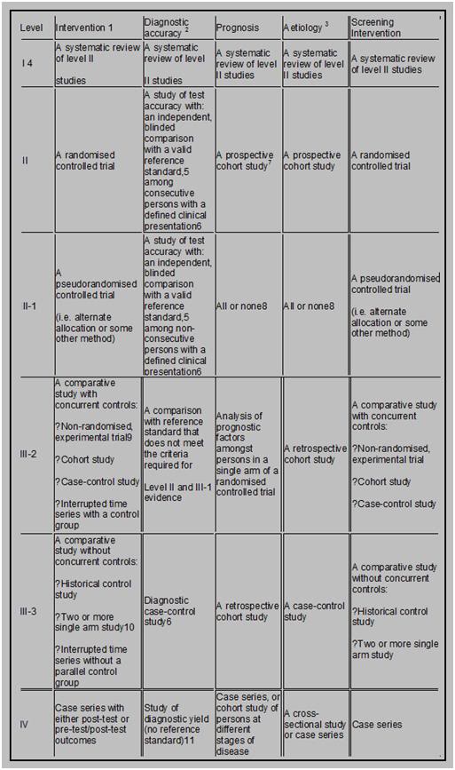
There’s a proven technique in animals for reaching the maximum lifespan; the longest time that animals of a given species have the inherent capacity to survive. It’s called calorie restriction, and there is a large body of animal data in an impressive range of species that says it works. There is even an ongoing project being conducted by the National Institutes of Aging (NIA) to evaluate calorie restriction in primates and it seems to be working in them, too. The calorie restricted Rhesus macaque monkeys have lower death rates, lower rates of age-associated degenerative diseases, and their overall health and activity level are dramatically better than is the case for the control animals, who are fed a diet that simulates the ad lib calorie intake by humans in the Developed World.

There’s just one catch, and that is that calorie restriction, to the extent necessary to get the individual to the maximum end of the lifespan envelope is, for most humans, a miserable experience. It is also one fraught with the potential for malnutrition and the development of eating disorders, such as anorexia nervosa and bulimia. But there’s another problem with calorie restriction in humans, and that’s that the end results are that you end up a blind, debilitated old crone or codger, and then you die.

Having said that, I don’t want to minimize or dismiss the probable very real advantages of calorie restriction in humans and they are that there is likely to be, on average, a 15-25 year extension of the healthy and reasonably productive lifespan, with a large decrease in most of quality of life eroding (and costly) degenerative diseases, such as diabetes, cardiovascular disease, osteoarthritis, dementia and very likely, tooth decay and gum disease. That’s impressive, even if it isn’t very practical for most people without some kind of pharmacological assistance.

There is also ongoing research to discover drugs that mimic the effects of calorie restriction on gene expression so that the benefits of the technique can be had without the attendant suffering and the very real risks of adverse effects on psychology and nutrition.[1] This is a promising area of research, and it will be covered here in considerable detail on an ongoing basis. However, this is not the time to start any discussion of  specific ‘evidence based’ technologies for extending healthy lifespan. Indeed, before we go any further, it is necessary to become familiar with the concept of evidence based medicine (EBM) (Figure 2).

Evidence Based Medicine


Figure 2:  Detailed Diagrammatic representation of the levels of evidence used in Evidence Based Medicine.

Evidence-based medicine (EBM), also called evidence-based practice (EBP) aims to apply the best available evidence gained from the scientific method to clinical decision making. It seeks to assess the strength of evidence of the risks and benefits of treatments, including the lack of treatment, and diagnostic tests.Evidence quality can range from meta-analyses and systematic reviews of double-blind, placebo-controlled clinical trials at the top of the pyramid (above), to conventional wisdom at the bottom.

The discrete types or levels of evidence I will be using in all my discussions here on Chronosphere are those of the Centre for Evidence Based Medicine (CEBM),  as set out in their “’Levels of Evidence’ Document” which is reproduced, below.

1. A summary of how evidence can be graded.

In simple terms, one way of looking at levels of evidence is as follows (the higher the level, the better the quality; the lower, the greater the bias):

or…

  • Category I:  Evidence from at least one properly randomized controlled trial.
  • Category II-1: Evidence from well-designed controlled trials without randomization.
  • Category II-2: Evidence from well-designed cohort or case-control analytic studies, preferably from more than one center or research group.
  • Category II-3: Evidence from multiple times series with or without intervention or dramatic results in uncontrolled experiments such as the results of the introduction of penicillin treatment in the 1940s.
  • Category III: Opinions of respected authorities, based on clinical experience, descriptive studies and case reports, or reports of expert committees.

[Source: Harris, R.P. et al. (2001). Current methods of the U.S. Preventive Services Task Force: a review of the process. American Journal of Preventive Medicine. April 20 (3 Supplement): 21-35.]

Not surprisingly, the place to start in looking for any reliable method(s) of life extension is at the top of the evidence pyramid, which consists of Systematic Reviews (including well designed meta-analyses) and Randomized Double Blind Clinical Studies.

Figure 3: The EBM pyramid made simple.
The majority of “amateur interventive gerontologists,” or active “life extensionists” who are pursuing lifespan extending therapies on themselves are usually both surprised and dismayed when looking at this pyramid (Figure 3). The first reason for this is that either all, or almost all, of the interventions they are using are at the very bottom of the pyramid. The second reason for the shock and dismay (and often disbelief) is that animal and in in vitro research rank below the ideas, editorializing and opinions of medical professionals, instead of at the top of the pyramid, where most activist life extensionists typically feel they should belong.

However, the fact is that very little animal, or in vitro research has any direct clinical applicability to humans.[2-4] This is not because government regulations or “greedy” pharmaceutical companies don’t want people to benefit from disease-curing or life extending drugs, but rather, because the vast majority of that research is either bad (junk) science or it fails to translate to humans.[4-10] Even when animal studies are well designed and carried out in relevant animal models of disease and show strongly positive results, mostly these findings fail to translate to humans. There are many reasons for this, but chief amongst them is that animals, despite their high ‘percentage’ of genetic overlap with humans, are really biochemically sufficiently different that the findings aren’t applicable to humans. The public are bombarded with numbers, such as chimpanzees are 96% to 98% genetically homologous with humans; cats: 90%, dogs: 82%, cows: 80%, rats: 69% and mice: 67% (http://www.ncbi.nlm.nih.gov/homologene).  These numbers get even more impressive when it is noted that 75% of mouse genes have equivalents in humans and 90% of the mouse genome can be matched to a comparable region on the human genome. In fact, recent research indicates that ~ 99% of mouse genes turn out to have analogs in humans.

Figure 4: If we were mice, most cancers would be treatable or cured, there would be effective drugs for stroke and cerebral ischemia, and a wide range of other conditions would have effective therapies. However, we are not mice.

So what’s the problem with the applicability of animal research to humans? Well, consider that at in 3 out of ten patients the drug prescribed for them fails to work. It’s not that the patient is non-compliant or just doesn’t get better; it is that the drug failed to have the anticipated therapeutic effect. Thus, in those patients, the drug was a waste of money and time; and to the extent that it may have adverse effects, a real danger. In fact, there are 2.2 million serious cases of adverse drug reactions (ADRs) and over 100,000 deaths each year in the US. That makes (ADRs) one of the leading causes of hospitalization and death in the US! Most people take ADRs and lack of therapeutic effect in the drugs they are prescribed (or purchase over the counter (OTC)) for granted. “Oh, that doesn’t work for me,” or “I can’t take that because…” are commonplace remarks. And they apply to people who use only “natural” or herbal remedies as much or more as they do those who use “synthetic” drugs.

The reason for these phenomena is very instructive about why animal research turns out to have so little applicability to humans. The cause of the huge variation in responsiveness to drugs in humans is genetic variation between individuals; even identical twins are not genetically identical, due to mutations and to variations in gene activation (epigenetic factors).[11] There are two types of genetic variations known to impact drug metabolism; copy number variation, which results from deletions, inversions, insertions and duplications in genes, and nucleotide variations, or single nucleotide polymorphisms (SNPs). It is estimated that approximately 0.4% of the genomes of unrelated people typically differ with respect to gene copy number. The nucleotide diversity (SNPs) between humans is about 0.1%, which is 1 difference per 1,000 DNA base pairs! Combine these two numbers and human genetic variation is estimated to be at least 0.5% or, if you prefer, 99.5% similarity between individuals.[11-13]

That seemingly trivial amount of genetic variation is responsible for the observed and well documented large disparity in response to therapeutic drugs observed within the human species. Even the SNPs (pronounced “snips”) have a profound effect on the response (or lack thereof) to therapeutic drugs and they are the sources of a major research effort to develop a catalog of SNPs that can be used as a diagnostic tool to predict and individual person’s drug response. This rapidly developing area of research is called pharmacogenomics and it has already seen clinical application in cancer chemotherapy, anticoagulant dosing, the treatment of Hepatitis C and psoriasis, and in seizure disorders.[14-20]

New Drug Development: May I Suggest Roulette, Instead?

A few more words need to be said about the drug development research and success. Leaving animal data aside, most human clinical trials to evaluate refinements of existing (and proven) drugs or therapies either fail, or result in active harm.[2, 21-23] The chances of a novel molecule making it from in vitro or animal testing to clinical use in humans are ~ 1,000 to 1. You’d be much, much better off playing straight-up roulette, where the odds against you are only 37 to 1. Even in studies or clinical trials where there are ample existing theory and prior in vitro, animal research and clinical trials data that were positive and point compellingly to a favorable outcome, trials often fail.

A good example of this with direct relevance to life extensionists is the saga of vitamin E in the treatment and prevention of atherosclerosis, and in particular, coronary artery disease.There are many animal experiments showing that vitamin E reduces or inhibits the development of atherosclerosis. Epidemiological studies in humans provided robust support to these data, since consumption of vitamin E in the diet was inversely associated with mortality from cardiovascular disease.[24, 25] And to the theoreticians and mechanists, there was the perhaps even more compelling fact that the free radical biochemistry implicated as being a primary factor underlying atherogenesis (oxidized low density lipoprotein (LDL)) is favorably impacted by the addition of vitamin E and similar chain breaking antioxidants to the diet in supraphysiological amounts.[26] The free radical theory of aging also supports the idea that vitamin E and other antioxidant molecules might reduce the incidence of degenerative disease, and perhaps retard aging. Further, in accordance with both theory and the animal data, administration of vitamin E to human volunteers reduced the level of lipid peroxidation, and in particular reduced the level of oxidized LDL.[27]
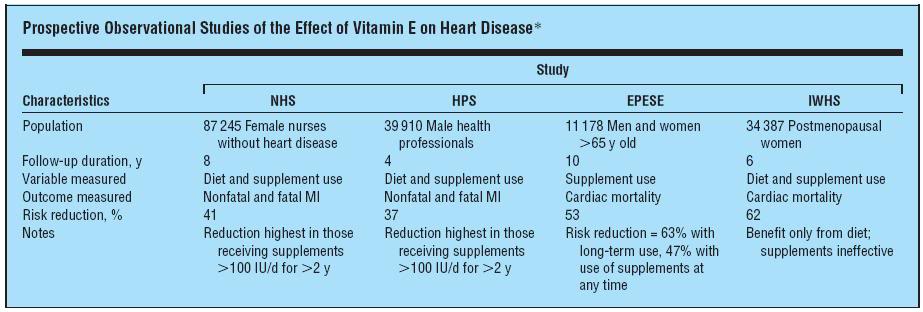
Figure 5:*NHS indicates Nurses’ Health Study; HPS, Health Professionals’ Follow-up Study; EPESE, Established Populations for Epidemiologic Studies of the Elderly; IWHS, Iowa Women’s Health Study; MI, myocardial infarction; and ellipses, none.
Several prospective studies in which vitamin E was given as a supplement, including the US Nurses’ Health Study[28] and the US Health Professionals’ Follow-up Study, found a 34% and 39% reduction (respectively), in the incidence of myocardial infarction, [29] More impressively still, the  Iowa Women’s Health Study found a 47% reduction in cardiac mortality.[30] These were not small studies published in obscure journals. They were very large trials (Figure 5) and they were published in the New England Journal of Medicine. So what’s the problem? The problem was that other researchers could not duplicate the results and so subsequent, carefully designed trials were conducted.

The largest and best designed of these was the a randomized, placebo-controlled Medical Research Council/British Heart Foundation (MRC/BHF) Heart Protection Study in which antioxidant vitamin supplementation was examined in 20,536 individuals with coronary disease, other occlusive arterial disease, or diabetes mellitus. The study participants were randomized to receive vitamin E (600 mg), vitamin C (250 mg), and beta carotene (20 mg) daily or matching placebo. Intention-to-treat comparisons of outcome were conducted among all participants. An advantage to this study was that critics of earlier failed trials pointed out that vitamin E can act as a pro-oxidant in the absence of vitamin C and that it has in vitro pro-oxidant activity in cell membrane lipids under some conditions. In vivo, vitamin C is the molecule which disposes of the water soluble radical species that can be generated by vitamin E and beta carotene was added to scavenge lipid soluble radicals.

The MRC/BHF study found no significant differences between the vitamin and placebo groups in all-cause mortality, or in deaths caused by vascular or nonvascular conditions. Nor were there any significant differences between groups in the incidence of nonfatal myocardial infarction or sudden cardiac death, nonfatal or fatal stroke, or coronary or non-coronary re-vascularization. In fact, the study found that the use of antioxidant vitamins did not produce any significant reductions in 5-year mortality from, or incidence of, any type of vascular disease, cancer, or other major outcome, compared with placebo.[31]

Other studies also showed no benefit [32-34] and there was even some suggestion of harm in the form of an apparent increase in mortality and morbidity from gastrointestinal and intracranial bleeding. In 2009, a metanalysis of vitamin E supplementation trials by Dotan, et al., using Markov model analysis showed that the vitamin E supplemented “virtual cohort” had 0.30 decrease in their quality-adjusted life year (QALY) (95%CI 0.21 to 0.39) compared to the non-treated “virtual cohort.”[35] QALY is a statistical measurement tool used to evaluate not just death or discrete injurious events, such as heart attack or hemorrhagic stroke, but rather measure these events, along with all deaths or debilities as a single entity, and report them in terms of how much loss or gain of functional life occurs in a given group. This work supports an earlier metanalysis showing increased all-cause mortality associated with vitamin E doses ~500 mg/day or more. When a metanalysis was done to look specifically for the effects of vitamin E on stroke it was found that vitamin E increased the risk for hemorrhagic stroke by 22% and reduced the risk of ischemic stroke by 10%.[1]

The metanalysis indicating that vitamin E supplementation (≥500 mg q.d.) is associated with an increase in morbidity and mortality is consistent with the known effect of vitamin E in such doses on bleeding time. Supraphysiologic vitamin E antagonizes vitamin K and causes platelet dysfunction resulting in an increased prothrombin time. It is almost axiomatic in medicine that any increase in bleeding time (anticoagulation) is associated with an increased incidence of clinically significant gastrointestinal (GI) and intracranial bleeding. For vitamin E to show benefit, it would be necessary for any increase in adverse effects to be offset by the benefits it conferred. For vitamin E, this was not the case, whereas for aspirin, which also increases bleeding time and causes an increased incidence of GI and intracranial bleeding, shows such strong benefit in the reduction of myocardial infarction that it is worth the associated risk in the appropriate patient population (i.e., those 50 or over and those with known cardiovascular disease).

This kind of “reversal of fortune” happens over and over again in medicine with respect to drugs as as to other treatment interventions and it is one of the well justified reasons why the astute clinician is very skeptical about putative therapies to treat disease that have not been scientifically vetted – preferably repeatedly, internationally and in well designed and executed trials. It is thus an unfortunate reality that no matter how compelling a therapy seems theoretically or in the laboratory, it still must be proven clinically. And it is even truer that the overwhelming majority of putative therapeutic interventions either fail to work, or injure or kill the patient. There is absolutely no reason to think that this will not be the case with putative life extension drugs.

It is also usually the case that taking multiple drugs, or polypharmacy as it is formally known, negatively shifts the risk to benefit ratio (especially in the ill the debilitated or the elderly). This is so because the biochemistry of living systems is not only enormously complex; it is also interdependent and self regulating. The vast majority of drugs, or supraphysiological doses of nutrients, will perturb multiple biochemical pathways and the more molecules administered, the more likely it becomes resulting adverse interactions will occur. It is, as the Taoist maxim cautions, virtually impossible “to do just one thing” when dealing with a complex and dynamic system. Alter one part of the system in a desirable way and there will likely be consequences in other parts of it – and the odds are high that they will not be favorable.[2]
Thus, in making decisions about which putative life extension therapies to use, the most rational course is to start with those where there is Level-1 evidence of benefit. That may not even seem possible, since there are no known lifespan extending drugs or treatments in humans, let alone ones that have undergone extensive, well designed and repeated clinical trials. Or are there? The answer to that question will be the subject of the next article in this series.

Footnotes


[1] While the 10% reduction in the risk of ischemic stroke may seem promising this is really constitutes no advantage since hemorrhagic strokes have a far higher mortality and morbidity rate than is the case for ischemic stroke.

[2] Like mutations, most arbitrary alterations to the biochemistry or gene expression of a complex living system are not likely to result in benefit.

 References

1.            Ingram D, Zhu, M, Mamczarz, J, Zou, S, Lane, MA, Roth, GS, deCabo, R.: Calorie restriction mimetics: an emerging research field. Aging Cell 2006, 5(2):97-108.

2.            Ikonomidou C, Turski L: Why did NMDA receptor antagonists fail clinical trials for stroke and traumatic brain injury? Lancet Neurol 2002, 1(6):383-386.

3.            Ozdemir FN, Akcay A, Elsurer R, Sezer S, Arat Z, Haberal M: Interdialytic weight gain is less with the Mediterranean type of diet in hemodialysis patients. J Ren Nutr 2005, 15(4):371-376.

4.            Whiteside GT, Adedoyin A, Leventhal L: Predictive validity of animal pain models? A comparison of the pharmacokinetic-pharmacodynamic relationship for pain drugs in rats and humans. Neuropharmacology 2008, 54(5):767-775.

5.            Harber LC, Armstrong RB, Ichikawa H: Current status of predictive animal models for drug photoallergy and their correlation with drug photoallergy in humans. J Natl Cancer Inst 1982, 69(1):237-244.

6.            Olson H, Betton G, Robinson D, Thomas K, Monro A, Kolaja G, Lilly P, Sanders J, Sipes G, Bracken W et al: Concordance of the toxicity of pharmaceuticals in humans and in animals. Regul Toxicol Pharmacol 2000, 32(1):56-67.

7.            Pound P, Ebrahim, S, Sandercock, P, Bracken, MB, et al.: Where is the evidence that animal research benefits humans?: http://www.ncbi.nlm.nih.gov/pmc/articles/PMC351856/pdf/bmj32800514.pdf. BMJ 2004, 328:514-517.

8.            Dixit R, Boelsterli UA: Healthy animals and animal models of human disease(s) in safety assessment of human pharmaceuticals, including therapeutic antibodies. Drug Discov Today 2007, 12(7-8):336-342.

9.            Caldwell J: Problems and opportunities in toxicity testing arising from species differences in xenobiotic metabolism. Toxicol Lett 1992, 64-65 Spec No:651-659.

10.          Wilbourn J, Haroun L, Heseltine E, Kaldor J, Partensky C, Vainio H: Response of experimental animals to human carcinogens: an analysis based upon the IARC Monographs programme. Carcinogenesis 1986, 7(11):1853-1863.

11.          Bruder CE, Piotrowski A, Gijsbers AA, Andersson R, Erickson S, Diaz de Stahl T, Menzel U, Sandgren J, von Tell D, Poplawski A et al: Phenotypically concordant and discordant monozygotic twins display different DNA copy-number-variation profiles. Am J Hum Genet 2008, 82(3):763-771.

12.          Sachidanandam R, Weissman D, Schmidt SC, Kakol JM, Stein LD, Marth G, Sherry S, Mullikin JC, Mortimore BJ, Willey DL et al: A map of human genome sequence variation containing 1.42 million single nucleotide polymorphisms. Nature 2001, 409(6822):928-933.

13.          Cargill M, Altshuler D, Ireland J, Sklar P, Ardlie K, Patil N, Shaw N, Lane CR, Lim EP, Kalyanaraman N et al: Characterization of single-nucleotide polymorphisms in coding regions of human genes. Nat Genet 1999, 22(3):231-238.

14.          Beaulieu M, de Denus S, Lachaine J: Systematic review of pharmacoeconomic studies of pharmacogenomic tests. Pharmacogenomics, 11(11):1573-1590.

15.          Beery TA, Smith CR: Genetics/genomics advances to influence care for patients with chronic disease. Rehabil Nurs, 36(2):54-59, 88.

16.          Cacabelos R, Hashimoto R, Takeda M: Pharmacogenomics of antipsychotics efficacy for schizophrenia. Psychiatry Clin Neurosci, 65(1):3-19.

17.          Johnson JA, Liggett SB: Cardiovascular pharmacogenomics of adrenergic receptor signaling: clinical implications and future directions. Clin Pharmacol Ther, 89(3):366-378.

18.          Schwab M, Schaeffeler E, Zanger UM, Brauch H, Kroemer HK: [Pharmacogenomics: hype or hope?]. Dtsch Med Wochenschr, 136(10):461-467.

19.          Kamal SM: Hepatitis C virus genotype 4 therapy: progress and challenges. Liver Int, 31 Suppl 1:45-52.

20.          Yoshida S, Sugawara T, Nishio T, Kaneko S: [Personalized medicine for epilepsy based on the pharmacogenomic testing]. Brain Nerve, 63(4):295-299.

21.          Wiendl H, Neuhaus O, Kappos L, Hohlfeld R: [Multiple sclerosis. Current review of failed and discontinued clinical trials of drug treatment]. Nervenarzt 2000, 71(8):597-610.

22.          Corman LC, Davidson RA: Why clinical trials fail: the hidden assumptions of clinical trials. South Med J 1992, 85(2):117-118.

23.          Krum H, Tonkin A: Why do phase III trials of promising heart failure drugs often fail? The contribution of “regression to the truth”. J Card Fail 2003, 9(5):364-367.

24.          Rimm E, Stampler, MJ, Ascherio, A, Giovannuci, E, Willett, GA, Colditz, WC.: Vitamin E consumption and the risk of coronary heart disease in men. N Engl J Med 1993, 328::1450-1455.

25.          Stampfer M, Hennekens, CH, Manson, JE, Colditz, GA, Rosner, B, Willett, WC.: Vitamin E consumption and the risk of coronary disease in women. N Engl J Med 1993, 328:1444-1449.

26.          Stampfer M, Rimm, EB: Epidemiologic evidence for vitamin E in prevention of cardiovascular disease. Am J Clin Nutr 1995, 62:1365S-1369S.

27.          Reaven P, Witztum JL.: Comparison of supplementation of RRR-alpha-tocopherol and racemic alpha- tocopherol in humans. Effects on lipid levels and lipoprotein susceptibility to oxidation. Arteriosclerosis, Thrombosis, and Vascular Biology 1993, 13:601-608.

28.          Stampfer M, Hennekens, CH, Manson, JE, Colditz, GA, Rosner, B, Willett, WC.: A prospective study of vitamin E consumption and risk of coronary disease in women. N Engl J Med 1993, 328:1444-1449.

29.          Rimm E, Stampfer, MJ, Ascherio, A, Giovannucci, E, Colditz, GA, Willett, WC.: Vitamin E supplementation and the risk of coronary heart disease among men. N Engl J Med 1993, 328:1450-1456.

30.          Kushi L, Folsom, AR, Prineas, RJ, Mink, PJ,Wu,Y, Bostick, RM.: Dietary antioxidant vitamins and death from coronary heart disease in postmenopausal women. N Engl J Med 1996, 334:1156-1162.

31.          Aizawa K, Shoemaker JK, Overend TJ, Petrella RJ: Metabolic syndrome, endothelial function and lifestyle modification. Diab Vasc Dis Res 2009, 6(3):181-189.

32.          Yusuf S, Dagenais, G, Pogue, J, Bosch, J, Sleight, P.: Vitamin E supplementation and cardiovascular events in high-risk patients. The Heart Outcomes Prevention Evaluation Study Investigators:  http://content.nejm.org/cgi/content/full/342/3/154. N Engl J Med 2000;, 342(3):154-160.

33.          Lonn E, Bosch, J, Yusuf, S, Sheridan, P, Pogue, J, Arnold, JM, et al.: HOPE and HOPE-TOO Trial Investigators. Effects of long-term vitamin E supplementation on cardiovascular events and cancer: a randomized controlled trial. JAMA 2005;, 293(11):1338 -1347.

34.          Vivekananthan D, Penn, MS, Sapp, SK, Hsu, A, Topol, EJ.: Use of antioxidant vitamins for the prevention of cardiovascular disease: meta-analysis of randomized trials. Lancet 2003, 1:2017 -2023.

35.          Dotan YP, I. Lichtenberg, D.  Leshno, M.: Decision analysis Supports the paradigm that Indiscriminate supplementation of vitamin E does more harm than good. Arterioscler Thromb Vasc Biol 2009, 29:1304-1309.


Posted in Gerontology | 17 Comments

The Kurzwild Man in the Night

Ray Kurzweil with a portrait of his father.
It’s as if you took a lot of very good food and some dog excrement and blended it all up so that you can’t possibly figure out what’s good or bad. It’s an intimate mixture of rubbish and good ideas, and it’s very hard to disentangle the two, because these are smart people; they’re not stupid.”

– Douglas Hofstadter, author of Gödel, Escher, Bach, on the books of Ray Kurzweil and Hans Moravec. [See Ross, Greg. "An interview with Douglas R. Hofstadter." American Scientist. Retrieved 2011-08-10.]

It is not very often that I see something that simultaneously evokes sympathy, anger and pity. I am a regular viewer of ABC’s “Nightline program which airs beginning at 2330 in most of the US. It’s part of my ‘wind-down ritual’ at the end of the day. Often, I’m reading, or otherwise engaged while the bits and bytes comprising the program make their way from geosynchronous orbit and chatter out of the television. The introduction to the 09 August program caught my attention, because it was to feature Ray Kurzweil, talking about practical immortality. Of course, I know who Kurzweil is – both of them. There is the maverick Edisonian inventor who brought us the Kurzweil Reader (and thus the CCD flatbed scanner and the text-to-speech synthesizer) and the Kurzweil who transformed digital musical instrumentation with his Kurzweil K250 music synthesizer. And then, well then there is the Ray Kurzweil who brought us the idea of the Singularity, and three books that expound scientifically bankrupt ideas for ‘do it yourself’ interventive gerontology: The 10% Solution for a Healthy Life Fantastic Voyage: Live Long Enough to Live Forever, TRANSCEND: Nine Steps to Living Well Forever.

And last, but by no means least, there is the Ray Kurzweil who made one of the creepiest movies I’ve ever seen, “The Singularity is Near,” which I viewed as a rough cut in a private screening in Europe. That film was the near perfect combination of suggested transgendered autoerotic pedophilia with narcissism of cosmic proportions. I watched it, immobilized as one is when witnessing a public beheading, or the torture of small animals in an Egyptian souk. I was immobilized in a way that only disbelief and shock immobilize you. An extended trailer of his latest documentary, Transcendent Man is available here: http://transcendentman.com/

The “Nightline” segment on Kurzweil opened as follows:

“Ray Kurzweil, a prominent inventor and “futurist” who has long predicted that mind and machine will one day merge, has been making arrangements to talk to his dead father through the help of a computer.

“I will be able to talk to this re-creation,” he explained. “Ultimately, it will be so realistic it will be like talking to my father.”

Kurzweil’s father, an orchestra conductor, has been gone for more than 40 years.
However, the 63-year-old inventor has been gathering boxes of letters, documents and photos in his Newton, Mass., home with the hopes of one day being able to create an avatar, or a virtual computer replica, of his late father. The avatar will be programmed to know everything about Kurzweil’s father’s past, and will think like his father used to, if all goes according to plan.

“You can certainly argue that, philosophically, that is not your father,” Kurzweil said. “That is a replica, but I can actually make a strong case that it would be more like my father than my father would be, were he to live.”
Said to look and sound like Woody Allen’s nerdier younger brother, Kurzweil has been working on predicting the future for decades. At age 17, he was invited to appear on the CBS show “I’ve Got a Secret” to demonstrate how a computer program he invented could compose music.

Kurzweil went on to invent optical scanners, machines that read for the blind and synthesizers. Still inventing today, Kurzweil has developed a reputation for himself from just making predictions, mostly about how fast our technology is advancing.”
The program continued to document Kurzweil’s plan to recreate his father, and he argues that this can be done by using documents, photographs and his own memory of the man. At one point, he even asserts that such an emulation would be “more like my father than my father, had he lived.”

Sympathy? Yes, I felt a great deal of sympathy because I too have lost those I have loved to death, and also suffered, and suffer still, because I lack the power to bring them back to life.

Anger? Yes, a fair bit of anger because what Kurzweil is proposing insults the intelligence of anyone who has even the sketchiest conception of what it is to be human. The idea that a person can be inferred from boxes of paper documents and photographs with technology, extant or foreseeable, let alone in Kurzweil’s possession now, is ludicrous. That Kurzweil’s insight into the nature of personhood, including his own, is so shallow and uni-dimensional goes a long way towards explaining the cluelessness with which he is pursuing his social engineering campaign to make radical life extension, cryonics and uploading socially acceptable.

The “Nightline” program was surprisingly respectful and matter of fact. Kurzwel has superb public relations people and the “Nightline” editors were amply stocked with photos, film clips and in short, a very impressive visual montage to accompany Kurzweil’s modest proposal for resurrection of the dead from letters, news clippings, old photos and presumably rent receipts and cancelled checks documenting visits to the dentist or the haberdasher.

But as even most of the most unreflective and superficial dullards understand, if only emotionally, a person is not and cannot be reconstructed from the empty wrappers of a life long ended. A few bars of melody, a scent, a fragment of a recorded voice, the taste of something long forgotten, all of these can, and do from time to time evoke in reflective and self aware people, streams of memories, and with those memories countless connections, relationships, thoughts sounds, sensations and yes, and very importantly, feelings. One of the things I found so appalling and so narcissistically selfish about the Kurzweil interview is that he is not really interested in having his father live again, rather he is only interested in having his personal experience of his father available for his self-gratification again. It doesn’t matter what his father thinks or feels, it only matters that the Avatar Father makes Kurzweil think and feel that he has been returned to life. The equation of an avatar of the person with the person himself is an utterly repellant thing, because at its root it is the penultimate in dehumanization; and I think that on some level Kurzweil must know this, since he is trying to persuade the rubes that it really is resurrection.

Consider this justifiably oft quoted sentence from Proust’s Remembrance of Things Past:

“And as soon as I had recognized the taste of the piece of madeleine soaked in her decoction of lime-blossom which my aunt used to give me (although I did not yet know and must long postpone the discovery of why this memory made me so happy) immediately the old grey house upon the street, where her room was, rose up like a stage set to attach itself to the little pavilion opening on to the garden which had been built out behind it for my parents (the isolated segment which until that moment had been all that I could see); and with the house the town, from morning to night and in all weathers, the Square where I used to be sent before lunch, the streets along which I used to run errands, the country roads we took when it was fine.”

That is the merest sampling of what a person is. And as beautiful and evocative of the complex tangle of memory, sensation, reaction and the recursion of all those things as that passage is, even a hundred million, or a billion like it would not describe the mind of the dullest human being who moves amongst us.

Actress Marilu Henner was featured on 60 Minutes because it’s a day she’ll never forget — just like every day in her life; pas, present and future.
If you still have any doubts about the staggering volume of information, not to mention the unique wetware on which it is processed, that comprises the human mind, consider the recent scientific verification that people exist who have “superior autobiographical memory,” or hyperthymesia.[1-3] These individuals have essentially complete audiovisual recall of almost every waking moment of their lives. They can “run the movie” of their life experience forward or backward in their head and extract information from what they “re-experience.” As actress Marilu Hennner, one of those identified with this trait remarked on the CBS documentary program “60 Minutes”:”It’s like putting in a DVD and it queues up to a certain place. I’m there again, so I’m looking out from my eyes and seeing things visually as I would have that day.” These are otherwise normal individuals who have no profound cuts in normal cognitive function which might be used to explain the extraordinary storage of such memory minutiae. The “60 Minutes” segment on hyperthymesia is compelling viewing, and it is available on line: http://www.cbsnews.com/video/watch/?id=7166313n

Given the flashes of such recall most of us experience momentarily and erratically in our lives, this phenomenon begs the question: are all of us recording and storing such a broad bandwidth of information? Is it that we are not storing it, or that we cannot, and for good reason, access it with such fidelity at will? The individuals who possess this capability all describe it as burdensome and at times traumatic – memories come unbidden, constantly triggered by cues in the everyday world around them. And with some of those memories come searing emotions. If we need an evolutionary reason for the stoppering-up of such a prodigious memory in dark, amber bottles, to be dispensed only in needful draughts, these people are living examples.

Kurzweil seems to be suffering from an all too common syndrome in highly successful mavericks who have a history of repeatedly proving the experts (as well as their critics) wrong. This course through life is much the same as fame – especially if it brings fortune with it, and thus the ability to surround oneself with people who either share your worldview, or who will (or actually do) agree with any idea or obsession that takes charge. Removed from the tempering focus that reality affords most people, it becomes easy to slip into a world where the line between your dreams and desires, and what is really possible, becomes blurred and then disappears altogether. Kurzweil appears to be well on his way there, if he hasn’t reach that final destination already, and that, well, that is just pitiful.

Many of Kurzweil’s ideas are crazy – a mixture of wishful thinking, inappropriate application of animal data to humans, and in the case of his resurrection scheme, poisonous and dangerous to cryonics on at least two levels. First, it is wrong – people are not scraps of paper, or even whole heaps of them. That is a demeaning idea at best, and a dangerous one at worst, if it is taken seriously. Second, while Kurzweil still commands respect, at some point the men in the media with the butterfly nets will come calling. Kurzweil’s anti-aging program is much more likely to shorten his lifespan and deplete the pocketbook of the average person, upon whom he urges its use, than it is to provide any medical benefit.

This kind of disconnected, narcissistic spiral carried out privately is a thing that evokes pity, and even shame in seeing it. Those of us who have been involved in life extension for 20, 30, or 40 years have seen it before; increasingly desperate and delusion belief that barely suggestively beneficial molecules in animal studies will confer decades of added life, and finally, the decline into frailty and death. As I watched the “Nightline” program, I realized that there is yet another advantage to cryonics that I had not previously considered, and that is the extraordinary dignity and courage with which most cryonicists confront the end of this life cycle. While many were ridiculed for their lack of realism for a lifetime, most were men and women who did what they reasonably could to live as long as possible now, made no exaggerated or unreasonable claims about cryonics – and in fact, regarded it and represented it as what it currently is – a long shot experimental procedure that may well not work, but for them was infinitely better than the alternative.

The extraordinarily accurate, generally matter of fact, and with few exceptions dignified coverage of Bob Ettinger’s passing into cryopreservation is an example. It’s a worthy example and the way we should strive to be seen. Kurzweil reportedly has cryonics arrangements with Alcor. I’m glad to hear that, because I think he is a fundamentally a very good and very decent man who shares our core values. He has improved and enriched the lives of countless people through his scientific and technological innovations. However, as I can tell you from experience, while many disabilities are now tolerated in our society, crazy and creepy are not amongst them.

References

1.            Cahill L, McGaugh JL: Modulation of memory storage. Curr Opin Neurobiol 1996, 6(2):237-242.

2.            Cahill L, McGaugh JL: A novel demonstration of enhanced memory associated with emotional arousal. Conscious Cogn 1995, 4(4):410-421.

3.            Parker ES, Cahill L, McGaugh JL: A case of unusual autobiographical remembering. Neurocase 2006, 12(1):35-49.


Posted in Cryonics Philosophy, Culture & Propaganda, Gerontology | 35 Comments