IV. EFFECTS OF CRYOPRESERVATION ON THE HISTOLOGY OF SELECTED TISSUES (Left Ventricle and Cerebral Cortex)
Left Ventricle
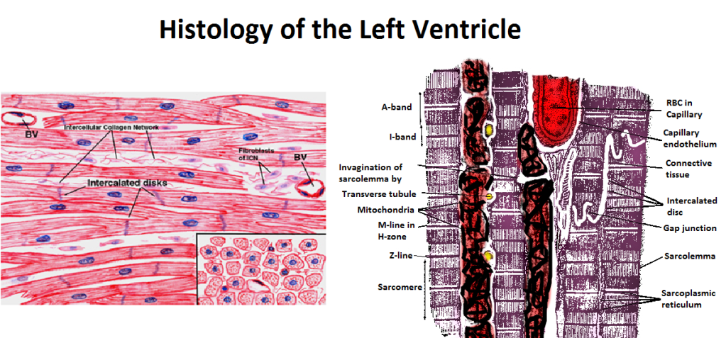 Figure 43: The myofibrils of each cardiac muscle cell are branched and contain a single nucleus. The branches interlock with those of adjacent fibers by adherens junctions which act to prevent scission of the cardiomycytes during the high-shear, forceful contractions of the heart. The muscle is richly supplied with mitochondria which are largely confined to the spaces between the fibrils. The fibrils are covered with a membrane, the Sarcolemma, which is frequently invaginated to form the Transverse tubules. These invaginations of the plasma membrane or sarcolemma, are called transverse tubules and they reach deep into the myofibrils and bring the action potential deep into the fibers. Specialized intercellular junctions, the Intercalated discs, facilitate rapid transmission of the electrical signals which initiate myocyte contraction. The myofibrils are formed by myosin and actin fibers aligned in a distinct pattern which is visible under light microscopy as the A-, H- and I- bands.
Yajima stain was used to prepare the Control (Figure 44), FGP and FIG cardiac tissue for light microscopy. The FGP cardiac muscle showed increased interstitial space, probably indicative of interstitial edema. In many areas the sarcolemma appeared to be separated from the cytoplasm of the myocyte and, occasionally, appeared to have disintegrated into debris in the interstitial spaces (Figure 45). The myofilaments appeared maximally relaxed with widened I-bands . The mitochondria were grossly swollen and contained numerous amorphous matrix densities. The sarcolemma was fragmented beneath an intact basement membrane and there was increased space between the capillary endothelium/basement membrane and intact areas of the sarcolemma of the cardiomyocytes. The cell nuclei were unremarkable.
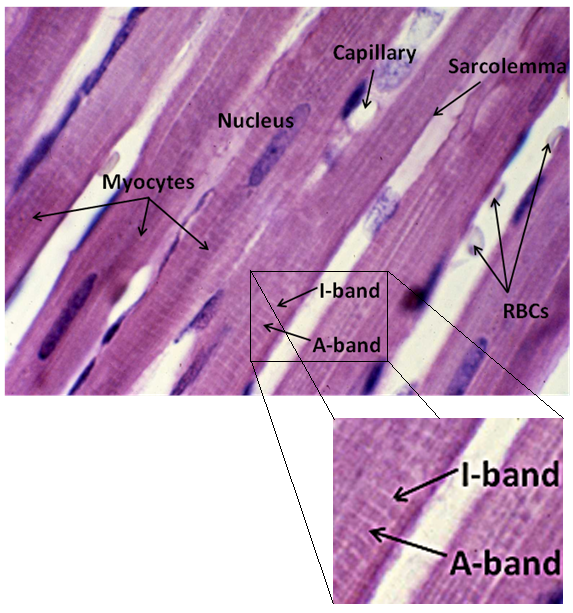
Figure 44: Control-1, Left Ventricle, Yajima, 100x. Control cardiac muscle demonstrated crisp, well defined membranes and the normal density and pattern of myofibril structure. Capillary endothelium appeared intact and the capillary basement membrane was well anchored to adjacent myocytes and appeared intact.
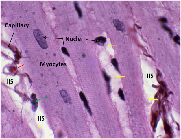
Figure 45: FGP-1 Left Ventricle, Yajima, 100x. In the FGP animals the myocardium exhibited increased interstitial space (IIS) as well as the presence of debris in the IIS which appeared to be disrupted sarcolemma (yellow arrows). The capillary basement membrane was often observed to be separated from the sarcolemma of the adjacent myocytes and endothelial cell nuclei were sometimes observed devoid of plasma membranes or cytoplasm (red arrow).The occasional naked myocyte nucleus could also be observed (green arrow).
The same changes were also present in the FIG group with the added presence of a “ragged” or rough appearance of the myofibrils where they were silhouetted against interstitial space (Figure 46). There also appeared to be holes or spaces, possibly as a result of edema, in the fabric of the myofibrils that were not present in the myocardium of either the control, or the FGP animals.
Most surprising was the general absence of contraction band necrosis in the FIG group, possibly as a consequence of the protective effect of reasonably prompt post-cardiac arrest refrigeration. No microscopic evidence of fracturing, either gross or microscopic, was noted in the myocardium of either the FGP, or the FIG groups.
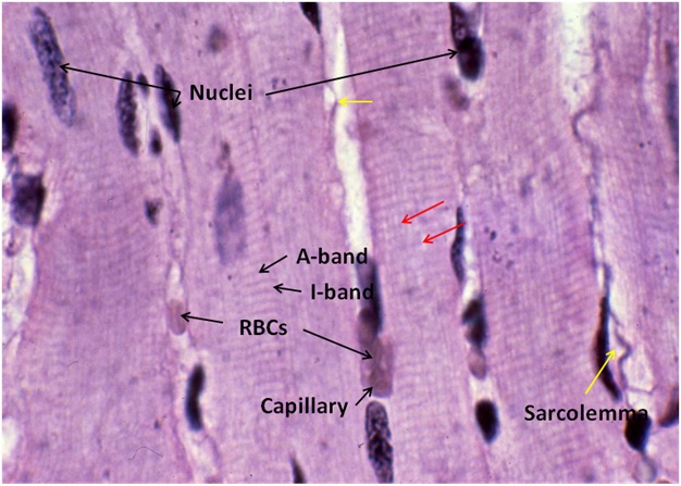
Figure 46: FIG-2 Left Ventricle, Yajima, 100x. Separation and fragmentation of the sarcolemma were observed in the FIG myocardium to a greater extent than that seen the in myocardium from the FGP animals (yellow arrow). Additionally, the fibers of myofibrils had a more ragged appearance and consistently displayed open spaces in the bands which were not seen in the myocardium of either the Control or the FGP animals (red arrows).

Figure 47: The myofibrils of both the FGP and FIG animals appeared maximally relaxed with a marked increase in the thickness of the I-band. Intact red blood cells (RBCs) were observed in the FIG animals and represent incomplete blood washout (red cell trapping) despite perfusion with large volumes of washout, cryoprotectant and fixative solution (~8-10 L) over a time course of ~140 minutes of perfusion.
Cerebral Cortex
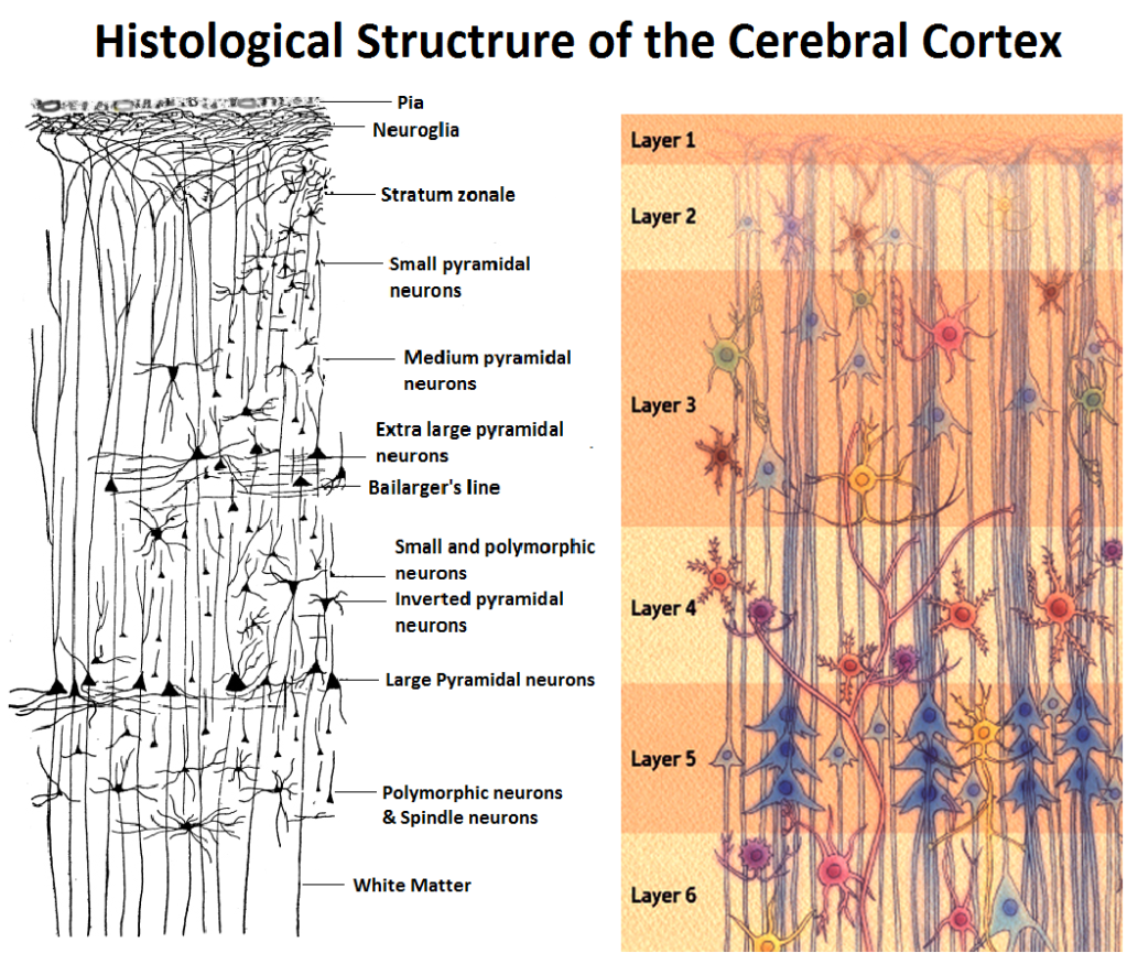 Figure 48: The cerebral cortex consists of six distinct layers, beginning with the first layer, the Molecular Layer (Stratum zonale), which consists of finely branched medullated and non-medullated nerve fibers. The molecular layer is largely devoid of neuronal cell bodies. Those neuronal cell bodies which are present are the cells of Cajal which possess irregular cell bodies and typically have four or five dendrite that terminate within the molecular layer and a long nerve fiber process, or neuraxon, which runs parallel to the surface of the cortical convolutions.
The second layer of the cortex consists of a layer of small Pyramidal cells with the apices of the pyramids being directed towards the surface of the cortex. The apex of the small Pyramidal cells terminates in a dendron, which reaches into the molecular layer, giving off several collateral horizontal branches. The final branches in the molecular layer take a direction parallel to the surface. Smaller dendrites arise from the lateral and basal surfaces of these cells, but do not extend far from the body of the cell. The neuronal axon (neuraxon) always arises from the base of the small Pyramidal cells and passes towards the central white matter, thus forming one of the nerve-fibers of the white matter. In its path, the neuraxon gives off a number of collaterals at right angles, which are distributed to the adjacent grey matter.
The third cortical layer consists of Pyramidal neurons which are characterized by the presence of cells of the same type as those of the preceding layer, but of a larger size. The nerve-fiber process becomes a medullated fiber of the white matter.
The fourth layer is comprised of Polymorphous neurons which are irregular in outline and give off several dendrites which branch into the surrounding grey matter. The neuraxons of the Polymorphous neurons give off a number of collaterals, and then become a nerve-fiber of the central white matter. Scattered through these three layers are the cells of Golgi, whose neuraxon divides immediately with the divisions terminating in the immediate vicinity of the Polymorphous neuron cell-bodies. Some cells are also found in which the neuraxon, instead of extending into the white matter of the brain, passes towards the surface of the cortex; these are called cells of Martinotti.
The fifth cortical layer contains the largest pyramidal neurons which send outputs to the brain stem and spinal cord and comprise the the pyramidal tract. Layer 5 is particularly well-developed in the motor cortex.
Layer 6 consists of pyramidal neurons and neurons with spindle-shaped cell bodies. Most cortical outputs leading to the thalamus originate in layer 6, whereas most outputs to other subcortical nuclei originate in layer 5.
The cortical blood supply is via the pia mater which overlies the cerebral hemispheres.
Bodian stain was used to prepare the control, FGP, and FIG brain tissue samples for light microscopy. Three striking changes were apparent in FGP cerebral cortex histology: 1) marked dehydration of both cells and cell nuclei, 2) the presence of tears or cuts at intervals of 10 to 30 microns throughout the tissue on a variable basis (some areas were spared while others were heavily lesioned), and 3) the increased presence (over Control) of irregular, empty spaces in the neuropil as well as the occasional presence of large peri-capillary spaces (Figures 54,56, and 57). These changes were fairly uniform throughout both the molecular layer and the second layer of the cerebral cortex. Changes in the white matter paralleled those in the cortex, with the notable exception that dehydration appeared to be more pronounced (Figure 55).
Other than the above changes, both gray and white matter histology appeared remarkably intact, and only careful inspection could distinguish it from control (Figures 52, 58, 59 and 60). The neuropil appeared normal (aside from the aforementioned holes and tears) and many long axons and collaterals could be observed traversing the field. Cell membranes appeared crisp, and apart from appearing dehydrated, neuronal architecture appeared comparable to control. Similarly, staining was comparable to that observed in Control cerebral cortex. Cell-to-cell connections appeared largely intact.
The histological appearance of FIG brain differed from that of FGP animals in that ischemic changes such as the presence of pyknotic and fractured nuclei were much in evidence and cavities and tears in the neuropil appeared somewhat more frequently. The white matter of the FIG animals presented a macerated appearance, in addition to exhibiting the rips or tears observed in the white matter of the FGP brains (Figure 61).
Both FGP and FIG brains presented occasional evidence of microscopic fractures.
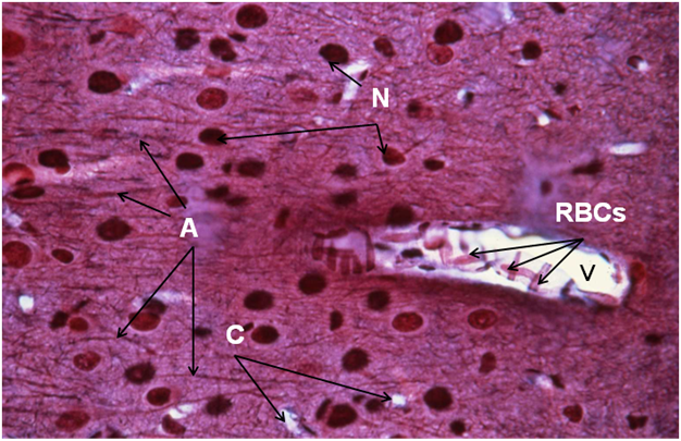
Figure 49: Control-1, 1st (molecular) cell layer, cerebral cortex, Bodian, 40x. Cells of Cajal (N) and a dense weave of axons (A) are visible. The tissue is perforated by numerous capillaries (C) and a small venue containing many red blood cells (RBCs).
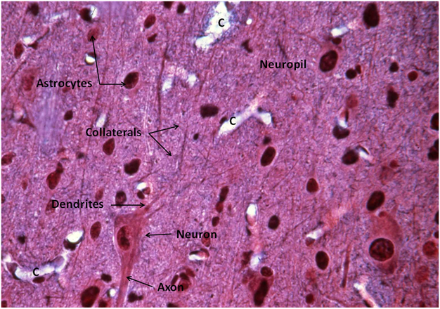
Figure 50: Control-1, 2nd cell layer, cerebral cortex, Bodian, 40x, showing a pyramidal neuron (N, lower left) multiple capillaries (C) and the interwoven connections of dendrites that comprise the neuropil.
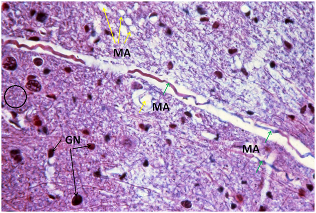
Figure 51: Control-1, white matter, cerebral cortex, Bodian, 40x. Myelinated axons (MA) appear both in cross section (yellow arrows) and laterally (green arrows). Unmyelinated axons are present inside the black circle. Glial cell nuclei (GN) are scattered throughout the tissue.
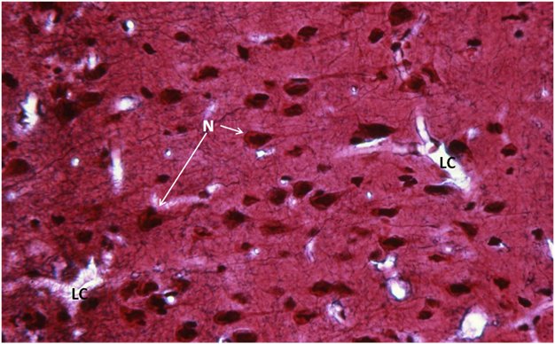
Figure 52: FGP-1, Cerebral Cortex, 1st cell layer, Bodian, 40x. Two large capillaries (LC) are present, one with a red blood cell present (right). Neurons (N, cells of Cajal) are present in normal density and the neuropil appears intact. This section appears indistinguishable from that of the Control animal.
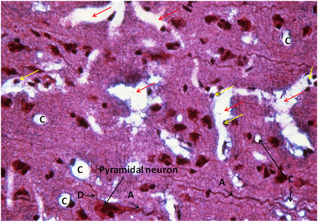
Figure 53: FGP-1, Cerebral Cortex, 2nd cell layer, Bodian, 40x. This area of FGP cerebral cortex shows injury typical of that seen in both FGP and FIG animals. There are a number of large tears in the neuropil (red arrows) approximately 10 to 30 microns across. A pyramidal neuron is present in the lower left of the micrograph and it appears somewhat dehydrated. There are a number of naked glial cell nuclei (yellow arrows), as well some nuclei with what appears to adherent cytoplasm visible at the margins of the tears in the neuropil.
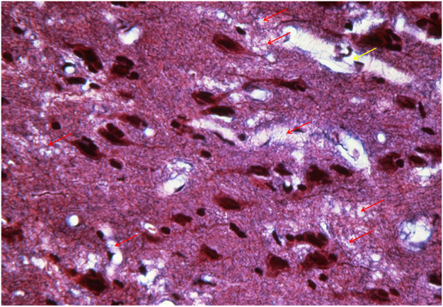
Figure 54: FGP-1, Cerebral Cortex, 2nd cell layer, Bodian, 40x. In this area of the 2nd layer of the cerebral cortex the neuropil presents a somewhat “moth eaten” appearance, with numerous tears and vacuoles in evidence (red arrows). One large tear appears to be a pericapillary ice hole (yellow arrow).
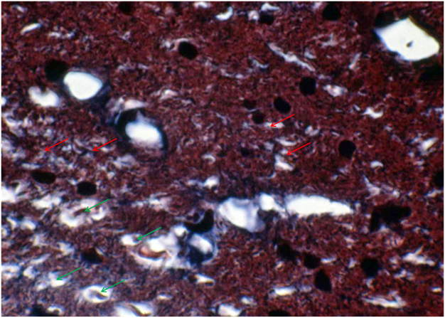
Figure 55: FGP-3, Cerebral Cortex, white matter, Bodian, 100x. There are numerous open spaces in the white matter that appear to be ice holes (red arrows). The density of the tissue appears markedly increased over that of the Control white matter, possibly as a result of glycerol-induced dehydration. This apparent dehydration is also evident in the increased density of the axoplasm seen in the myelinated axons (green arrows).
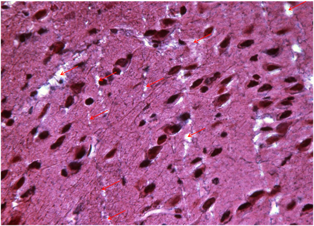
Figure 56: FIG-3, Cerebral Cortex, 1st cell layer, Bodian, 40x. Extraordinarily normal appearing Molecular layer of the FIG cerebral cortex. The neuropil appears intact with the exception of what appear to be scattered tears or ice holes (red arrows).
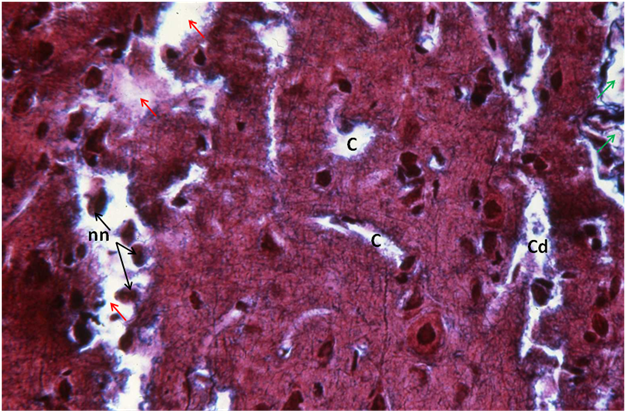
Figure 57: FIG-2, Cerebral Cortex, 1st cell layer, Bodian, 40x. Large tears are evident (red arrows) and naked glial cell nuclei and fragmented cytoplasm are apparent (nn). Several intact capillaries are in evidence (C) as well as what appears to be two capillaries that have been separated from the neuropil and appear largely surrounded by open (pericapillary) space (green arrows). A mass of debris appears to occupy some of the luminal space of what appears to have been a capillary (Cd).
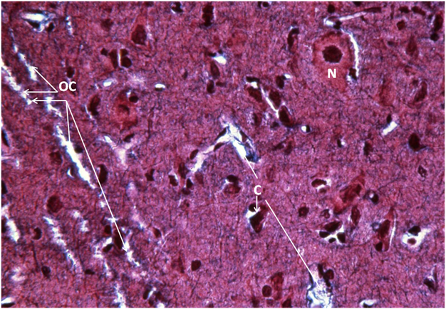
Figure 58: FIG-2, Cerebral Cortex, 2nd cell layer, Bodian, 40x. Remarkably intact neuropil with several capillaries, including several capillaries sectioned oblique to the plane of the tissue (OC). A neuron (N) with what appears to be a crisp plasma membrane is present at the upper right of the micrograph.
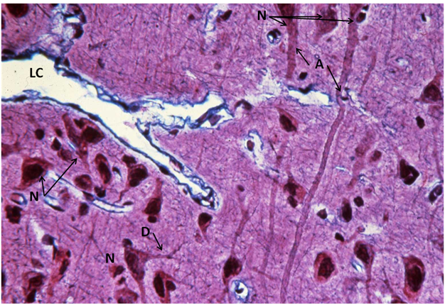
Figure 59: FIG-2, Cerebral Cortex, 2nd cell layer, Bodian, 40x.Normal appearing cerebral cortex in an FIG animal. There are multiple intact neurons with normal appearing dendrites (D) and axons (A). An intact large capillary (LC) is present and appears free of red cells.
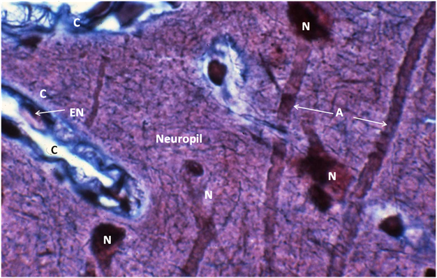
Figure 60: FIG-2, Cerebral Cortex, 2nd cell layer, neuropil, Bodian, 100x. Normal appearing layer 2 of the cerebral cortex with intact neurons (N), axons (A), and neuropil. A capillary (C)with intact endothelial cells and an endothelial cell nucleus (EN) is also visible (left, center).
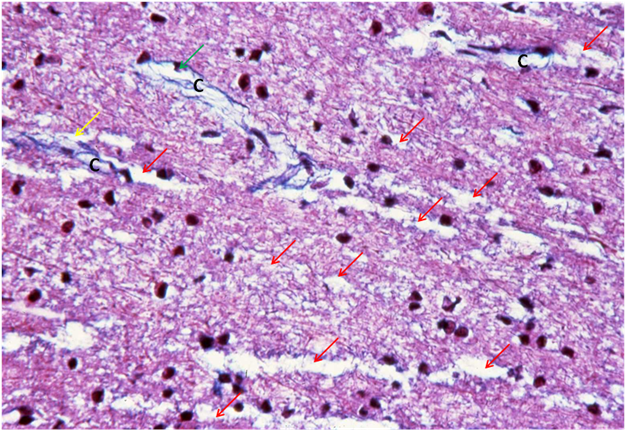
Figure 61: FIG-2, Cerebral Cortex, white matter, Bodian, 40x. Severely injured white matter typical of that seen in FIG animals. The tissue presents a macerated appearance (black circles) with numerous rips and tears, possibly as a result of ice formation (red arrows). The capillaries (C) are separated from the tissue parenchyma (yellow arrow) and what appears to be a naked endothelial cell nuclei projected into the intraluminal space of one capillary (green arrow).
END OF PART 3
